Scleroderma is a chronic autoimmune disease in which antibodies are produced to the body's own cells. The exact causes of the development of the disease have not been fully studied. There are predisposing factors for the occurrence of scleroderma, the presence of which increases the possibility of the formation of foci of sclerosis. Autoimmune disease is classified depending on the degree of disease activity, as well as the location of the pathological focus. Scleroderma can affect the skin and spread to internal organs. Clinical symptoms of the disease are varied. Autoimmune pathology is often mistaken for other diseases due to the similarity of symptoms.
Diagnosis of scleroderma is not difficult. For this purpose, laboratory and instrumental research methods are used. Based on diagnostic data, individual therapy is selected. Currently, scleroderma is considered an incurable disease. However, regular use of medications can minimize the possibility of exacerbation of the disease. In order to control the course of the disease, preventive measures have been developed, including recommendations on lifestyle, work schedule and other indicators. The prognosis for scleroderma depends on the activity of the process. The earlier the pathology can be diagnosed, the more favorable the prognosis for life. In the international classification of diseases ICD-10, scleroderma has code M34.
The Yusupov Hospital deals with all types of diagnosis and treatment of scleroderma. Highly qualified personnel, using modern equipment, quickly identify an autoimmune disease and prescribe the required amount of therapy.
Causes
The exact causes of scleroderma have not yet been determined. Experts identify predisposing factors, the presence of which increases the risk of autoimmune disease. These include:
- Genetic predisposition. The presence of scleroderma in relatives significantly increases the likelihood of developing the disease in future generations. There is a theory according to which a defect in the HLA-9QA1 gene is responsible for the formation of an autoimmune disease. Genome mutations can be congenital or acquired. The following factors contribute to the development of a genetic defect:
- Radiation exposure.
- Prolonged exposure to ultraviolet rays.
- Taking certain medications. For example, cytostatics.
- Viral or bacterial infection.
- Exposure to high and low temperatures.
- Inflammatory process. Biologically active substances produced during the inflammatory reaction can provoke tissue and vascular changes. Vascular permeability increases, and the inflammatory process intensifies. As a result, the prerequisites are created for the formation of chronic inflammation. The risk of scleroderma increases with the addition of other provoking factors.
- Immune disorders. Scleroderma is an autoimmune disease in which the body's own cells are perceived as foreign. Immune reactions are triggered by connective tissue growth factor.
- Infectious diseases. There is no consensus regarding the involvement of infectious agents in the development of scleroderma. Experts do not identify a specific pathogen that provokes autoimmune processes. It was noticed that in some patients with diagnosed scleroderma, cytomegalovirus fragments were detected.
- Environmental factors. Experts highlight a theory according to which there is a relationship between exposure to environmental factors and the occurrence of scleroderma. If there is a genetic predisposition, exposure to environmental factors increases the risk of developing an autoimmune disease. The main provoking factors include:
- Chemical compounds. For example, silicon, mercury, benzene, toluene, epoxy resin.
- Nutritional supplements. Among them are L-tryptophan, mazindol, fenfluramine.
The mechanism of scleroderma occurrence is associated with the development of an immune response against the body's own cells. As a result, an inflammatory process is formed, thinning the skin and blood vessels. Circulating complexes settle on internal organs, causing their damage. Scleroderma is accompanied by the formation of sclerotic changes with the formation of scar tissue.
Causes of systemic scleroderma
The disease systemic scleroderma begins as a result of the appearance of excess collagen in the skin and other tissues of the human body. Collagen is a type of protein that forms the basis of skin and connective tissue.
The reasons for this phenomenon have not been fully established, but many scientists believe that our immune system is “to blame” for this. As a result of an unknown malfunction, it triggers a mechanism that stimulates excessive production of collagen (connective tissue protein).
Risk factors for the development of systemic scleroderma may include:
- gender of the person (women get this pathology 4 times more often than men);
- race (people with dark skin suffer from this disease much more often than white-skinned people);
- external factors that provoke the onset of the disease (hypothermia, exposure to chemicals, certain types of medications, prolonged stress, etc.).
Symptoms
Scleroderma is characterized by damage to various organs and systems. In this regard, it is accompanied by a variety of clinical symptoms. The severity of signs of the disease depends on the degree of activity of the disease and its form. The following symptoms are common to all types of scleroderma:
- Pain syndrome in muscles and joints.
- Weakness, sudden loss of strength.
- Headache, dizziness.
- Shortness of breath and heartburn may occur.
According to the degree of prevalence of the sclerotic process, 2 forms of the disease are distinguished:
- Systemic. Scleroderma is accompanied by the spread of growth of foci of sclerosis in the thickness of the skin, as well as internal organs. In this regard, their damage and functional disorders occur.
- Limited. Sclerotic lesions are located in the skin, rarely in muscles and bones. The process affects blood vessels. Limited scleroderma is classified depending on the shape of the lesion. In accordance with this, they distinguish:
- Plaque scleroderma. The most common type of disease. The foci of sclerosis are small in size. As the disease progresses, they grow. The skin in the area of the pathological focus becomes dense.
- Linear scleroderma. This form of autoimmune disease is localized in the scalp. Linear scleroderma occurs most often in children. The only focus of sclerosis is gradually growing. Outwardly, it resembles a scar.
- Bullous scleroderma. Occurs in combination with another form of the disease. The clinical picture is characterized by the formation of blisters on the skin surface.
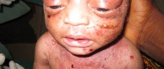
- Limited scleroderma with unilateral progressive facial atrophy. Severe form of the disease. It can occur as an independent pathology or in combination with another type of disease. Foci of sclerosis affect half of the face. In this case, skin atrophy, hair loss, eyelashes, eyebrows, and damage to nerve fibers occur. As a result, the face takes on an asymmetrical appearance. Most often, limited scleroderma with unilateral progressive facial atrophy occurs at a young age.
- White spot disease. Characterized by the formation of small white spots. Most often, this form of scleroderma is localized in the neck, torso, and mucous membranes. A distinctive feature of white spots is a pink edging.
Depending on the location of the sclerosis focus, damage to the following organs and tissues is distinguished:
- Leather. The development of scleroderma of this localization occurs in three stages:
- Edema. Biologically active substances provoke dilation of blood vessels. As a result, the fingers, hands, and feet increase in volume due to pastosity. Itchy skin and a bluish tint may occur. The duration of the edema stage depends on the degree of activity of the process. On average it is several weeks.
- Seal. As a result of the proliferation of connective tissue, normal cells are replaced by pathological ones. Foci of sclerosis are defined as a dense, cold, hard surface.
- Atrophy. The final stage of skin scleroderma. In this case, normal cells are completely replaced by pathological ones, as a result of which the affected area changes color. The skin in this area becomes dry and cracks form when exposed to any traumatic factor. Skin scleroderma is most often localized in the arms, forearms, chest, abdomen, legs, and feet.
- Vessels. Damage to blood vessels in scleroderma is expressed in the form of Raynaud's syndrome. In this case, a spasm of the arteries occurs, causing characteristic symptoms. The development of the syndrome occurs in three phases:
- Paleness of fingers. A provoking factor (for example, cold) causes a spasm of blood vessels. This leads to microcirculatory disorders. As a result, the skin will acquire a pale color, which becomes noticeable by a clear border with the healthy area.
- Turning blue. Fingers acquire a bluish color as a result of the accumulation of hemoglobin. At the same time, a sufficient amount of nutrients and oxygen does not reach the affected area.
- Hyperemia. After the spasm of the blood vessels is eliminated, a sharp release of blood occurs to the affected area. This leads to redness of the fingers. In addition to changes in the color of the fingers, Raynaud's syndrome is accompanied by numbness and a tingling sensation.
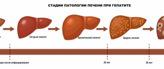
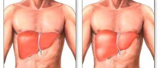
- Liver. The spread of connective tissue with the formation of foci of sclerosis provokes the development of liver cirrhosis. The main symptom of the disease is jaundice. It occurs due to the accumulation of large amounts of bilirubin. Over time, itching of the skin and acholic feces appear.
- Lungs. The most dangerous localization of scleroderma is the lungs. As a result of the formation of pathological foci, sclerosis of the capillaries and alveoli occurs. This provokes gas exchange disorders in the lungs. In addition, pulmonary artery pressure may increase. Similar changes are characteristic of pulmonary hypertension. Clinically this is expressed by the appearance of the following symptoms:
- Dyspnea
- Feeling short of air
- Pain in the chest area and right hypochondrium.
- Pain syndrome. The pain is localized behind the sternum.
- Shortness of breath. The symptom appears during physical activity.
- Feelings of rapid heartbeat.
- Headache, dizziness.
- Heart rhythm disturbances. Arrhythmia appears.
- Arterial hypertension.
- Headache, dizziness.
- Heaviness in the head.
- Unsteady gait.
- Swelling of the lower extremities.
- Brief loss of consciousness.
- Ascites. It is formed as a result of the accumulation of fluid in the abdominal area.
- Pleurisy. The accumulation of fluid in the lung area occurs as a result of intoxication with decay products.
- Endocarditis. Fluid in the heart occurs due to kidney failure.
- Anemia, thrombocytopenia.
- Pain syndrome. The localization of pain may vary. At the same time, it intensifies with movement, and also appears at rest.
- Stiffness of movements. The symptom occurs due to severe pain.
- Amyotrophy.
- Flexion contracture. It is formed as the pathological process progresses.
- Erectile disfunction.
- Frequent urge to urinate. The symptom occurs due to a decrease in the volume of the bladder as a result of sclerosis of its tissues.
- Pain syndrome. Foci of sclerosis cause a decrease in the function of the vaginal glands, which is expressed in vaginal dryness. This causes pain during sexual intercourse.
- Decreased libido.
- Early onset of menopause.
- Increased gas formation, bloating.
- Unstable chair. Diarrhea alternates with constipation.
- Pain syndrome in the abdominal area.
- Nausea, vomiting. In this case, blood may appear in the vomit.
- Stool incontinence.
- Impaired passage of food. Swallowing is difficult due to narrowing of the esophagus.
- Reflux. A reverse reflux of food occurs. Heartburn appears.
The above symptoms are not specific to scleroderma. Similar manifestations occur in other diseases of the gastrointestinal tract. Therefore, it is important to seek medical help if you have the first signs of pathology for timely diagnosis.
To designate the most common lesions in scleroderma, there is CREST syndrome. It includes:
- C – Calcinosis – Calcinosis. Calcinosis is dense subcutaneous formations that are deposits of calcium salts. In some cases, they form ulcers that contain bacterial agents that can cause infection of soft tissues and bones (osteomyelitis).
- R – Raynauld's phenomen – Raynaud's phenomenon. Raynaud's phenomenon occurs due to spasm of peripheral blood vessels and is manifested by impaired blood circulation in the fingers and toes. Characterized by a change in the color of the fingers when exposed to low temperatures.
- E – Esophageal dysmotility – impaired mobility of the esophagus. Due to damage to the smooth muscles of the esophagus, the swallowing process is disrupted, patients experience involuntary regurgitation and heartburn.
- S – Sclerodactyly – sclerodactyly. Sclerodactyly is a pathological condition in which the skin of the fingers is thickened, the subcutaneous tissue is atrophied, and the terminal phalanges are enlarged. Against the background of all these changes, the mobility of the fingers is impaired, and squeezing the hand becomes much more difficult.
- T – Telangiecasia – spider veins. Due to damage to the capillaries and other small blood vessels of the skin, small red-blue dots, similar in shape to stars, appear on the face, upper half of the body and on the mucous membranes of the mouth, eyes and genitals.
| Affected organ | Mechanism of action | Main symptoms |
| Liver | The spread of connective tissue with the formation of foci of sclerosis provokes the development of liver cirrhosis. Jaundice occurs due to the accumulation of large amounts of bilirubin. | 1. Jaundice. 2.Skin itching. 3.Aholic feces. |
| Lungs | As a result of the formation of pathological foci, sclerosis of the capillaries and alveoli occurs. This provokes gas exchange disorders in the lungs. In addition, pulmonary artery pressure may increase. Similar changes are characteristic of pulmonary hypertension. | 1. Shortness of breath 2. Feeling of lack of air 3. Pain in the chest area and in the right hypochondrium. |
| Kidneys | Scleroderma most often affects blood vessels, including the kidneys. Such changes lead to impaired renal function and the development of renal failure. Blood filtration deteriorates, which is accompanied by the accumulation of decay products with the development of intoxication syndrome. Sclerotic kidney damage causes an increase in blood pressure. This sign is considered favorable, as it indicates the normal functioning of the cardiac system. | 1. Arterial hypertension. 2. Headache, dizziness. 3.Heaviness in the head. 4. Unsteady gait. 5.Swelling of the lower extremities. 6.Short-term loss of consciousness. 7.Ascites. It is formed as a result of the accumulation of fluid in the abdominal area. 8. Pleurisy. The accumulation of fluid in the lung area occurs as a result of intoxication with decay products. 9.Endocarditis. Fluid in the heart occurs due to kidney failure. 10.Anemia, thrombocytopenia. |
| Heart | Connective tissue replaces functional tissue. As a result, the function of the heart, which is responsible for pumping blood, is disrupted. This leads to insufficient blood supply to blood vessels, tissues and the heart. Venous stagnation of blood occurs. | 1. Pain syndrome. The pain is localized behind the sternum. 2. Shortness of breath. The symptom appears during physical activity. 3. Feeling of rapid heartbeat. 4.Headache, dizziness. 5. Heart rhythm disturbance. Arrhythmia appears. |
| Bones and muscles | Damage to the musculoskeletal system is considered one of the most common manifestations of scleroderma of internal organs. | 1. Pain syndrome. The localization of pain may vary. At the same time, it intensifies with movement, and also appears at rest. 2. Stiffness of movements. The symptom occurs due to severe pain. 3. Muscle atrophy. 4. Flexion contracture. It is formed as the pathological process progresses. |
| urinary system | The spread of the sclerotic process rarely affects the genitourinary system. As a result of damage to blood vessels, microcirculation in the groin area is disrupted. | 1. Erectile dysfunction. 2.Frequent urge to urinate. The symptom occurs due to a decrease in the volume of the bladder as a result of sclerosis of its tissues. 3. Pain syndrome. Foci of sclerosis cause a decrease in the function of the vaginal glands, which is expressed in vaginal dryness. This causes pain during sexual intercourse. 4.Decreased libido. 5.Early onset of menopause. |
| Gastrointestinal tract | As a result of the proliferation of connective tissue, peristalsis is disrupted. Foci of sclerosis lead to impaired absorption of nutrients. This leads to a sharp loss of body weight and the development of vitamin deficiency. | 1. Increased gas formation, bloating. 2.Unstable stool. Diarrhea alternates with constipation. 3.Pain syndrome in the abdominal area. 4. Nausea, vomiting. In this case, blood may appear in the vomit. 5. Stool incontinence. 6. Impaired passage of food. Swallowing is difficult due to narrowing of the esophagus. 7.Reflux. A reverse reflux of food occurs. Heartburn appears. |
| Form | Characteristic | Photo |
| Plaque scleroderma | The most common type of disease. The foci of sclerosis are small in size. As the disease progresses, they grow. The skin in the area of the pathological focus becomes dense. | |
| Linear scleroderma | This form of autoimmune disease is localized in the scalp. Linear scleroderma occurs most often in children. The only focus of sclerosis is gradually growing. Outwardly, it resembles a scar. | |
| Bullous scleroderma | Occurs in combination with another form of the disease. The clinical picture is characterized by the formation of blisters on the skin surface. | |
| Limited scleroderma with unilateral progressive facial atrophy | Severe form of the disease. It can occur as an independent pathology or in combination with another type of disease. Foci of sclerosis affect half of the face. In this case, skin atrophy, hair loss, eyelashes, eyebrows, and damage to nerve fibers occur. As a result, the face takes on an asymmetrical appearance. Most often, limited scleroderma with unilateral progressive facial atrophy occurs at a young age. | |
| White spot disease | Characterized by the formation of small white spots. Most often, this form of scleroderma is localized in the neck, torso, and mucous membranes. A distinctive feature of white spots is a pink edging. |
Focal scleroderma
Over the past decade, the understanding of systemic connective tissue diseases has significantly expanded, among which scleroderma ranks second in frequency. The disease is characterized by systemic progressive damage to connective tissue with a predominance of fibrosclerotic and vascular changes of the type of obliterating endarteritis with widespread vasospastic disorders [7, 12].
Despite the lack of official statistical data, it can be argued that there are more and more patients with an autoimmune disease such as focal scleroderma and this disease is more aggressive [15]. This may be due to non-compliance with clinical examination standards and treatment periods [11].
Discussions about the relationship between systemic scleroderma (SSc) and limited scleroderma (LSD) continue. According to some authors, OSD and SSD are varieties of the same pathological process, which is confirmed by the presence of visceropathies in OSD, the unidirectionality of metabolic changes, the commonality of pathohistological changes in the skin in both forms of the disease, as well as cases of transformation of a localized process into progressive systemic sclerosis [3, 12, 14 , 18]. Other researchers include only SSD in the group of “diffuse connective tissue diseases”, believing that OSD and SSD are two sharply different diseases in their clinical picture, course and prognosis. However, it is not always possible to draw a clear boundary between focal and systemic processes. Clinical observations have shown that skin lesions as one of the first signs of diffuse scleroderma are observed in 61% of cases, and descriptions of the transformation of a limited process, in particular lichen sclerosus, into systemic scleroderma give reason to assume the unity of these two forms. As evidenced by the results of examination of patients with limited scleroderma, the unfavorable course of the disease with the transition to a systemic process is mainly contributed to by 4 factors:
- onset of the disease before the age of 20 or after 50 years;
- multiple plaque or linear forms of the disease;
- localization of lesions involving the skin of the face or areas above the joints of the limbs;
- severity of cellular immunity deficiency, disimmunoglobulinemia, increase in large dispersed circulating immune complexes and antilymphocyte antibodies [2, 3, 12, 16].
OSD, like SSD, affects females more often; for example, girls are more than 3 times more likely than boys to get sick, and women aged 40–55 years make up 75% of patients with scleroderma [21]. The disease can occur at any age, even in newborns, usually starting without any subjective sensations or disturbances in the general condition. Due to the tendency of the growing organism to spread pathology and to pronounced vascular reactions in children, this disease often has a tendency to cause extensive damage, although in the early stages it can manifest itself in single foci.
The pathogenesis of scleroderma is associated mainly with hypotheses of metabolic, vascular and immune disorders. The occurrence of OSD is also influenced by disorders of the autonomic nervous system and neuroendocrine disorders. It is customary to consider limited scleroderma as a kind of autoimmune disease, which is based on autoimmune and inflammatory reactions to various antigens. V. A. Vladimirtsev et al. (1982) believe that an increased level of collagen proteins, being a source of active antigenic stimulation, creates a background against which, with a genetic predisposition, autoimmune reactions are realized. The emerging vicious circle of mutual influence of lymphoid and collagen-synthesizing cells leads to the progression of the fibrotic process [6]. Established disorders of humoral and cellular immunity in patients with scleroderma were more often recorded in women. Cellular immunity in women, in contrast to its humoral component, is less active compared to men. A decrease in cellular immunity, especially its suppressor component, with an increase in the activity of humoral immunity leads to the fact that women develop an autoimmune process much more often than men. There is a connection between scleroderma and pregnancy and menopause [21]. In recent years, studies have appeared on the participation of estrogens and progesterone, as well as some other hormones in the reactions of synthesis of collagen and other components of connective tissue. Of particular pathogenetic importance in scleroderma are changes in microcirculation, which are most pronounced during menopause. They are based primarily on lesions of the walls of small arteries, arterioles and capillaries, proliferation and destruction of the endothelium, and intimal hyperplasia [3, 5, 12, 16, 20]. The role of heredity in the development of OSD is still being debated. According to Furst A. (2004), the native Indians of Oklahoma are 8 times more likely to suffer from scleroderma than other residents of the United States. Black people are also more susceptible to this disease; they are more likely to get sick in childhood and have a more widespread process compared to white people. However, studies conducted by the same author found that only 6% of twins have scleroderma at the same time, and this is not a high enough incidence among twins to support a purely genetic etiology of the disease.
The contradictions in the literature data are probably due to the fact that the nature and severity of immunological, endocrine and metabolic changes largely depend on the course of the disease as a whole and on the degree of damage individually [8].
To this day, many researchers continue to support the infectious theory of scleroderma. The development of scleroderma may be associated with diseases such as influenza, tonsillitis, scarlet fever, and pneumonia. Some authors consider widespread scleroderma as a late manifestation of borreliosis (syn.: ixodic tick-borne borreliosis, Lyme disease), which is confirmed by the determination in some patients (especially plaque and scleroatrophic forms) of a high titer of immunoglobulin antibodies to Burgdorffer's borrelia and a strikingly rapid improvement after treatment of the disease with penicillin. S. Bucher, based on the results of immunological studies and spirochete-like structures found in frozen biopsies, considered this to be confirmation of the spirochete theory of the occurrence of scleroderma [9, 18]. Observations have established various skin manifestations of Lyme borreliosis: plaque form of scleroderma (98%), Pasini-Pierini atrophoderma (80%), anetoderma and chronic atrophic acrodermatitis (100%) and rarely - lichen sclerosus [4, 9, 16, 18] .
In contrast to the data presented, many researchers tend to regard cases of limited scleroderma with a high titer of antibodies to Borrelia and the detection of spirochetes as borreliosis occurring under the guise of limited scleroderma, and sclerosis of the skin as pseudosclerotic changes, but in no case as manifestations of true scleroderma. According to N. S. Potekaev et al. (2006), the pathogenetic connection of OSD with Lyme disease, as well as Pasini-Perini atrophoderma and Perry-Romberg syndrome, is only assumed [17]. To confirm the presence of Lyme disease in a patient with scleroderma lesions, it is advisable to determine specific antibodies in the sera of patients using indirect immunofluorescence reaction (IDIF), polymerase chain reaction (PCR), as well as identifying borrelia in skin biopsies from lesions using the silvering method [9, 18].
Despite the variety of theories of the occurrence of OSD, none of them reveals the initial cause and interaction of factors in the pathogenesis of the scleroderma process. The most interesting are the studies of some indicators of calcium metabolism conducted by Bolotnaya L.A. et al. (2004). Based on the results obtained, the authors concluded that changes in calcium and magnesium detected at all stages of OSD are of pathogenetic significance. The degree of these disorders is directly dependent on the activity, form and duration of dermatosis. A defect in the functions of cell membranes can cause the accumulation of calcium in various cells of patients with OSD and enhance the synthetic activity of fibroblasts, constriction of microvasculature, and stimulation of lymphocytes. Hypomagnesemia, detected in these patients, contributes to the destabilization of cell membranes and may be one of the reasons for the accumulation of calcium in erythrocytes, as well as cause dysfunction of a number of enzymes [4].
There is no single generally accepted classification of OSD; in our opinion, the classification of S.I. Dovzhansky (1979) is more acceptable, in which all clinical forms of OSD are most fully represented:
- Plaque (discoid): a) indurative-atrophic; b) superficial “lilac”; c) knotty, deep; d) bullous; d) generalized.
- Linear: a) “saber strike” type; b) ribbon-like, strip-like; c) zosteriform.
- 3. White spot disease.
- 4. Idiopathic atrophoderma Pasini-Pierini.
Excessive fibrosis formation and impaired microcirculation form the clinical picture of the disease and the features of its manifestations. Among all the clinical types of OSD, the most common form is plaque. Plaque scleroderma is characterized by the formation of a small number of rounded lesions. The lesions go through three stages in their development: spots, plaques and atrophy. The disease begins imperceptibly with the appearance of one or several lilac-pink round spots of various sizes. Gradually, the center of the spots turns pale and begins to thicken; the lesion eventually turns into a very dense plaque of a characteristic yellowish-white (“ivory”) color with a smooth, shiny surface. Along the periphery of the plaques, a lilac corolla remains for some time, due to which their growth occurs and by which one can judge the activity of the process. The hair on the plaques falls out, the skin pattern is smoothed out, sweating and sebum secretion stops; the skin on the affected area cannot be folded. In this state, the lesions can remain indefinitely, and then gradually undergo atrophy [2, 10, 19].
A more rare type of limited scleroderma is strip-shaped (linear), usually observed in children. The difference from plaque scleroderma lies only in the outlines of the lesions - they look like stripes and are usually located on the extremities and along the sagittal line on the forehead (resembling a scar from a saber strike).
Another type of scleroderma is lichen sclerosis (scleroderma guttate, white spot disease, Tsumbusha's lichen alba). It is believed that it may be an atrophic form of lichen planus, or kraurosis; The independence of dermatosis cannot be ruled out. However, a combination of plaque scleroderma and lichen sclerosus is often observed, which indicates the unity of these clinical forms. Rashes with lichen sclerosus are represented by small scattered or grouped whitish spots, sometimes with a liquid tint, 0.5–1.5 cm in size, most often localized on the skin of the torso and neck, as well as on any part of the skin. The disease often develops in girls and young women in the genital area. There are common forms of lichen sclerosus and atypical variants; bullous and telangiectatic [2, 10, 19]. The bullous form is characterized by the formation of blisters with a dense tire and serous contents. The blisters may burst, revealing erosions, or shrink into a dense serous crust. Bubbles indicate the progression of the atrophic process, and if erosions and ulcers subsequently form in their place, the process is difficult to treat. In the telangiectatic form, telangiectasias form in areas of whitish atrophy of the skin [12].
Lichen sclerosus of the vulva (LSV) is considered a rare disease, but in children the disease is not as rare as it appears from foreign literature. Most children (70%) become ill before the age of 10–11 years, i.e. before the onset of puberty. SALV is considered a disease of unknown etiology and pathogenesis; some authors note the participation of a hormonal factor in its pathogenesis. In particular, E. A. Burova (1989) points to the leading role of dishormonal disorders in the pituitary gland - adrenal glands - ovaries system. The clinical picture of SALV is represented by the formation of small scleroatrophic foci of a whitish-grayish color, sometimes with a pearlescent tint, shine, pinpoint depressions, follicular keratosis, and a lilac edge. Atrophic changes are most pronounced when localized in the vulva area. Due to low estrogen levels, girls with SALV are characterized by later puberty and menstrual dysfunction [2, 5, 10, 19].
Pasini-Pierini atrophoderma is characterized by a few spots, which are located mainly on the back and are usually large in size (up to 10 cm or more) and often irregular in outline. The disease is a transitional form between plaque scleroderma and skin atrophy. This variety is usually seen in young women. The rash is in the form of bluish-violet spots with a smooth, slightly sunken center, but without the phenomenon of finger sinking or hernial protrusion. Sometimes a lilac ring is visible around the spot. A characteristic feature of this form of OSD is a prolonged absence of compaction at the onset of the disease. In some cases, pigmentation is clearly expressed. Simultaneously with the clinical manifestations of Pasini-Pierini atrophoderma, typical manifestations of OSD can be observed [2, 10, 19]. Although there is an opinion that atrophoderma is an independent disease, it is apparently more correct to consider it as a clinical type of OSD, especially since in some cases atrophy and hyperpigmentation precede the development of sclerosis, which nevertheless appears on atrophoderma plaques only after a few years. The difference between idiopathic atrophoderma and plaque scleroderma is that with atrophoderma, the skin of the trunk is affected mainly, and not the face and limbs, and the process itself develops over a long period of time (over several years), the lesions are plaques with almost no compaction, bluish-brown in color without a purple ring around the periphery. Complete regression of atrophoderma is not observed, while the focus of plaque scleroderma may disappear completely (with timely treatment) or mild atrophy or persistent pigmentation may remain [2, 10, 19].
One of the rare manifestations of scleroderma is Parry-Romberg facial hemiatrophy - a disease characterized by progressive atrophy of only one half of the face, manifested by dystrophic changes in the skin and subcutaneous tissue, and to a lesser extent - in the muscles and facial skeleton. The general condition of patients, as a rule, remains satisfactory; the main complaint is a cosmetic defect in the facial area. According to the literature, women predominate among patients. In most cases, the disease develops between the ages of 3 and 17 years. Left-sided and right-sided localization of the process is equally often noted. As a rule, the disease has a long, chronic course. The active stage lasts mainly up to 20 years, in some observations - up to 40 years. The first signs of the disease are local changes in the skin of the face, which soon acquires a yellowish or bluish tint. Skin thickening in the lesions gradually develops. Subsequently, in places of compaction, the skin atrophies; over time, atrophic changes progress with the involvement of subcutaneous fatty tissue and facial muscles in the process. The most pronounced and frequent signs of skin damage are its sharp thinning, wrinkling, hyperpigmentation of a diffuse or focal nature. In atrophied areas of the skin there is no hair growth. In patients, not only the skin suffers, but also the underlying soft tissues, which, as a rule, leads to severe deformation of the face in the form of significant asymmetry of its right and left halves, most pronounced when the disease debuts in early childhood. Bone structures are also affected if hemiatrophy occurs before the end of their growth. Some patients experience atrophy of half of the tongue. There are clinical observations of the development of progressive facial hemiatrophy as a manifestation of the terminal stage in patients with an aggressive course of stripe scleroderma on the face. The literature provides data on the results of examination of patients with scleroderma, 16.7% of whom subsequently developed facial hemiatrophy. Such cases give reason to assume that Romberg hemiatrophy is an unfavorable variant of the course of OSD [1].
Diagnosis of limited scleroderma can present certain difficulties, especially in the initial stage of the disease. Differential diagnosis of OSD is carried out with vitiligo, kraurosis, undifferentiated form of leprosy, Shulman syndrome.
At the beginning of the development of plaque scleroderma, when the induration is not yet pronounced and there is only a discolored spot, the process may resemble vitiligo or a depigmented spot in undifferentiated leprosy. With vitiligo, the spots have a clearer border, which is clearly visible in the presence of a hyperpigmented zone. The surface of the spots is smooth, without signs of atrophy and peeling. Vitiligo spots persist for quite a long time without thickening.
In undifferentiated leprosy, changes in the skin are characterized by patchy rashes. The latter can be erythematous, of various shades (from pink to bluish) and hypopigmented. In the area of the spots, pain, tactile and temperature sensitivity is reduced.
It is more difficult to differentiate linear scleroderma from a linearly located keloid-like nevus. A distinctive feature may be the detection of a keloid-like nevus in the first months of life and its long-term existence without pronounced changes over many years.
Kraurosis of the vulva can to some extent resemble scleroderma, in particular lichen sclerosus, since in this disease the surface of the affected areas is dry, shiny, and dense. However, with kraurosis of the vulva, there is intense itching and telangiectasias. Subsequently, atrophy of the labia minora and majora, leukoplakia and, often, cancer develop. Kraurosis of the penis manifests itself in the form of chronic atrophy and wrinkling of the glans penis and the inner layer of the foreskin, while sclerotic changes in the foreskin and glans penis cause phimosis and narrowing of the urethral opening. In contrast to kraurosis of the vulva, with lesions of the penis there is no itching and the disease is not complicated by cancer.
Shulman syndrome (syn.: eosinophilic fasciitis, diffuse fasciitis with hypergammaglobulinemia and eosinophilia). This disease is understood as diffuse scleroderma-like thickening of the skin, thickening of the muscle fascia, and its infiltration with eosinophils, lymphocytes and plasma cells. The lesions are most often localized on the extremities and lead to flexion contractures. Differential signs characteristic of Shulman syndrome are the absence of a violet rim around the area of compaction and skin atrophy, as well as the presence of pain and eosinophilia in the peripheral blood [2, 10, 19].
When localizing the focus of scleroderma on the face, you need to remember about such a rare type of tumor as the scleroderma-like form of basalioma, in which the nodule, pathognomonic for basalioma, slowly increases in size, transforms into a dense, slightly raised ivory-colored plaque with a waxy sheen in the central part in which telangiectasias are visible. The boundaries of the lesion are sharp, the outlines are round or irregular, the size is from 1 to 3 cm or more. This form is difficult to diagnose if you ignore the nodules present on the periphery of the lesion, pathognomonic for basal cell carcinoma.
Treatment. Therapy for OSD should be multi-course and multi-component. With an active process, the number of courses should be at least 6, with an interval of 1–2 months; if the process has stabilized, the interval between courses of treatment increases to 4 months; for residual clinical manifestations and for preventive purposes, therapy is carried out 2-3 times a year with drugs that improve microcirculation.
When the process is active, the following groups of drugs should be included in the treatment of OSD:
- Penicillin antibiotics - recommended course of 15–20 million units. If you are intolerant to penicillin, it can be replaced with fusidic acid, oxacillin, ampicillin, amoxicillin, which must be prescribed under the guise of antihistamines. The first 3 courses of treatment must include antibiotics.
- An important drug in the treatment of the disease is Lidaza, which contains hyaluronidase, which breaks down hyaluronic acid, which is the cement of connective tissue. In addition, Lidaza increases tissue permeability and facilitates the movement of fluid in the interstitial spaces. Lidase can be replaced with aloe or placenta extract, Collalysin, Actinohyal, Longidase.
- Vascular agents. First of all, these are nicotinic acid preparations, which have a vasodilating effect, improve carbohydrate metabolism, and have hypocholesterolemic activity (reduce triglycerides and lipoproteinemides). Xanthinol nicotinate (Teonikol, Complamin) is a combination of theophylline and nicotinic acid, dilates peripheral blood vessels, improves cerebral circulation, and reduces platelet aggregation. It is important to know that xanthinol nicotinate is not recommended for use in the first trimester of pregnancy, for stomach ulcers; it also cannot be combined with antihypertensive drugs. Also the drugs of choice are Trental, Mildronate. When choosing a medicinal drug, one should not forget about herbal remedies that improve microcirculation. A certain therapeutic effect was obtained from injections of Madecassol (a preparation from Cintella asiatica) within 2-4 weeks of use. Madecassol has a less pronounced antifibrotic, but positive vascular effect. Experimental studies have established the suppression of the biosynthesis of collagen and components of the main substance of connective tissue, and the slowdown of fibrosis formation under the influence of the drug. Madecassol is most effective in patients with widespread OSD, in which simultaneous administration of the drug in tablets and ointment form is indicated. Also recommended among herbal medicines are Piascledine, Aescusan, Berberine, 1 tablet each. 3 times a day for 1–2 months.
- Calcium ion antagonists. Drugs in this group have the specific ability to inhibit the penetration of calcium ions into myofibrils and thereby reduce the activity of myofibrillar ATPase. They cause relaxation of muscle fibers and reduce resistance in the coronary and peripheral vessels. Calcium ion antagonists improve coronary blood flow and oxygen supply to the heart, dilate peripheral vessels and cause a slight decrease in systemic blood pressure. Drugs in this group include nifedipine (Corinfar, Phenigidine, Calcigard retard), verapamil. Bolotnaya L.A. (2004) used the example of cinnarizine (Stugeron) to prove the clinical effectiveness of calcium antagonists in the treatment of OSD. “Natural physiological calcium blockers” also include preparations containing magnesium (Magne B6) [3].
For lichen sclerosus, it is recommended to add Retinol palmitate 100,000 units per day to treatment, and topical ointments “Solcoseryl”, “Actovegin”, creams with vitamin E, F.
For single lesions, you can limit yourself to prescribing vitamin B12 in suppositories and phonophoresis with Lidase, Ronidase, trypsin, chemotrypsin (No. 7–10). Local treatment of OSD should consist of applications of external agents and physiotherapy. In topical therapy for OSD, the following ointments are usually used: Heparin, Heparoid, Troxovasin, Butadionic, Teonicol. The drugs of choice for topical therapy are Dimexide, Unithiol, Ronidase, trypsin, chymotrypsin, Lidaza, which can be used in the form of applications or injected into the lesions using electro- and phonophoresis. Ronidase is used externally by applying its powder (0.5–1.0 g) to a cloth moistened with saline solution. Apply a napkin to the lesion, fixing it with a bandage for half a day. The course of applications continues for 2–3 weeks. It is recommended to prescribe ultraphonophoresis with Cuprenil and Hydrocortisone to the lesions. Magnetic therapy, vacuum decompression, and low-intensity laser therapy are also used for OSD. At the end of the course of therapy, massage of the lesions can be added. When the activity of the process declines, hydrogen sulfide and radon baths are used [10, 19, 20].
Current trends towards reducing the volume of drug therapy promote the introduction of drugs that combine several therapeutic effects. Systemic polyenzymes, which are a stable mixture of enzymes of plant and animal origin, rutin, have such a multifactorial effect on the body. The rationale for using Wobenzyma (in tablets and as an ointment) in the treatment of OSD was its effect on collagen metabolism, the ability to suppress the formation and breakdown of pathological immune complexes, increase the cytotoxic activity of macrophages, improve microcirculation due to its effect on platelets and rheological properties of blood, maintain normal functioning of endogenous enzymes. Starting dose 5 tablets. 3 times a day during the main therapy, then according to indications: 3–4 tablets. 3 times a day.
In recent years, hyperbaric oxygenation (HBO) has become widely used in the treatment of various pathologies, promoting a more intensive enrichment of tissues with oxygen, which at elevated pressure can also have an antimicrobial effect, especially against anaerobic microorganisms. HBO increases the metabolic activity of mitochondria and their ability to regenerate, normalizes lipid oxidation, increases the level of oxygen utilization by tissues due to the activation of aerobic processes in lesions, and improves microcirculation. A number of authors indicate the advisability of using HBOT for scleroderma [3, 13].
Many authors recommend plasma replacement drugs dextran (Dextran, Reomacrodex) [3, 10, 16]. However, in our opinion, such therapy should be used for common, rapidly progressive forms of OSD.
We did not note a significant effect from treatment with hormones, anabolic steroids and quinoline drugs.
In conclusion, I would like to emphasize that the treatment of patients with OSD must be approached individually, depending on the stage of the process, prevalence, and the presence of concomitant diseases. It is necessary to explain to the patient the advisability of long-term therapy, thorough examination and preventive treatment.
Literature
- Belousova T. A., Kolmogorova I. V., Mokina E. V. Romberg’s facial hemiatrophy: a modern view of the problem // Russian Journal of Skin and Venereal Diseases. 1999, no. 5. pp. 20–23.
- Berenbein B. A., Studnitsin A. A. Differential diagnosis of skin diseases. M.: Medicine, 1989. 672 p.
- Bolotnaya L. A., Serbina I. M. Modern pathogenetic therapy of scleroderma // “International Medical Journal”. 1999, no. 3. pp. 56–58.
- Bolotnaya L. A., Shakhova F. B., Serbina I. M. New in the pathogenesis and therapy of limited scleroderma // Bulletin of Dermatogy and Venereology. 2004, no. 2. pp. 31–34.
- Burova E. A. Lichen sclerosus of the vulva in children, clinical features, pathogenesis, treatment // Dis. ...cand. honey. Sci. M., 1989. 110 p.
- Vladimirtsev V. A., Avdeeva Zh. I., Guseva N. G. et al. Study of the cellular immune response to type 1 collagen in patients with systemic scleroderma // Issues. rheumat. 1982, no. 1. pp. 33–38.
- Guseva N. G. Systemic scleroderma: clinical picture, diagnosis, treatment // Ros. magazine leather and veins diseases. 2002, no. 4. pp. 5–15.
- Dovzhansky S.I. Clinical and immunological parallels in limited and systemic scleroderma // Ros. magazine skin and veins bol. 2002, no. 4. pp. 26–29.
- Domaseva T.V., Babkin A.V. Spectrum of skin atrophy in Lyme borreliosis // Eighth All-Russian Congress of Dermatovenerologists. Abstracts of scientific works, part 1. Dermatology. Moscow, 2001. P. 176.
- Ivanov O. L. Skin and venereal diseases // Handbook. M.: Medicine, 1997. 352 p.
- Korobeynikova E. A., Martynova L. M., Anisimova A. V. Clinical aspects of limited scleroderma // Ros. magazine skin and veins bol. 2004, no. 3. pp. 27–29.
- Kryazheva S.S., Boldyreva M.V. Telangiectatic form of lichen sclerosus // Russian Journal of Skin and Venereal Diseases. 1996, no. 6. pp. 27–29.
- Kryazheva S.S., Sapronova T.I., Bulokhova L.M. Hyperbaric oxygenation in complex therapy of plaque scleroderma // Russian Journal of Skin and Venereal Diseases. 1998, no. 4. pp. 39–41.
- Kryazheva S.S., Sapronova T.I., Bulokhova L.M. On the problem of transformation of limited cutaneous forms of scleroderma into systemic ones // Russian Journal of Skin and Venereal Diseases. 1998, no. 6. pp. 10–13.
- Kubanova A. A., Tikhonova L. I. Dermatovenereology in Russia. Reality and prospects // Bulletin of dermatology and venereology. 2004, no. 2. pp. 4–11.
- Ponomarev A. A., Kulikov E. P., Karavaev N. S., Fedoseev A. V. Rare cutaneous-visceral syndromes. Ryazan, 1998. 648 p.
- Potekaev N. S., Potekaev N. N. Lyme disease and skin lesions caused by it // Bulletin of Dermatology and Venereology. 2006, no. 6. pp. 3–9.
- Samsonov V. A., Olisova M. O., Milonova T. I. On the relationship between Lyme disease and focal scleroderma // Bulletin of Dermatology and Venereology. 1996, no. 1. pp. 8–9.
- Skripkin Yu. K. Skin and venereal diseases. A guide for doctors in 4 volumes. T. 2 / Edited by Yu. K. Skripkin. M.: Medicine, 1995. 544 p.
- Smirnov A.V., Glavinskaya T.A. Modern ideas about the pathogenesis and possibilities of therapy for limited scleroderma // Nizhny Novgorod Medical Journal. 1997, no. 3. pp. 73–82.
- Furst A. Scleroderma: a fascinating, troubling disease // Advanced Practice Nursing journal. 2004. 4 (2).
Yu. A. Gallyamova , Doctor of Medical Sciences, Associate Professor of the Russian Medical Academy of Postgraduate Education , Moscow
Diagnostics
Scleroderma is characterized by a chronic course and constant progression of the process. In this regard, it is necessary to conduct dynamic monitoring for timely adjustment of treatment. Comprehensive diagnosis of the disease includes:
- General blood analysis. Allows you to determine the presence of an inflammatory process. At the same time, the concentration of leukocytes and ESR in the blood increases. Scleroderma may be accompanied by a decrease in the number of red blood cells with the development of anemia.
- Blood chemistry. Determination of the level of creatine, urea, bilirubin, alanine aminotransferase and aspartate aminotransferase is necessary to assess the function of internal organs. An increase in the concentration of C-reactive protein indicates the presence of an inflammatory reaction in the body. This indicator is used to determine the degree of disease activity.
- General urine analysis. The inflammatory process in the body affects the quality of urine. During the study, an increase in the concentration of protein and leukocytes is determined. A sediment of red blood cells is characteristic.
- Determination of CXCL4 level. An increase in the concentration of this biologically active substance occurs when lung tissue is damaged with the formation of pneumofibrosis or pulmonary hypertension.
- Determination of NT-proBNP level. Brain natriuretic peptide is produced during excessive exercise. Determination of this factor is necessary to assess the degree of pulmonary hypertension.
- ECG. It is carried out to diagnose changes in the heart muscle. If pathological conditions are detected, additional appointment of echocardiography is recommended.
- Ultrasonography. A painless and accessible method of instrumental research. Prescribed to assess the condition of internal organs. Ultrasound of the kidneys, abdominal organs and heart can determine the initial changes associated with scleroderma.
- FVD. Measuring the vital volume of the lungs is necessary to determine the degree of damage to the lung tissue. This study is recommended to be carried out every 6 months for timely diagnosis of sclerotic changes.
- Biopsy. A fragment of skin or lungs is sent for histological examination. This is necessary for differentiating scleroderma from other systemic diseases. The biopsy shows signs of chronic inflammation, tissue fibrosis with collagen deposition.
Diagnosis of scleroderma is impossible without determining the concentration of autoantibodies. Their increase in the blood is possible before the appearance of clinical symptoms of the disease. Autoantibodies are used for the primary diagnosis of scleroderma, as well as for monitoring the course of the disease.
- ANA. Antinuclear bodies are diagnosed in almost all cases of scleroderma. They are immunoglobulins that attack cell nuclei.
- Anti-Scl-70yu. Detection of a high level of antibodies to topoisomerase I increases the risk of damage to lung tissue with the development of pneumofibrosis. These autoantibodies are diagnosed in 30% of cases of the disease.
- Anti-RNA polymerase I and III. Responsible for the progression of skin symptoms of the disease. Antibodies to RNA polymerase I and III are detected when the risk of renal failure increases.
- Anti-RNP. Autoantibodies of this group are detected in the presence of several autoimmune diseases. Antibodies to ribonucleoproteins increase significantly when muscles and lungs are affected.
- Anticentromere antibodies. The definition is necessary to assess the severity of vascular damage.
More than 30 years ago, criteria for scleroderma were developed, based on the clinical picture of the disease. Depending on the frequency of occurrence, they were divided into large and small. The diagnosis of scleroderma is established if one major and at least two minor criteria of the disease are present.
| Large criteria | Small criteria |
| The only criterion for this group is sclerosis of the skin. The process spreads throughout the body. | The skin of the fingers thickens, which causes limited mobility and stiffness of movement. This process is called sclerodactyly. |
| Scleroderma causes ulcerative or scarring lesions on the palmar surface of the nail phalanges. | |
| The proliferation of connective tissue leads to lung damage with the formation of fibrosis. |
The Yusupov Hospital has the latest equipment for accurate diagnosis of scleroderma. The hospital staff has many years of experience researching and treating this disease. Highly qualified rheumatologists at the clinic prescribe an individual course of diagnostics, allowing you to select the required amount of treatment.
Diagnosis of systemic scleroderma
To confirm the diagnosis, a rheumatologist may prescribe the following examinations:
- general blood analysis;
- general urine analysis;
- blood chemistry;
- rheumatic tests (may show increased amounts of certain antibodies);
- skeletal muscle biopsy;
- electromyography.
In addition, other tests may be required to determine the state of the heart, lungs, and gastrointestinal tract.
1 General blood test
2 Biochemical blood test
3 Diagnosis of systemic scleroderma in MedicCity
Treatment of scleroderma
Currently, scleroderma is an incurable disease. However, modern therapy allows him to remain in long-term remission. The amount of treatment depends on the degree of activity of scleroderma, damage to internal organs, and clinical manifestations. The following groups of drugs are used to treat autoimmune diseases:
- Enzymes. The mechanism of action of enzyme drugs is associated with their ability to block the growth of connective tissue, thereby reducing sclerotic processes. In addition, enzymes improve the elasticity of the skin. Administration of this group of drugs is possible in the form of injections or transdermally using electrophoresis. The course of treatment is determined by the attending physician depending on the severity of clinical symptoms and the activity of the pathological process.
- Immunosuppressants. Due to the fact that scleroderma is an autoimmune disease, it is necessary to prescribe drugs that block the activity of the immune system. For this purpose, immunosuppressants are used. These include:
- Rituximab. Scleroderma is accompanied by the formation of antibodies to one's own cells. Rituximab blocks B lymphocytes, which are involved in their formation.
- Methotrexate. The action of the drug is based on inhibition of immune cell division.
- Cyclophosphamide. It is the most common drug of the group. It is actively used in the treatment of many autoimmune diseases. The mechanism of action of cyclophosphamide is associated with a slowdown in the rate of proliferation of connective tissue.
- Cyclosporine. Used as a suppressor of T-lymphocytes involved in the development of diffuse scleroderma.
- Calcium channel blockers. Drugs in this group block the supply of calcium, which is involved in the processes of muscle contraction. Thanks to this, spasm of vascular smooth muscles is reduced.
- Phosphodiesterase inhibitors. The action of the drugs is associated with blocking an enzyme involved in the contraction of blood vessels. Medicines in this group are used for pulmonary hypertension.
- ACE inhibitors. The mechanism of action of the drug is associated with blocking renin. Medicines have an antihypertensive effect and also protect kidney tissue from pathological effects.
The choice of course of therapy is made by the attending physician based on diagnostic studies. The Moscow Yusupov Hospital has been treating scleroderma for many years. Highly qualified rheumatologists select individual therapy according to the degree of disease activity. Treatment is carried out in accordance with the latest international recommendations for the treatment of chronic autoimmune diseases.
Treatment
This autoimmune pathology has not yet been eliminated - such a cure does not currently exist. All actions are aimed at stabilizing the patient’s condition and prolonging his life. Treatment of scleroderma for this purpose includes:
- taking various medications: antifibrotic, anti-inflammatory drugs, enzymes, etc.;
- surgical intervention. It is necessary to restore the mobility of the limbs, to eliminate Raynaud's syndrome, as well as to correct cosmetic imperfections (mainly on the face);
- physiotherapy. This includes laser blood irradiation (low-intensity), as well as PUVA therapy sessions;
- nutrition correction. A diet for scleroderma involves choosing healthy foods that are as rich as possible in vitamins and microelements.
Patients suffering from this pathology are advised to give up bad habits and move towards a healthy lifestyle.
Forecast
Life expectancy and prognosis for scleroderma are determined by the nature of its course. In accordance with this, the following types of disease are distinguished:
- Spicy. Scleroderma is characterized by rapid development and early onset of severe clinical symptoms. In this case, the prognosis for recovery and life is considered unfavorable. As a rule, the acute course of the pathology leads to multiple organ damage a year after the onset of the disease. Acute scleroderma is considered the most dangerous and severe form of the disease.
- Subacute. The autoimmune disease develops quickly, however, tissue sclerosis occurs more slowly than in the acute course. This provides a more favorable prognosis. The success of therapy depends on the degree of activity of scleroderma.
- Chronic. The development of the disease occurs slowly. In this regard, the prognosis becomes favorable.
Scleroderma is an incurable disease. Compliance with treatment and lifestyle recommendations allows you to maintain long-term remission. Preventive measures include:
- Regular medical examination. This is necessary when signs of exacerbation appear, as well as in the case of a stable condition to control the disease over time.
- Taking medications. Self-adjustment of the dose of prescribed medications is not allowed without consultation with your doctor.
- Avoiding hypothermia and fatigue. A prolonged stressful situation can provoke an exacerbation of scleroderma. Therefore, it is necessary to observe a work schedule with rest breaks.
- Maintaining a rational and balanced diet. There is no specific diet for scleroderma. However, it is recommended to include foods rich in vitamins, minerals, and microelements in your daily diet. The menu limits the amount of salt and water, as well as vitamin C due to its stimulating effect on the growth of connective tissue.
Compliance with the above recommendations will help minimize the number of exacerbations of scleroderma. When the first signs of the disease appear, you must seek medical help for diagnostic measures. In this way, the development of complications and severe scleroderma can be prevented.
Discussion
The presented patients demonstrate the difficulties of the diagnostic search due to the clinical picture atypical for scleroderma. In patient G., the skin rashes were distinguished by an annular shape, unusual for plaque scleroderma, and were similar to the atrophic form of lichen planus or granuloma annulare. In addition, these rashes turned out to be resistant to the prescribed standard therapy.
Other clinical cases of deep scleroderma show us the peculiarities of the scleroderma process in the lower extremities, which in their debut simulated the clinical manifestations of vasculitis, lupus erythematosus or toxicoderma, which led to a delayed correct diagnosis. Treatment prescribed to patients without taking into account data from previously performed pathohistological studies led to a pronounced spread of the pathological process into the deeper layers of the dermis and subcutaneous fat.
Treatment of scleroderma in Moscow
Autoimmune diseases require timely diagnosis and correct treatment. You can get examined in Moscow at the Yusupov Hospital. The clinic has been identifying and treating any rheumatological diseases for many years. The hospital has the latest equipment that allows us to carry out the diagnostic measures necessary to clarify the presence of scleroderma. Based on the data obtained, highly qualified personnel conduct a course of individual treatment, selected in accordance with the stage of the disease. You can make an appointment with a specialist, as well as ask any question you have by phone or on the official website of the Yusupov Hospital.
Clinical observations
To illustrate the difficulties of diagnosing some forms of localized scleroderma, we present three of our own clinical observations.
Patient G.
Patient G.,
58 years old, contacted the consultative and diagnostic department of the State Budgetary Healthcare Institution of the KKVD of the Ministry of Health of the Republic of Kazakhstan in July 2021 with complaints of rashes on the skin of the chest and back.
From the anamnesis:
considers himself sick for about six months, when after prolonged sun exposure round, hyperemic spots with a white peripheral edge appeared. She noted a weak centrifugal growth of elements. I didn’t see a doctor and didn’t receive treatment.
Objectively:
on the skin of the body in the chest and back area, ivory-colored lesions are visualized, 1.5–2.0 cm in size, ring-shaped, with a smooth surface and a waxy sheen (Fig. 1 A–D). A faint pale lilac corolla is observed along the periphery of the lesions. The elevation and density of the edges of the elements are noted by palpation. There are no subjective sensations.
During the examination:
general blood test: erythrocytes - 4.7 × 10¹²/l, hemoglobin - 121 g/l, color index - 0.8, leukocytes - 6.9 × 10⁹/l, erythrocyte sedimentation rate (ESR) - 4 mm/h.
General urine analysis is within normal limits.
Biochemical blood test: total bilirubin - 14.0 µmol/l, glucose - 5.5 mmol/l, cholesterol - 5.0 mmol/l, total protein - 69 g/l, aspartate aminotransferase (AST) - 22 U/l, alanine aminotransferase (ALT) - 18 U/l.
From the internal organs: no pathology was detected.
Based on the anamnesis and clinical picture, a preliminary diagnosis was made: Lichen sclerosus? Lichen planus?
A biopsy was performed to clarify the diagnosis.
from a pathological focus located on the skin of the back (Fig. 1 E, F). Biopsy results: hyperkeratosis. The epidermis tends to atrophy, the papillae are smoothed over a large area. Areas of vacuolar degeneration of cells of the Malpighian layer. Under the epidermis there are areas of homogenization and basophilia of collagen fibers. In the upper parts of the dermis, the capillaries are spasmodic, moderate perivascular and diffuse histiolymphocytic infiltrates with plasma cells, an admixture of neutrophils and mast cells. In the reticular layer there is swollen, homogenized, and in places fragmented collagen. Sweat glands are located inside the dermis. Conclusion: the morphological picture, taking into account clinical data, is more consistent with scleroderma.
An immunological examination was performed, the patient was consulted by a rheumatologist, and systemic scleroderma was excluded.
Based on the clinical picture and pathohistological examination (PHI), the patient was given a final diagnosis:
Plaque scleroderma.
Treatment recommended
: tissue regeneration stimulators, vascular drugs (pentoxifylline), multivitamins with microelements, external therapy (topical glucocorticosteroids, topical anti-inflammatory drugs).
During treatment, slight positive dynamics were observed.
Patient N.
Patient N.,
65 years old, contacted the consultative and diagnostic department of the State Budgetary Healthcare Institution of the KKVD of the Ministry of Health of the Republic of Kazakhstan in June 2021 with complaints of tightness in the area of the right shin and pain when walking.
From the anamnesis:
has been ill since 2008, when swelling and pain appeared in the area of the right shin. I contacted a dermatologist at my place of residence, who made a preliminary diagnosis: Scleroderma? Vasculitis? and recommended a skin biopsy. PGI conclusion: histological signs of scleroderma, initial stage. Next, the patient consulted an angiosurgeon. A diagnosis of thrombophlebitis was made, for which she periodically received treatment (diosmin, acetylsalicylic acid, diclofenac, warfarin, heparin-containing ointment) with little effect. She did not receive treatment for scleroderma.
Objectively:
on the skin of the lower third of the anteromedial surface of the right leg, mild hyperpigmentation and deformation of the underlying tissues are observed (Fig. 2 A, B). The skin is difficult to fold. On palpation, pronounced density and tenderness of the tissues are determined. Subjectively - a burning sensation and pain when walking.
During the examination:
general blood test: erythrocytes - 4.7 × 10¹²/l, hemoglobin - 128 g/l, leukocytes - 7.1 × 10⁹/l, ESR - 14 mm/h.
General urine analysis is within normal limits.
Biochemical blood test: total bilirubin - 14.0 µmol/l, glucose - 5.8 mmol/l, cholesterol - 5.6 mmol/l, total protein - 74 g/l, AST - 22 U/l, ALT - 18 U/l.
Fluorography of the chest - without pathology.
Consultation with a therapist: stage 1 obesity. BMI=31.9 kg/m2. Osteochondrosis of the lumbar spine. From the internal organs: no pathology was detected. Based on the history and clinical picture, a preliminary diagnosis
: Focal scleroderma? Vasculitis?
biopsy was performed to clarify the diagnosis.
from a pathological focus located on the skin of the medial surface of the right leg (Fig. 2 C, D). Biopsy results: epidermis of normal thickness. Vacuolar degeneration of cells of the Malpighian layer. In some places, the content of melanin in the basal layer of the epidermis is increased. The dermis is thickened. Superficial vessels are spasmed, their walls are thickened. In all parts of the dermis and subcutaneous fat there is swollen, thickened, and in places homogenized collagen. The walls of the vessels are sharply thickened due to mucoid swelling, some are compressed by sclerotic tissue. Mild perivascular histiolymphocytic infiltrates with an admixture of neutrophils and fibroblasts. Sweat glands and islands of adipose tissue are located inside the dermis. Conclusion: the morphological picture, taking into account clinical data, is more consistent with scleroderma.
An immunological examination was performed, the patient was consulted by a rheumatologist, and systemic scleroderma was excluded.
Based on the clinical picture and PGI data, the patient was given a final diagnosis
: Focal scleroderma, subcutaneous form.
Treatment recommended
: tissue regeneration stimulators, vascular drugs (pentoxifylline), multivitamins with microelements, external therapy (topical glucocorticosteroids, topical anti-inflammatory drugs).
During treatment and upon its completion, the patient noted positive dynamics of the pathological process - a decrease in infiltration and atrophy.
Patient K.
Patient K.
49 years old, contacted the consultative and diagnostic department of the State Budgetary Healthcare Institution of the KKVD of the Ministry of Health of the Republic of Kazakhstan in June 2021 with complaints of rashes and thickening of the skin in the shin area, and minor periodic itching.
From the anamnesis:
was ill for more than 5 years, when, after the temperature rose to 38 °C, several hyperemic dense rashes appeared on the skin of the legs. She noted muscle pain. I was also concerned about swelling in the area of the right upper eyelid. The patient was diagnosed with: Vasculitis? Lupus erythematosus? After treatment with “vascular” drugs, remission occurred. In May 2021, similar rashes appeared. I contacted a dermatologist at my place of residence, who recommended a skin biopsy with preliminary diagnoses: Erythema multiforme exudative? Toxicoderma? PGI conclusion: idiopathic atrophoderma Pasini-Pierini. The patient was treated independently with methyluracil ointment; the skin pathological process progressed.
Objectively
: on the skin of the middle third of the anterior surface of the left shin and the upper third of the lateral surface of the right shin there are 2 nodes 3x4 cm, covered with pink-brown skin with signs of atrophy (Fig. 3A). Palpation revealed pronounced tissue density. Subjectively - periodic itching during exacerbation.
During the examination:
general blood test: erythrocytes - 5.12×10¹²/l, hemoglobin - 142 g/l, hematocrit - 45.70%, leukocytes - 9.60×10⁹/l, neutrophils - 85.80%, lymphocytes - 7.80 %, monocytes - 5.00%, eosinophils - 0.60%, basophils - 0.10%, ESR - 37 mm/h.
General urine analysis is within normal limits.
Biochemical blood test: total bilirubin - 13.30 µmol/l, glucose - 5.8 mmol/l, cholesterol - 10.31 mmol/l, total protein - 71.43 g/l, AST - 10.00 U/l , ALT - 13.00 U/l, urea - 4.30 mmol/l, creatinine - 81.00 µmol/l, antistreptolysin-O - 27.70 U/ml (normal - 0.00–200.0 U/ml ml), C-reactive protein - 19.36 mg/l (normal - 0.00–8.00 mg/l), rheumatoid factor - not detected, C4 complement component - 0.22 g/l (normal - 0. 12–0.52 g/l).
Immunological examination: ANA antinuclear antibodies - 1>1:160 (negative), type of luminescence - speckled, cryoglobulins - 0.050 (0.00–0.02), antibodies to native DNA - doubtful (negative), circulating immune complexes - total fraction — 0.080 units wholesale pl. (0.040–0.100). Antinuclear IgG antibodies to the SS-A60++ antigen.
Fluorography of the chest - without pathology.
Consultation with a rheumatologist: no convincing evidence for a systemic process has been identified.
From the internal organs: no pathology was detected.
Based on the history and clinical picture, a preliminary diagnosis
: Focal scleroderma? Secondary calcification?
biopsy was performed to clarify the diagnosis.
from a pathological focus located on the skin of the lateral surface of the right leg (Fig. 3B). Biopsy results: epidermis of normal thickness. Areas of vacuolar degeneration of cells of the Malpighian layer. In some places, the content of melanin in the basal layer of the epidermis is increased. The dermis is thickened. In the upper parts of the dermis, the walls of blood vessels are thickened. Perivascular mild lymphohistiocytic infiltrates. The reticular layer contains swollen, locally fragmented collagen. The number of fibroblasts is increased. Sweat glands and islands of adipose tissue are located inside the dermis. In the lower parts of the dermis, with a transition to subcutaneous fat, sclerotic changes are revealed in the form of pronounced thickening, homogenization and hyalinosis of collagen fibers. The walls of the vessels are thickened, some are hyalinized, perivascular moderate lymphohistiocytic infiltrates with an admixture of neutrophils. Conclusion: the morphological picture, taking into account clinical data, is more consistent with deep scleroderma.
Based on the clinical picture and PGI data, the patient was given a final diagnosis
: Focal scleroderma, subcutaneous form.
Recommended treatment:
stimulators of tissue regeneration, vascular drugs (pentoxifylline), multivitamins with microelements, absorbable (bovhyaluronidase azoximer) and external therapy (topical glucocorticosteroids, topical anti-inflammatory drugs).
During treatment and upon its completion, the patient noted positive dynamics of the pathological process - a decrease in infiltration and atrophy.