Stages of disease development
The pathogenesis of a brain abscess involves four stages of its development:
- Early brain inflammation (1-3 days). The patient develops encephalitis, a limited inflammation of brain tissue. The important thing is that at this stage it is still quite possible to reverse the disease. The inflammatory process can end spontaneously or upon completion of antibacterial therapy.
- Late stage (4-9 days). This stage occurs when the protective functions of the patient’s body are weakened or due to incorrectly chosen treatment tactics. Therefore, the inflammation begins to progress - the cavity filled with pus begins to increase in size.
- Early encapsulation (10-13 days). This stage of inflammation is characterized by necrosis of the central part of the brain, as well as the formation of a capsule that limits the further spread of pus.
- Late encapsulation (starting from day 14). Starting from the second week after activation of the inflammatory process, the patient is diagnosed with a clear collagen capsule filled with pus and surrounded by a zone of gliosis. The further development of inflammation depends on the reactivity of the patient’s body, the virulence of the flora, and proper treatment. Often at this stage there is an increase in the volume of purulent content and the formation of new foci of inflammation.
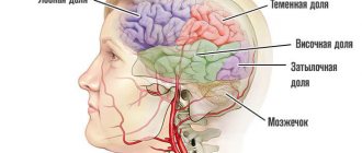
General cerebral symptoms
General cerebral symptoms arise due to sudden jumps in intracranial pressure. The most common symptom of the pathology is headache accompanied by vomiting. The patient may experience problems with vision: often, against the background of an abscess, optic neuritis develops, and congestive discs appear in the fundus. The clinical picture of the disease also includes mental disorders, inhibition of thought processes, lethargy, weakness, and apathy. In the case of intracranial hypertension, epileptic seizures may occur. Most patients also experience persistent drowsiness, and in the most severe cases, coma may occur.
Diagnostics
In order to accurately diagnose the location of the brain and cerebellum abscess, computed tomography and magnetic resonance imaging of the brain are performed.
When performing a computed tomography scan, a thin, smooth wall of the abscess is revealed, which has regular contours. Magnetic resonance imaging can also determine the abscess capsule. If it is not possible to carry out the above types of research, pneumoencephalography or radioisotope scintigraphy of the brain can be performed. Abscess of the brain and cerebellum should be distinguished from purulent meningitis, non-purulent local encephalitis, serous leptomeningitis (arachnoiditis), purulent labyrinthitis. With meningitis, there is a high temperature and tachycardia (increased heart rate), and with an abscess, the body temperature is low-grade and there is severe bradycardia. Patients with meningitis are excited, and with an abscess they are drowsy and lethargic; there is a characteristic “congestive nipple” in the fundus. You can distinguish between meningitis and an abscess by examining the cerebrospinal fluid: with meningitis there will be high pleocytosis, and with an abscess there will be moderate pleocytosis and an increase in the amount of protein. With arachnoiditis, there are no changes in the cerebrospinal fluid. Unlike encephalitis, with a brain abscess there are no symptoms of intoxication.
Course of a brain abscess
As for the course of the disease, it often has a very violent and acute onset, which is characterized by focal and hypertensive manifestations. The inflammatory process almost always develops against a background of elevated temperature. In rare cases, the onset of the disease may be less pronounced and resemble the clinical picture of meningitis. However, with minimal symptoms and normal temperature, the first stage of the disease is extremely rare.
After 5-30 days, the disease passes into the next, latent, stage, which is characterized by either a complete absence of any symptoms, or minimally expressed signs of the disease. The patient may complain of severe and regular headaches, mental retardation and vomiting. The duration of this stage varies: in some patients it lasts a couple of days, while in others it lasts several years. Then, due to the influence of some factor (for example, infection), this stage ends and the patient’s symptoms begin to actively progress. The most severe and life-threatening consequence of a brain abscess is its rupture, which usually leads to death.
Clinical picture
The clinical picture depends on the location of the abscess and its size, the stage of development of the pathological process, and the reaction of surrounding tissues.
There are 4 stages during the disease: initial, latent, overt and terminal. The initial stage lasts one to two weeks and is called the encephalic stage of brain abscess. It is characterized by symptoms such as lethargy, high body temperature, headache, nausea and vomiting.
The duration of the latent stage is on average 2 weeks. It is characterized by 4 groups of symptoms.
The first group includes manifestations that are characteristic of suppurative processes: lack of appetite, lethargy, stool retention, bad breath, exhaustion, “coated” tongue, changes in the composition of the blood characteristic of the inflammatory process.
Make an appointment right now!
Call us by phone or use the feedback form
Sign up
The second group includes general cerebral symptoms: headache, bradycardia (decrease in heart rate below 60 beats), stiff neck, positive Kernig and Brudzinski symptoms.
The third group presents symptoms of disturbances in the activity of conduction systems and subcortical nuclei: hemiparesis (paresis of one half of the body) and hemiparalysis, paresis of the facial nerve of the central type, convulsive seizures, paresis of the oculomotor nerve, pyramidal symptoms (Babinsky, Oppeheim, etc.).
The fourth group of symptoms is very important for determining the localization of the process and reflects cluster symptoms. If amnestic and sensory aphasia (speech disorder) appears in right-handed patients, this indicates that the abscess is localized in the left temporal lobe of the brain. If patients experience ataxia (impaired coordination of movements) and dizziness, then most likely the abscess is located in the right temporal lobe and spreads to the pathway that connects the right temporal lobe and the left cerebellum. Mental disorders of an emotional and personal nature may also be observed, which are manifested by euphoria or a depressive state, lack of a critical attitude towards one’s illness, psychomotor agitation, negativism (opposition and contradiction to other people). When the right temporal lobe is affected, an important symptom is a violation of the visual fields - hemianosmia, with loss of the same halves of vision in each eye.
Friends! Timely and correct treatment will ensure you a speedy recovery!
With a cerebellar abscess, there is a decrease in muscle tone on the affected side. The main symptom is ataxia, which is usually detected when walking and performing coordination tests (digital-digital, finger-to-digital, digital-to-digital, adiadochokinesis (inability to perform rapid, alternating movements), study in the Romberg position). Falls and misses on the part of the affected cerebellum are characteristic. Very indicative is nystagmus (involuntary movement of the eyeballs), directed upward and in both directions, often multiple.
A blood test reveals moderate neutrophilic leukocytosis and increased ESR. With an uncomplicated abscess, cerebrospinal fluid leaks under high pressure. Most often it is light and has a moderate shift: slight pleocytosis (increase in the number of cells) to a level of 100-200 cells in 1 μl. and a moderate increase in protein. If an abscess breaks into the subarachnoid space, the cerebrospinal fluid becomes purulent. With a cerebellar abscess, when taking a spinal tap, the fluid must be released slowly and in small portions to avoid wedging the brain stem into the foramen magnum.
At the terminal stage of development of the disease, dislocation symptoms manifest themselves: anisocoria (unequal pupil sizes), limited gaze, impaired breathing rhythm, loss of consciousness. The duration of this stage of abscess of the brain and cerebellum is several days and ends with the death of the patient. In this case, there is increasing swelling of the brain, paralysis of centers of vital importance, or an abscess breaking into the ventricles of the brain. Quite rarely, the breakthrough of an abscess into the ventricles of the brain and purulent ventriculitis (inflammation of the ventricles of the brain) result in a favorable outcome.
Diagnosis of brain abscess
Timely comprehensive diagnosis of brain abscess is important in its further treatment. To make a diagnosis, a neurologist uses medical history and examination results of the patient, as well as information obtained during instrumental and laboratory tests. The following methods are used to diagnose the disease:
- General blood analysis. The disease is usually indicated by test results such as an increase in ESR and pronounced leukocytosis. At the stage of capsule formation around the abscess, a normal or slightly increased number of leukocytes is observed in the patient’s blood.
- CT scan. The accuracy of detecting an abscess using this technique depends on the stage of the pathology. In the early stages, an abscess is very difficult to detect. At the stage of encephalitis, CT may reveal an area of reduced density that has an uneven shape. At this stage, the contrast agent accumulates unevenly - often only in the peripheral areas. It is much more accurate to diagnose the disease at a late stage in the development of encephalitis.
- Magnetic resonance imaging. This is a more accurate and effective method of diagnosing an abscess, which allows it to be detected at an early stage. Since the technique is considered the most informative, treatment can be prescribed based on its results even without bacteriological tests.
- Echoencephaloscopy. This diagnostic method is usually prescribed if for some reason MRI and CT cannot be performed. Using this study, it is possible to detect displacement of brain structures, which indicates compression of its tissues by an abscess.
- Bacteriological research. This technique involves taking a puncture of pus from the abscess for examination. A detailed examination of the pus helps to identify the causative agent of inflammation, which then makes it possible to select the most appropriate tactics of drug therapy.
- X-ray of the skull. This technique is used to detect the source of infection that caused the abscess.
- Craniography. Prescribed to detect symptoms of intracranial hypertension.
Acute purulent otitis and brain abscess
O.V. Stratieva (From lectures for doctors)
Otogenic intracranial complications arise as a result of the penetration of infection from the ear cavities into the brain cavity. The number of patients with otogenic intracranial complications in relation to the total number of patients with diseases of the middle and inner ear ranges from 1 to 15% and tends to decrease. However, the problem of otogenic intracranial complications remains relevant. Over the past three years, we have operated on more than 30 patients with otogenic intracranial complications. The most common were: thrombosis of the sigmoid and transverse sinuses, epidural abscess of the middle and posterior cranial fossae, perisinous abscess, otogenic meningitis, abscess of the temporal lobe of the brain; cerebellar abscess was observed less frequently. The features of the modern clinical picture of otogenic intracranial complications are characterized by a case from our practice.
Case from practice.
G—a, 68 years old, was admitted to the ENT center with complaints of headache, dizziness, nausea, weakness in the left leg, fever up to 37.5 degrees, and malaise.
It is known that the patient had been ill for a month when pain in the right ear and purulent discharge from the ear suddenly appeared. She treated herself with dry heat. After two weeks, the condition improved. The ear pain and suppuration stopped. However, inadequacy, lethargy and headache appeared in the patient’s behavior, which forced the patient’s relatives to seek advice from an otolaryngologist.
On admission: the patient is conscious. There is no pain behind the ear. The eardrum is dull, there is no perforation, there is no pus. The neurologist found a deviation to the left during the finger-nasal test, and smoothness of the left nasolabial fold. In the fundus there is atherosclerosis of the retinal vessels.
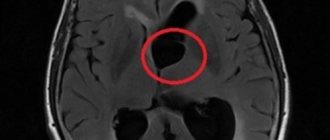
A computed tomography scan of the brain on the right in the temporo-parietal region reveals extensive round formations with a capsule up to 2.1 cm in diameter, a density of 19 to 24 units N, with contrast - up to 65 units N. Additionally, magnetic resonance imaging was performed (Fig. 1 , 2), where a purulent process was established in the right temporal pyramid with a bifocal brain abscess and thrombosis of the cavernous sinus.
Otolaryngologists and neurosurgeons performed two operations: craniotomy with brain decompression, opening and removal of the temporal lobe abscess, and extended mastoidectomy with exposure of the cranial fossae and sinuses of the brain. During the operation, osteomyelitis of the pyramid of the temporal bone, the wall of the auditory canal and the tympanic cavity was discovered. The patient was in intensive care for four days, then underwent rehabilitation at the ENT center for 14 days, and was then discharged home in satisfactory condition.
SUMMARY
Today, in comparison with the period from 1946 to 1980. XX century, the number of intracranial complications due to otitis media decreased significantly. The use of powerful antibiotics has significantly improved the quality of treatment, but at the same time has complicated diagnosis. If previously manifest forms with clear symptoms prevailed, today almost all otogenic intracranial complications develop covertly. Also, in recent years, diagnostic difficulties have been caused by a combination of several forms of intracranial complications.
Differential diagnosis of brain abscess
Since the majority of symptoms of brain abscess are not specific, differential diagnosis plays an important role. If the doctor has doubts during diagnosis, he may prescribe MR spectroscopy. This technique is carried out in order to differentiate a brain abscess from tumors of the cerebral hemispheres. It is based on different contents of lactate and amino acids in tumors and purulent accumulations.
As for other diagnostic methods, they are considered less informative. For example, signs such as an increase in C-reactive protein in the blood, chills, an increase in ESR, and leukocytosis may indicate a variety of inflammatory processes. Blood cultures from an abscess are often sterile.
Treatment of brain abscess
Treatment for a brain abscess usually involves both drug therapy and surgery. Doctors select the most optimal treatment tactics based on the results of diagnosing the disease, as well as the general health of the patient. The stage of the disease is also taken into account. For example, in the early stages of abscess formation, conservative treatment can be used. If an abscess has already formed, and a dense capsule has formed around it, neurosurgical intervention cannot be avoided.
- Drug treatment
- Surgery
Drug treatment of brain abscess involves the prescription of antibiotics, decongestants and anticonvulsants. Since the inflammatory process is provoked by bacteria, treatment of the disease necessarily involves their destruction. The combination of penicillin and chloramphenicol has been considered the most standard and commonly used treatment regimen for brain abscess for decades.
Penicillin was prescribed to treat the disease because it can kill streptococci and most other bacteria that can cause brain abscess. Chloramphenicol was used because of its ability to easily dissolve in adipose tissue and destroy anaerobic bacteria.
Today doctors are slightly adjusting this scheme. For example, cefotaxime is prescribed instead of penicillin, and metronidazole is prescribed instead of chloramphenicol. Typically, the doctor prescribes antibiotic therapy several weeks before surgery. The duration of taking antibiotics can be about 6-8 weeks.
Patients whose brain abscess occurs due to immunodeficiency are also prescribed amphoreticin. If the abscess has resolved, the patient will need to take a course of fluconazole for ten weeks. Drugs such as sulfadiazine and pyrimethamine are commonly included in the treatment regimen of patients with HIV.
Of great importance in the treatment of the disease is the correct identification of the causative agent of infection using an antibiogram. However, there are cases when the culture turns out to be completely sterile. Therefore, in such situations, empirical antibiotic therapy is prescribed.
In addition to antibiotics, medications are also prescribed to help reduce swelling. For example, glucocorticoids are used for this purpose. However, the prescription of these drugs is indicated only in case of a positive result from antibacterial therapy. They can reduce the severity of a brain abscess and reverse the development of the capsule around it. However, the opposite effect is also possible, when glucocorticoids activate the spread of inflammation beyond the boundaries of the lesion. To eliminate convulsive manifestations, phenytoin is usually prescribed.
If a brain abscess is diagnosed in the late stages, and a dense capsule has already formed around it, it is impossible to do without surgery. To treat the disease, puncture aspiration and removal of the abscess are most often used.
As for puncture aspiration, it is advisable to prescribe it in the early stages of the pathology. In this case, antibacterial therapy should be carried out simultaneously. Indications for this procedure may also include multiple abscesses, a deep location of the abscess, the stage of cerebritis and a stable neurological condition of the patient. To ensure that the procedure is performed as accurately as possible, the doctor resorts to stereotactic biopsy and intraoperative ultrasound.
Needle aspiration has one important drawback - in most cases, a repeat procedure may be required after it is performed. In difficult cases, complete removal of the abscess is prescribed. This technique is also prescribed if they want to avoid a possible relapse of the disease. It is advisable to remove an abscess for the following indications: if antibacterial therapy or puncture aspiration are not effective, with a superficial abscess and a well-formed capsule around it.
If multiple abscesses were discovered in a patient during diagnosis, then first you need to drain the inflammation to prevent pus from breaking into the ventricular system of the brain. If neurological disorders increase or there is no positive dynamics on MRI and CT, a repeat operation may be prescribed.
Treatment
Therapy for the disease must be comprehensive. Most often, surgical intervention is used in combination with adequate combination antibacterial therapy. Antibacterial therapy can last several weeks. Subsequently, a long period of rehabilitation is required. Depending on the resulting neurological defect, one should resort to neuroprotective therapy, massage, exercise therapy, and sometimes, when symptomatic epilepsy develops, the selection of antiepileptic drugs is necessary.
Prognosis for brain abscess
The outcome of the disease depends on whether the doctor was able to identify the causative agent of the abscess from culture. It is extremely important to do this, since then it will be possible to determine the sensitivity of bacteria to antibiotics and select the most appropriate treatment regimen. The prognosis for the health of a patient with a brain abscess also depends on the number of purulent accumulations, the patient’s health status, and the correct treatment tactics.
The risk of various complications with a brain abscess is very high. Namely, about 10% of all cases of the disease end in death, and 50% result in disability. In addition, most patients may develop epileptic syndrome after treatment, a condition characterized by the occurrence of epileptic seizures.
Doctors give less favorable prognoses to patients who have been diagnosed with subdural empyema. In this case, the patient does not have a clear boundary of the purulent focus due to the high activity of the infectious agent or the body’s insufficient resistance to it. Lethal cases with subdural empyema reach 50%.
The most dangerous form of brain abscess is considered to be fungal empyema, which is accompanied by immunodeficiency. This disease has practically no cure, and the number of deaths with it is about 95%. In turn, epidural empyemas have a more favorable prognosis and are almost never accompanied by complications.
Prevention of brain abscess
There are no effective methods for preventing brain abscess. However, with the help of several preventive measures, the risk of the disease can be significantly reduced. In particular, in the case of traumatic brain injury, the patient must receive adequate surgical care.
The disease can also be prevented by timely elimination of foci of infection (pneumonia, boils), treatment of purulent processes in the inner and middle ear, as well as the paranasal sinuses. Good nutrition also plays a great role in the prevention of brain abscess.