Medium-sized AVM of the lower cerebellum
Alteriovenous malformations (AVMs) include congenital, non-hereditary defects of the vascular system, in which there are no normal capillary networks between the artery and vein, and arterial blood is discharged directly into the venous system. Over time, aneurysms form arteriovenous malformations .
AVM most often occurs in the brain, but it can also occur in the spinal cord. One or more aneurysms may be found in association with an AVM. AVM begins to develop during the fetal stage. It grows unpredictably with age.
What is AVM?
Normally it is transported from the heart through large arteries leading to the main parts of the body. Gradually, the arteries branch, become smaller and transform into capillaries with a diameter of only one cell (approximately 5 microns). The network of capillaries is called the vascular bed; here the blood gives oxygen and nutrients to the tissues and, in turn, receives metabolic products. The blood then returns through the veins to the heart. With an AVM, the arteries intertwine with the veins, forming a pathological tangle rather than a vascular bed. In this case, a vascular shunt with high internal pressure, or fistula, is formed. Veins are not able to withstand the pressure of blood coming from the arteries. As a result, the vein dilates, the venous wall stretches and, in many cases, forms a “sac”, later called an aneurysm. The blood inside the aneurysm accumulates and does not circulate, so the surrounding tissues lack blood supply. If blood pressure continues to be high, the vein or aneurysm itself will eventually rupture, causing bleeding.
The first Soviet AVMs
In 1945 - 1946 under the leadership of L.I. Gutenmacher developed the first electronic analogue machines with repeating solutions. But since 1949, a team of Soviet developers, headed by V.B. Ushakov and V.A. Trapeznikov, invented a number of DC AVMs. This year began the history of the development of analog computer technology in the USSR. The use of operational amplifiers, which operated on the principle of automatic control systems with deep negative feedback, made it possible to carry out accurate modeling of mathematical operators, as well as parallel processing of information in real time when solving systems of differential equations.
Beginning of development
In 1949, the electrical modeling laboratory of ITMiVT and the department of the chief designer of the Penza plant SAM B.A. Matkin, an electron tube integrator ELI-12 was released. It was intended to solve a system of 12th order differential equations with constant coefficients and constant right-hand sides. The process of solving the problem was automatically repeated, as a result of which the solution results were displayed, measured and photographed on the CRT screen.
After finalizing the circuits and design of the electron tube integrator, as well as preparing for industrial production, the plant began mass production of ELI-12-1.
On its circuit-design basis, the ELI-14 integrator was developed, on which sixth-order differential equations were solved. There were 6 decisive blocks. For the manufacture of connecting tubes, neoleukorite was used, which was distinguished by increased insulating properties.
In 1949–1950 at NII-885 by a team led by V.B. Ushakov created the first AVMs, which were called direct current integrators. In 1949 - “IPT-1”, “IPT-2” and “IPT-3”, in 1950 - “IPT-4” and “IPT-5”, mass-produced. These machines were intended for solving linear differential equations with constant and variable coefficients. Thanks to operational amplifiers, AVMs provided solutions to the most important problems in various scientific and technical fields (aviation, rocket science, space research, defense industry, etc.).
The first AVMs based on vacuum tubes were created by the combined efforts of two teams: NII-855 MRP USSR and IAT USSR Academy of Sciences. Serial production of AVMs was organized at the Moscow, Penza and Chisinau factories of calculating and analytical machines and a number of other radio industry factories.
Developed in 1952–1953 under the leadership of V.B. Ushakov, AVMs received the name “direct current simulators” (MCT). Serial AVM "MPT-9" was intended for solving linear differential equations, "MPT-11" - for solving nonlinear differential equations.
"MN-8"
In 1955, the first Soviet high-power precision AVM “MN-8” was developed. It was the largest continuous nonlinear electronic mathematical machine. The development team was headed by V.B. Ushakov. The machine was supposed to solve differential equations of the sixteenth and higher order (32 integrating blocks). In the case of the sixteenth order, half of the linear part was used.
The electronic modeling installation consisted of 13 sections. Tasks were typed using cord connections on the switching fields of the linear part. By means of a pair of control panels, two different tasks were simultaneously solved. The maximum duration of the integration process reached 10,000 seconds, and the power consumption was 25 kW.
“MN-8” carried out up to 48 operations of summing 264 terms, 48 multiplications by a constant coefficient, 36 multiplications by a variable coefficient, 12 exact multiplications of the desired quantities. It was also possible to set 10 nonlinear dependencies of a function on one variable, 40 nonlinear dependencies of a signature type, 9 nonlinear dependencies of typical characteristics (backlash, limitations, dead zones).
The machine had 400 operational amplifiers with individual automatic zero stabilization and an improved control circuit. Through powerful amplifiers with economical output, interaction with real equipment took place. The control circuit had additional controls.
Only three foreign models could compare with the Soviet model: - the American electrical modeling unit “Typhoon”, released in 1951; - English electrical modeling installation "Trydac" - Three Dimensional Analogue Computer, created in the period 1950 - 1954. It was an electronic-hydraulic simulator, with the help of which projectile control was studied; - American installation “Convair”, released in 1954.
A special feature of MH-8 was that the duration of the processes studied by the installation on a natural time scale could be either small (equal to several seconds) or very significant.
The integrating block circuit accelerated the expansion process; it made it possible to automatically change the time scale simultaneously for all such installation blocks by 10 times.
MN-8 used precision multiplication units. They made it possible to obtain a static accuracy of the multiplication operation of about ±0.01%. The number of blocks in the machine made it possible to obtain 12 operations of exact multiplication of variables.
“MN-8” included 48 precise blocks for entering variable coefficients, which practically reproduced the graph of the variable coefficient using the piecewise linear approximation method. It also included 40 special nonlinear blocks for performing nonlinear dependencies of the signature type. This improvement significantly expanded the capabilities of the machine.
The Soviet electrical modeling installation had an improved control circuit. An electronic time meter was built on the basis of a decimal digital counter. With its help, the work of all functional blocks was synchronized. The solution process could be repeated periodically or stopped at predetermined periods of time.
The MH-8 used diode-triode electronic circuits for universal blocks, which were intended to reproduce nonlinear functions of one variable with an accuracy of a set of functions of ±0.2.
The block made it possible to dial very steep function edges, as well as functions with sharp breaks.
The simulated system of equations was typed in accordance with the block diagram for solving the problem on the switching fields that were located in the sections of the linear blocks of the installation. The main lines between sections made it possible to use blocks of adjacent sections in one task. The MH-8 did not have removable patch fields.
Registration and control of output values were carried out using six high-speed electronic potentiometers, an electronic digital voltmeter, a cathode beam indicator and other measuring instruments.
The MH-8 had 14 racks. The design of the device used small-sized blocks, so it was relatively compact. The circuit of the electrical modeling installation contained many high-quality electrical parts (they significantly increased the accuracy of the work). For example, about 8000 germanium planar diodes were used in the power section circuit.
"MN-8" was mass-produced by the Penza plant SAM.
"MH-9"
In 1958, the MH-9 electronic simulator was released.
It was intended to study the dynamics of the main parts of the clock mechanism. The development was led by chief designer I.M. Witenberg. The MH-9 had a bench design. Using switches and handles located on the front panels of the machine, operating modes were set.
"MN-9" solved systems of ordinary linear equations with variable coefficients. The device consisted of 5 blocks of a summing amplifier, 40 blocks of constant coefficients and 9 blocks of a nonlinear function of one variable. “MN-9” had 28 amplifiers with centralized automatic setting of zeros according to a circuit with capacitors and are characterized by the presence of an electronic circuit for automatic switching of machine blocks in function of the desired variable.
The MH-9 electronic simulator was not mass-produced.
"MN-10"
In 1957, a new development of Soviet specialists was released - a small-sized nonlinear lampless modeling unit “MN-10”.
It was the first analog computer with a circuit made entirely of semiconductors. Using the installation, problems that were described by ordinary nonlinear differential equations were solved and studied. For example this: , where i=1, 2,…, 6.
Among other things, the installation made it possible to solve differential equations that contained up to 6 nonlinear dependencies of the type of function on one variable or the product of two variables. The work could be carried out on a natural time scale. The results of the tasks were demonstrated on an electron beam indicator of the I-5 or I-4 type.
The MH-10 included 24 small-sized DC operational amplifiers. They performed operations of integration, differentiation, summation and large-scale transformations. Also 4 diode cells, which were used in circuits that reproduce typical nonlinear dependencies of the type of hysteresis loop, dry friction moment, dead zone and limitation.
The device occupied an area of 0.3 m2 and weighed 75 kg. The range of changes in the installation variables ranged from 30 V to +30 V. The duration of the integration process was 200 seconds.
The MN-10's power came from the ESV-10 unit included in its kit. And the mains power was supplied by single-phase alternating current with a voltage of 220 V and a frequency of 50 Hz. Power consumption was 100 W.
In the 70s, the installation was modernized and began to be mass-produced.
"MN-10M"
The MN-10M analog computer was developed by specialists from the Penza plant. The model belonged to small-sized desktop machines of low power, with the help of which real dynamic systems were studied using the method of mathematical modeling. Also, “MN-10M” was intended for the integration of ordinary differential equations of no higher than the tenth order.
The dimensions of the machine were 460 × 615 × 445 mm, and the weight was 50 kg. The MH-10M included 24 operational amplifiers, as well as a set of 12 feedback links.
Thanks to a set of operating blocks, it was possible to carry out various operations: - integration operations with simultaneous summation (up to 10); — inversion or summation operations (up to 24); — setting on voltage dividers (up to 60 constant coefficients); — reproduction of unambiguous continuous nonlinear functions of one variable with simultaneous summation of several variables; — an operation of multiplication or division with simultaneous summation of several variables; — reproduction of up to 6 typical nonlinear dependencies of the type of dead zone, limitation, dry friction; — conditional jump operations (up to 4).
More complex tasks were solved in parallel by two or three MN-10M machines. The result of the task could be observed and measured using a machine dial gauge or an external recording device (DRP, EPP-09, loop oscilloscope). They were not included in the vehicle.
Interface with external equipment was provided. The MN-10M power supply came from the ESV-4 unit, which was included in its kit. And the mains power was supplied by single-phase alternating current with a voltage of 220 V and a frequency of 50 Hz. Power consumption was 250 W. The circuitry of the machine and power supply was entirely built on semiconductor elements.
The machine solved ordinary differential equations. The range of variable values was from - 25 V to + 25 V. The duration of the integration process was 200 seconds.
The MN-10M block included an electronic stabilized rectifier (ESV-4), an analog communication computer (AVM), communication channels with external equipment, with the second and third machines.
More than 100,000 AVMs were manufactured in the first 20 years. From simple to the most powerful, like MH-8. At first, machines were used mostly as independent tools for mathematical modeling of dynamic objects in real time. But around the 60-70s, progress in the field of digital electronics required joint processing of information by AVM and digital computer.
There are four types of AVM
- Arteriovenous malformation is an abnormal plexus of blood vessels in which blood from the arteries enters directly into the veins, bypassing the capillaries, which is characterized by the absence of a capillary bed and high pressure.
- Cavernous hemangioma is an abnormal collection of dilated capillaries; as a rule, veins and feeding arteries are absent; characterized by low blood pressure.
- Venous malformation is an abnormal collection of dilated veins in the shape of wheel spokes; characterized by the absence of feeding arteries and low pressure; usually does not require treatment because it rarely bleeds.
- Capillary telangiectasia - abnormal capillaries with increased surface area (similar to cavernous hemangioma); pressure is very low.
AVMs can form in any part of the body; symptoms correspond to location. In the brain, arteriovenous malformations can be localized on the surface of the brain (cortical), inside brain tissue (hypothalamus, basal ganglia or brainstem), and also inside the dura mater - the dense protective layer of the brain. AVMs located within the membrane are called arteriovenous fistulas (AVFs). Normally, blood moves through the venous vessels of the brain and is transported to the venous sinuses located in the dura mater of the brain, after which it moves to the heart muscle. In AVF, the arteries are connected directly to veins or sinuses. In addition to the arteriovenous fistula of the lining of the brain, the most common are carotid-cavernous fistulas (shunts between the carotid artery and the venous sinus). Spinal AVMs form either on the surface of the spinal cord (extramedullary) or inside it (intramedullary). There are four types of such AVMs:
- Type 1 – (most common) arteriovenous tunica fistula. This AVF usually has a single feeding artery, and symptoms are caused by increased venous pressure.
- Type 2 - (glomus) is localized intramedullary, consists of a very dense tangle of vessels in a small area of the spinal cord.
- Type 3 – (juvenile) extensive AVM, consisting of anomalous vessels and localized intra- and extramedullary.
- Type 4 – (located within the sheath) extramedullary AVF on the surface of the spinal cord
Cavernoma of the brain and spinal cord
Cavernoma of the brain and cerebellum is also known as cavernous hemangioma . These are congenital and usually non-hereditary vascular cavernous malformations of veins located in the brain or spinal cord (spine).
Unlike AVMs (arteriovenous malformations), blood flow in a cavernoma is quite weak. Since the walls of the cavernoma are weak, blood from them slowly leaks into the surrounding tissue.
Cavernomas can vary in size from one millimeter to several centimeters. Thanks to modern diagnostic methods (CT, MRI), cavernomas have recently been diagnosed more often than before. Because of their low blood flow, these defects are not visible on angiograms.
Cavernomas develop insidiously and, according to preliminary estimates, about half a percent of the population has cavernomas of the central nervous system. Most cavernomas are discovered between the ages of 20 and 40 due to the following symptoms : headache, epileptic seizures, neurological deficit (weakness of the limbs) or small but sometimes numerous cerebral hemorrhages.
The risk of cavernoma bleeding is about 1-2%. Bleeding from a cavernoma is less common than from an aneurysm or arteriovenous malformation (AVM). Cavernomas located in the brainstem and cerebellum tend to bleed more easily and cause more damage than those located in the brain.
Symptoms
AVMs have different symptoms depending on the type and location. Common symptoms are migraine-like headaches and seizures. However, most AVMs do not cause symptoms until they rupture and begin bleeding.
Common signs of cerebral AVMs:
- sudden attack of headache, vomiting, neck stiffness (patients often complain of “the worst pain of my life”)
- convulsions
- migraine-like headaches
- unusual tinnitus (whistle or ringing) caused by pulsating blood
General signs of spinal AVMs:
- sudden severe back pain
- weakness in the legs or arms
- paralysis
Where can the malformation be located and how does it manifest itself?
Arteriovenous malformations can occur in:
- brain;
- spinal cord;
- internal organs.
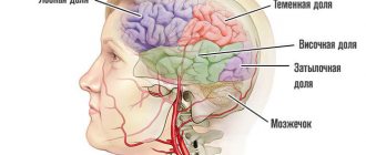
Vascular malformation of the brain manifests itself depending on the location of the lesion:
- Damage to the frontal lobe is manifested by slurred speech, decreased intellectual abilities, headaches, decreased ability to work, less commonly, stretching of the lips in a “tube,” and seizures.
- When the cerebellum is damaged, there is a lack of coordination of movements, muscle hypotonia (weakness), horizontal nystagmus (involuntary eye movements), falling during normal walking, instability in the Romberg position (in a standing position, stretch your arms forward and close your eyes).
- When the temporal lobe is involved in the pathological process, severe headaches, deterioration in speech perception occur (patients hear well, but do not understand the essence of what they say), pulsation in the temporal region, convulsive seizures, and a decrease in visual fields.
- If arteriovenous malformation occurs at the base of the brain, it manifests itself as: strabismus, one- or two-sided blindness, paralysis (complete loss of movement) of the lower or upper extremities. The movements of the eyeballs are impaired.
The largest arteriovenous malformations occur in the posterior hemispheres of the brain.
When the spinal cord is damaged, the following symptoms occur:
- with a superficial location, a pronounced pulsating formation is noted;
- pain in the spinal column, especially in the thoracic or lumbar region, can radiate (give) to the thoracic region, upper and lower extremities;
- decreased muscle tone;
- convulsive twitching of the arms or legs;
- it is possible to disrupt the functional activity of organs located in the pelvis. This manifests itself in severe cases as fecal or urinary incontinence;
- increased fatigue, especially when walking or exercising.
From internal organs, arteriovenous malformations can occur between the aorta and the pulmonary trunk (two large vessels emerging from the heart muscle) - patent ductus arteriosus (white heart defect). This condition can be provoked by chromosomal diseases, for example, Down syndrome; rubella suffered by the mother while carrying a child; prematurity of the baby. Normally, it overgrows no more than eight weeks, in rare cases up to fifteen. This condition manifests itself:
- an increase in the size of the heart muscle;
- shortness of breath;
- “machine noise”, which can be heard in the second or third intercostal spaces;
- increased heart rate (tachycardia);
- slow growth and development of the child;
- cyanosis of the skin;
- in severe cases, swelling of the upper or lower extremities, hemoptysis, impaired functioning of the heart, difficulty breathing. Spontaneous cardiac arrest may occur.
Arteriovenous malformation arising in the kidneys manifests itself:
- pain in the lumbar region on one or both sides;
- hematuria (the appearance of blood in the urine);
- an increase in blood pressure that is difficult to correct with antihypertensive drugs.
With liver damage, which occurs very rarely, specific symptoms cannot be detected. They occur only with concomitant bleeding. Pallor of the skin, weakness, fainting (syncope), dizziness, stool may turn black (melena), and in severe cases, blood pressure drops.
If a vascular malformation affects the lungs, it manifests itself:
- headaches;
- pulmonary hemorrhages in 10 - 15% of cases;
- increased amount of carbon dioxide in the blood;
- increased susceptibility to thrombosis.
AVMs cause the following injuries to the brain or spinal cord:
- When an AV malformation ruptures, there is bleeding in the brain or in the space between the brain and the skull (subarachnoid hemorrhage). Small AVMs (less than 3 cm) are more likely to rupture than large ones. Hemorrhage can lead to a stroke.
- If the AV malformation grows, it increases pressure on the brain tissue, causing seizures and hydrocephalus. Such complications are usually typical for large AVMs.
- AV malformations can cause oxygen starvation of surrounding brain tissue. Since the AVM vessels lack capillaries, brain cells do not receive enough oxygen from the blood and begin to die.
The risk of bleeding from an AV malformation is estimated to be 2–3% per year. Mortality due to first bleeding is approximately 10-30%. If a hemorrhage occurs, the likelihood of a repeat incident during the first year increases 9 times. Before deciding to have surgery, patients often want to know what the long-term likelihood of bleeding is. Here's a simple formula for this assessment: % bleeding risk = 105 - age For example, a 25-year-old man with a diagnosed AVM has an 8 out of 10 chance (i.e., an 80% chance) of having at least one bleed at some point. A number of factors influence these indicators, in particular the location and type of AVM. It is best to consult your doctor to discuss these risks as they apply to your specific case.
Why does it occur?
The issue of genetic predisposition to this pathology has been discussed for a long time, but no reliable evidence has been found. It is a congenital disease. The processes of vascular disruption that lead to this pathology occur in the first or second month of intrauterine fetal formation. Currently, predisposing factors are considered:
- uncontrolled use of medications, especially those affecting the fetus (with a teratogenic effect);
- past illnesses that occur in the first trimester of pregnancy, of a viral or bacterial nature;
- rubella disease during pregnancy;
- smoking and drinking alcohol during pregnancy. Not all girls, even when “in position”, give up these habits;
- ionizing radiation;
- pathology of the uterus;
- severe poisoning with chemicals or other toxic agents;
- intrauterine fetal injuries;
- exacerbation of a chronic pathology in a pregnant woman (diabetes mellitus, glomerulonephritis, bronchial asthma, etc.)
As a result, the vascular bundles connect incorrectly, become intertwined, and arteriovenous malformation occurs. When it forms to a significant size, cardiac output increases, the walls of the veins hypertrophy (compensatory enlargement), and the formation takes on the image of a large pulsating “tumor.”
How is the diagnosis made in Israel?
Seeing a doctor about an AVM is usually associated with either its rupture or the intention to begin treatment for an AVM that was previously discovered. In any case, the doctor reviews symptoms and other medical problems, family history, a list of medications taken on an ongoing basis, and also conducts a physical examination. Next, the patient will be referred for additional tests to determine the type and size of the AVM, its location and interaction with other structures.
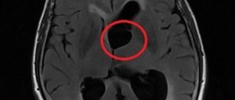
- Computed tomography (CT) is a non-invasive technique that takes detailed X-ray images of the brain to detect the presence of blood inside or outside the brain tissue. A relatively new modification of CT is called CT angiography. In this case, contrast is injected into the general bloodstream to visualize the cerebral arteries. The combination of standard CT and angiography makes it possible to assess the condition of brain tissue and blood vessels.
- Magnetic resonance imaging (MRI) is a non-invasive test that uses a magnetic field and radio frequency waves. Its type, magnetic resonance angiography (MRA), allows you to obtain detailed images of the vascular bed in a non-invasive way.
- Angiography is also used as an invasive procedure, when a catheter is inserted into a peripheral artery and passed into the vessels of the brain. After this, contrast is injected into the blood and a series of x-rays are taken.
Treatment of brain AVM
Treatment consists of surgical removal of pathological vessels. Classical open excision of a malformation is used mainly only in emergency cases, when a vessel ruptures, when it is necessary not only to remove it, but also to aspirate the resulting hematoma.
In case of uncomplicated malformation, one can resort to angioembolization, when an embolic substance is injected into the pathological vessel, its lumen is permanently closed and the vessel functionally disappears.
The most modern method is radiosurgery, during which the malformation is irradiated, leading to complete closure of the lumen of the vessel.
Radiosurgery
During this procedure, precisely focused radiation is directed at the abnormal vessels. Accurate focusing requires several hours of preparation; The irradiation itself takes another hour. The patient is discharged on the same day. Over the next 6 to 24 months, the vessels gradually close and are replaced by connective tissue. The advantages of this method are that the skull is not opened and the procedure is painless. The disadvantages of the method are that the best results are obtained only with small AVMs, and the therapeutic effect is achieved after a long time. During this time, the risk of hemorrhage remains. In a recent long-term experiment conducted in one of the large clinics in America, 73% of patients showed excellent or good results after radiosurgery. In these patients, the risk of future bleeding was reduced to almost zero.
Endovascular therapy
Treatment in Israeli clinics is carried out in the angiographic unit of the X-ray department by an experienced physician specialist in the field of neurovascular procedures. During this procedure, a catheter is used to inject an adhesive or other blocking material directly into the arteriovenous malformation to stop the flow of blood through the area. The patient is under general anesthesia. A catheter is inserted through a small incision in the groin and passed through a series of blood vessels directly to the artery that supplies the AVM. A blocking material (coil or medical adhesive) is then injected to narrow this artery and stop the flow of blood through the AVM. The procedure time may vary and the patient will remain in the hospital for several days for observation. This method is less invasive than surgery and can be used to treat deep and inoperable AVMs.
Surgery
Under general anesthesia, part of the skull is opened, called a craniotomy. Arteriovenous malformation is carefully detected. Using a laser and other instruments, abnormal vessels are cauterized. Once the AVM has emptied and shrunk, it is removed from the brain tissue. The hospital stay usually does not exceed 5-7 days, followed by short rehabilitation. The type of craniotomy depends on the size and location of the AVM. In general, the decision about surgical treatment is made depending on the general health of the patient. The advantage of surgery is that recovery is immediate if the AVM is removed. Disadvantages include the risk of bleeding, damage to surrounding brain structures, and stroke.