Lymphogranulomatosis or Hodgkin's disease is a pathological condition of malignant etiology, which is characterized by the presence of giant Reed-Sternberg cells. They are identified by microscopic examination of the tissues of the affected lymph nodes. Most often, the disease affects the structures of the lymphatic system located under the lower jaw, in the supraclavicular region and mediastinum. According to medical statistics, Hodgkin lymphoma most often affects male patients between the ages of twenty and thirty years or after sixty.
The hematology department of CELT invites you to undergo diagnosis and treatment of LMG in Moscow. Our clinic is multidisciplinary and has been operating in the capital’s paid medical services market for the third decade. Our hematologists have a modern diagnostic and treatment facility that allows them to quickly and accurately make a diagnosis, as well as carry out treatment in accordance with international standards. To find out the cost of a consultation, go to the “Services and Prices” section. Despite the fact that we regularly update price lists, do not forget to check the numbers with our operators in order to avoid misunderstandings.
Hodgkin's disease: causes
The etiological factors provoking the development of lymphogranulomatosis have not been established to date. Experts put forward versions, considering hereditary, viral and immune theories, but none of them is generally accepted, since they do not provide comprehensive explanations. The viral gene is detected in more than half of the cases; in addition, a connection between HF and infectious mononucleosis has been identified, which may speak in favor of a viral etiology. Adherents of the hereditary theory pay attention to the presence of a familial form of the disease, immunological - to the possibility of transfer of lymphocytes from mother to fetus and the subsequent development of the disease.
In addition, the reasons may lie in the effects of toxins, ionizing radiation, medications, etc. The most important sign of a malignant process in LGM is the presence of giant Reed-Sternberg cells, their mononuclear precursors - Hodgkin cells, T-lymphocytes, eosinophils and plasma cells. The neoplasm itself develops from a single focus located directly in the lymph nodes and can give metastases, which provoke changes in the lungs, gastrointestinal tract and bone marrow of the patient.
Medicines
Photo: abc.net.au
Medicines for lymphogranulomatosis are chemotherapy drugs and symptomatic medications. There are several effective polychemotherapy protocols. One of them is DAL-HD-90, which provides a five-year survival rate of 91-98% of patients. The longer the remission, the more favorable the prognosis.
Now the efforts of doctors and researchers are aimed at minimizing the side effects of such treatment. For example, replace procarbazine with an analogue to reduce the incidence of azospermia in men (the incidence of this complication is 40-60%). In order to prevent the development of secondary tumors as a result of chemotherapy, doctors regularly diagnose the patient's condition.
In parallel with chemotherapy, hyperhydration of the body is often carried out using glucose-saline solutions. For severe infections (sepsis, chickenpox, pneumonia), the course of treatment is interrupted. In other cases, antibacterial, antifungal or antiparasitic treatment is prescribed.
Lymphogranulomatosis: classification
The classification of a neoplasm is based on a number of parameters, from prevalence to speed of development.
| Type/form/stage of LCH | Distinctive features |
| By prevalence | |
| Local | Only one group of lymph nodes is affected by the tumor. |
| Generalized | The problem affects the spleen, skin, stomach, lungs and liver of the patient. |
| According to the speed of development | |
| Spicy | The development of a neoplasm from the initial to the last stage takes only a few months. |
| Chronic | The disease develops over several years, alternating exacerbations and remissions. |
| According to the morphological structure of the tumor | |
| Lymphohistiocytic | It is diagnosed in 15% of all cases of lymphogranulomatosis in male patients under the age of 35 years. Detected in the early stages and successfully treated. Mature lymphocytes are detected in the structure; giant cells are rare. |
| Nodular sclerotic | The most common form of the disease is diagnosed in almost half of the cases in women of reproductive age. It is distinguished by the presence of fibrous cords, due to which the lymphoid tissue is divided into nodes. The structure reveals giant and lacunar cells. |
| Mixed cell | This form accounts for a third of all cases. They are diagnosed in pediatric and elderly patients, most often male. The structure is represented by different cells, including giant cells, lymphocytes, fibroblasts, and plasma cells. |
| Suppressing lymphoid tissue | The rarest form: detected in 5% of cases, usually in elderly patients. The structure completely lacks lymphocytes, but many giant clients are found. |
| By stage | |
| First/local | Either one group of lymph nodes or one organ of the immune system is affected. |
| Second/regional | Two or more groups of nodes located on one side or one organ and its nodes are affected. |
| Third/generalized | The nodes on both sides and one organ of the immune system, the spleen, or both are affected. |
| Fourth/disseminated | One or more organs of the immune system (lungs, gastrointestinal tract, pleura, liver, bone marrow) and lymph nodes are affected. |
Stages
According to one of the popular and widespread classifications, there are four stages of Hodgkin lymphoma (and zero). We will not describe their symptoms, but how the patient feels at each stage:
- 0th. A person feels and behaves as if he is absolutely healthy.
- I. The patient can lead a completely adequate lifestyle, but he is slightly weakened, so physical labor is limited.
- II. Severe fatigue is already noticeable, the person cannot take care of himself, it is difficult for him to move. The patient may spend about half the day in bed.
- III. At this stage, the patient lies a lot (more than half a day) and needs constant care. It is difficult for him to be active and perform even usual actions.
- IV. At this stage, patients are the sickest. They constantly lie or sit, get tired extremely quickly and can even communicate with difficulty and for a short time.
The doctor will explain to the patient what processes occur in the body at each stage of Hodgkin lymphoma - there are many manifestations and their details. It all starts with damage to the lymph nodes or some organs (for example, the spleen) - and then the tumor begins to spread, gradually taking over the body.
Lymphogranulomatosis: symptoms
The symptomatic complexes characteristic of Hodgkin's disease are represented by intoxication of the body, lymphadenopathy, the appearance of foci of proliferation in the spleen, bone marrow and other organs of the immune system. In the initial stages, nonspecific symptoms of LGM are possible:
- Regular increase in temperature up to 39°C;
- Feeling of weakness and fatigue;
- Intense sweating at night;
- Dramatic weight loss with the same diet.
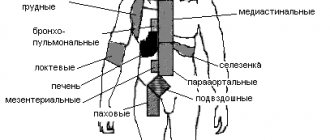
Lymphadenopathy (enlarged lymph nodes) is the first sign of the disease. Very often, patients identify them on their own on the neck, groin, armpits, and thighs. They are distinguished by their density and mobility, their palpation does not cause pain, there are no adhesions with each other or with the skin.
| Affected area | Clinical manifestations |
| Mediastinal nodes |
|
| Retroperitoneal nodes | Painful symptoms in the abdominal area, which occur in attacks or last constantly, as well as swelling of the legs. |
| Lungs |
|
| Bone marrow |
|
| Gastrointestinal tract |
|
Sources
- Hodgkin's lymphoma. Clinical recommendations. / Rumyantsev A.G., Poddubnaya I.V., Savchenko V.G., Kaprin A.D. – 2021.
- Federal clinical guidelines for the diagnosis and treatment of Hodgkin lymphoma. /Myakova N.V. – 2014.
- Lymphogranulomatosis in children: a case study // V. A. Lebedeva, Z. K. Zeinulina, D. K. Zhumadillaeva. – 2003.
- Treatment of Hodgkin's lymphoma. / Bartosh I.A. – 2021.
- Modern treatment of Hodgkin's lymphoma (lymphogranulomatosis). /Demina I.A. – 2002.
Hodgkin's disease: diagnosis
Before diagnosing a patient with LBM, CELT hematologists conduct comprehensive studies. The main criterion confirming lymphogranulomatosis is the presence of Reed-Sternberg cells in a sample extracted from the affected lymph nodes. In order to obtain it, the following methods are used:
- Lymph node biopsy;
- Diagnostic thoracoscopy;
- Examination of the abdominal cavity with a laparoscope.
In the presence of the disease, laboratory blood tests reveal an increased ESR, decreased levels of lymphocytes, platelets and eosinophils. If there is a suspicion of involvement of the bone marrow in the pathological process, the patient may be prescribed:
- Puncture of bone marrow tissue;
- Trepanobiopsy of bone substance.
Instrumental diagnostic techniques:
- X-ray of the sternum and abdominal cavity; Ultrasound scan or computed tomography of the peritoneum, mediastinum.
Forecast
The stage of the disease is of greatest importance in the prognosis for lymphogranulomatosis. Patients with stage 4 disease have a 75% five-year survival rate, while patients with stages 1-2 have a 95% survival rate. The prognosis for signs of intoxication is poor. Early signs of an unfavorable course of the disease are “biological” indicators of activity.
Biological indicators of activity include:
- alpha-2-globulin more than 10 g/l,
- haptoglobin more than 1.5 mg%,
- an increase in ESR in the general blood test of more than 30 mm/h,
- increase in fibrinogen concentration more than 5 g/l,
- cerruloplasmin more than 0.4 extinction units.
If at least 2 of these 5 indicators exceed the specified levels, then the biological activity of the process is determined.
Lymphogranulomatosis: treatment
The development of treatment tactics for lymphogranulomatosis by CELT hematologists is based on diagnostic results and individual patient indications. The stage of the disease must be taken into account, and an integrated approach is practiced. The methods used by our specialists allow us to completely cure the disease.
| Treatment method | When is it shown? |
| Radiation therapy | Indicated in the first (local) stage. Before it, the affected node and spleen are often removed. Affected and healthy lymph nodes are irradiated. Impact on the latter is necessary as a preventive measure. |
| Chemotherapy | Indicated at the last stage to destroy tumor cells or maintain a period of remission. |
| Their combination | In the second and third stages, introductory “chemotherapy” is first prescribed, aimed at irradiating the affected nodes, then all the rest are irradiated as much as possible. The patient needs to undergo maintenance procedures for two to three years. |
Treatment results may include complete or partial remission. The first is the complete disappearance of lymphogranulomatosis and its absence within thirty days, the second is the elimination of subjective symptoms, reducing the size of the nodes by half within thirty days.
The Department of Hematology at CELT accepts candidates, doctors and professors of medical sciences. Their practical and scientific work experience is more than twenty-five years. You can make an appointment with them online or by contacting our information line operators. If necessary, you can make an appointment with specialists at any of our departments and undergo the necessary procedures. No less experienced and qualified specialists work in the gynecological department of our clinic, where you can undergo the procedure of uterine artery embolization.
At CELT you can consult a hematologist.
- Initial consultation – 3,500
- Repeated consultation – 2,300
Make an appointment
By making an appointment with a hematologist, you can get a comprehensive consultation. The doctor is competent to treat various blood diseases, most of which can be identified in the early stages and prescribe timely treatment to cope with the disease quickly and easily.
How to treat lymphogranulomatosis?
The main method of treating patients with lymphogranulomatosis is combined chemoradiotherapy, which varies in intensity depending on the volume of the tumor mass, that is, the total number of tumor cells in all affected organs.
In addition, the following factors influence the prognosis:
- massive lesion of the mediastinum;
- diffuse infiltration and enlargement of the spleen or the presence of more than 5 foci in it;
- tissue damage outside the lymph nodes;
- damage to lymph nodes in three or more areas;
- an increase in ESR more than 50 mm/h in stage A and more than 30 mm/h in stage B.
To treat patients with an initially favorable prognosis, 2 to 4 courses of chemotherapy are used in combination with irradiation of only the affected lymph nodes. In the group with an intermediate prognosis, 4-6 cycles of polychemotherapy and irradiation of the affected areas of the lymph nodes are used. In patients with an unfavorable prognosis of the disease, 8 courses of polychemotherapy and irradiation of areas with a large array of affected lymph nodes are carried out.