Osgood-Schlatter disease can present as a painful lump in the area below the kneecap during childhood and adolescence as puberty begins. Osgood-Schlatter disease occurs most often in children who participate in sports, especially sports such as running, jumping, or sports that require rapid changes in movement trajectories, such as football, basketball, figure skating and gymnastics.
And although Osgood-Schlatter disease is more common in boys, the gender gap narrows as girls become more involved in sports. Osgood-Schlatter disease affects more adolescents who play sports (by a ratio of one to five). The age range of incidence has a gender factor, as girls experience puberty earlier than boys. Osgood-Schlatter disease usually occurs in boys between 13 and 14 years of age and in girls between 11 and 12 years of age. The disease usually goes away on its own as bone growth stops.
General information
Osgood-Schlatter disease is a specific disease of the musculoskeletal system, namely the knee joints, characterized by dystrophic damage to the tibia in the area of its tuberosity.
Such aseptic destruction of bone tissue occurs against the background of permanent or acute trauma and usually affects only young people at the stage of intensive skeletal development. Clinically, the disease is manifested by swelling of the knee joint, the formation of a kind of growth (bump) under it and pain in its lower part, which occurs during normal physical activity (running, squats, etc.) or even without it.
This pathology was first described in 1878 by the French surgeon O. M. Lannelong under the name “Apophysitis of the tibia,” and in 1903, thanks to the work of the American orthopedist R. B. Osgood and similar works of the Swiss surgeon K. Schlatter (Schlatter), it appeared its more detailed nosography. Wikipedia defines this painful condition with the term “Osteochondropathy of the tibial tuberosity,” and the international classification assigned it the ICD-10 code – M92.5 “Juvenile osteochondrosis of the tibia and fibula.” Despite this, in medical practice this disease is still most often referred to as “Osgood-Schlatter disease” or simply “Schlatter disease”.
Surgical treatment
Surgical intervention is used if, after completion of the ossification process and conservative therapy, a pineal protrusion remains, which causes pain and impairs the function of the joint. Also, an indication for surgery is a cosmetic defect.
The operation consists of fixing bone fragments to the tibial tuberosity if the fragment is large. Or these fragments are deleted.
Removal is performed openly, through a small incision in the area of the tibial tuberosity
In our clinic we use arthroscopic surgical treatment.
Clinical example. Patient M., 34 years old
appearance of the knee before surgery
X-ray before surgery
appearance of the knee joint
before surgery
X-ray after surgery
Advantages of the arthroscopic method of surgery:
- minimal invasiveness of the operation.
- good cosmetic result (the operation is performed through two skin punctures).
- achieving good results in almost 100% of cases.
- minimal number of complications.
- reduction of postoperative rehabilitation period.
The advantages of treating Osgood-Schlatter disease with us:
- diagnosis of the disease is carried out using modern equipment by a highly qualified specialist;
- making an accurate diagnosis, treating this pathology of the knee joint using a conservative or surgical method (using modern arthroscopic equipment);
- complete preoperative and postoperative management;
- preventing the occurrence of complications in the form of inflammation and relapses of the “bump”;
- patient consultation at all stages of treatment and recovery.
Don't waste your time and money! Don't risk your health!
Contact a qualified orthopedist at the first symptoms of the disease. In our clinic we will help you quickly get rid of your illness.
Pathogenesis
The mechanism of occurrence and further development of Osgood-Schlatter syndrome is directly related to the patient’s age and physical activity. According to statistics, in the vast majority of cases, doctors diagnose Schlatter's disease in children and adolescents in the age group from 10 to 18 years, while young people involved in sports suffer from it 5 times more often than their peers leading a passive lifestyle . The same reason for more intense physical activity explains the fact that this osteochondropathy mainly affects boys.
As is known, two large bones are involved in the formation of the human knee joint - the femur (above the knee) and the tibia (below the knee). In the upper part of the last of them there is a special area (tuberosity), to which the quadriceps femoris muscle is attached by means of a tendon. It is this part of the bone that is responsible for its growth in childhood and adolescence and is therefore particularly susceptible to various injuries and damage. During active physical activity, in some cases, the knee joint is subject to a large load and the quadriceps muscle is overstrained, which leads to stretching or tearing of the tendon and a lack of blood supply in this area. As a result of such a traumatic effect and a decrease in nutrition in the area of the tibial tuberosity, gradual necrotic changes develop in it, up to the death of individual parts of its core.
In addition, any injury to the knee joint or constant impact on its musculoskeletal structure (for example, jumping) can cause cracks and microfractures of the tibial tuberosity, which the growing body tries to quickly compensate for by the growth of new connective tissue. As a result of this, a person develops a bone growth (bump), typical of Osgood-Schlatter osteochondropathy, that forms just below the knee. Such a pathological process usually involves one leg, but bilateral involvement of the lower extremities is also possible.
Treatment of Schlatter's disease
- Creating limb rest is immobilization with a plaster cuff, which in some cases is not at all necessary; Nevertheless, rest is necessary, up to complete exemption from physical education lessons.
- Physiotherapy - electrophoresis with calcium and procaine (alternately), lidocaine (to relieve inflammation); with cocarboxylase, aminophylline; ozokerite, paraffin, mud
- Shock wave therapy
- Surgical treatment is indicated for significant fragmentation and persistent pain - fixation of the tuberosity to the tibia with a bone graft (required extremely rarely).
- It is mandatory to keep the knee in a bandage as the quadriceps muscle can be torn off.
- Monarch Disease Ontology release 2018-06-29sonu - 2018-06-29 - 2018. https://wikidata.org/wiki/Track:Q55345445″>
Classification
In the orthopedic environment, this pathology is usually classified according to the degree of its severity and the severity of the observed external and internal symptoms. Regarding this, there are three degrees of Schlatter’s disease, namely:
- initial – visual manifestations in the form of a lump-like growth under the knee are absent or minimal, pain in the area of the knee joint is episodic, mild and occurs mainly at the time of physical activity on the leg;
- an increase in symptoms - swelling of the soft tissues around the affected knee appears, a lump becomes visually visible directly below it, pain syndrome manifests itself during the period of loads on the leg and for a certain period of time after them;
- chronic - a lump-like formation is clearly visible under the knee, which is most often surrounded by swelling, discomfort and pain in the joint is persistent and is observed even at rest.
Causes
There are two main physical activity-related underlying causes of Osgood-Schlatter disease in adolescents and children:
- direct injuries to the tissues of the knee joint (subluxations and dislocations, sprains, bruises, fractures);
- systematic microtraumas (external and internal) of the knee joint that occur as a result of intense sports or other activities associated with excessive physical stress on the lower extremities.
The greatest risk factors for Schlatter's disease in adolescents and children are:
- football, basketball, handball, hockey, volleyball, tennis;
- track and field athletics, acrobatics, gymnastics;
- judo, kickboxing, sambo;
- skiing, sports tourism, figure skating, cycling;
- ballet, sports and ballroom dancing.
Treatment
Osgood-Schlatter disease usually resolves on its own, and symptoms disappear after bone growth has completed. If the symptoms are severe, then treatment includes medication, physiotherapy, and exercise therapy.
Drug treatment involves prescribing pain relievers such as acetaminophen (Tylenol, etc.) or ibuprofen. Physiotherapy can reduce inflammation and relieve swelling and pain.
Exercise therapy is necessary to select exercises that stretch the quadriceps muscle and hamstrings, which reduces the load on the area where the patellar tendon attaches to the tibia. Hip strengthening exercises also help stabilize the knee joint.
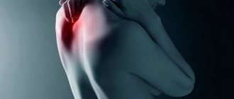
Symptoms of Osgood-Schlatter disease
The severity of the negative manifestations of this pathology in different patients may differ depending on the nature of the injuries received, the degree of physical activity and the personal characteristics of the body.
At the beginning of the development of the disease, the patient begins to experience vague pain in the knee area, which usually appears after or during physical activity on the affected limb. As a rule, such pain is not yet associated with an internal pathological process and therefore there are quite few visits to the doctor during this period.
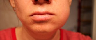
Over time, pain symptoms begin to increase, are localized in one place and can appear not only during physical activity, but also at rest. At the same time, swelling caused by edema appears around the affected knee, and a lump-like growth appears just below it. During this period of illness, it becomes increasingly difficult for the patient (especially the athlete) to perform his usual exercises, and sometimes even natural leg movements. The greatest intensity of pain is observed in the body position - kneeling.
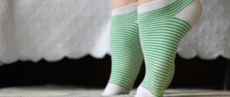
Photo of a “bump” in Osgood-Schlatter disease
In addition, the patient may experience other negative symptoms:
- tension in the leg muscles (mainly the thigh muscles);
- limited mobility of the knee joint;
- outbreaks of sharp “shooting” pain in the knee area, arising when it is overstrained;
- severe morning swelling in the upper or lower part of the knee, which forms the day after physical activity.
When you independently palpate the affected knee, points of pain are felt, as well as smoothness of the contours of the tibia. The texture of the knee joint is felt as densely elastic, and a hard lump-like formation is felt under the swollen soft tissues. The general well-being of the patient, despite the accompanying pain and pathological processes in the knee, does not change significantly. The skin over the affected joint does not turn red, temperature indicators remain normal.
In most clinical cases, this disease occurs in a measured chronic form, but sometimes its wave-like course can be observed with periods of sudden exacerbation and relative calm. Without medical intervention and with continued physical activity, negative symptoms can persist for many months and worsen against the background of further mechanical damage to the knee joint. However, the manifestations of the disease gradually disappear on their own over 1-2 years, and by the time the period of bone tissue growth ends (approximately 17-19 years) they usually eliminate themselves. Before treating Osgood-Schlatter, the need for such therapy should be comprehensively and individually assessed, since in some cases it may be inappropriate.
Literature review
A group of German scientists studied the effectiveness of radial shock wave therapy in patients with Schlatter's disease. The study included 14 patients with recurrent course of this disease. After completing the course of shockwave therapy, all patients noted a significant improvement. The pain at rest was completely relieved, and motor activity was restored. Only 2 patients had moderate pain when walking up stairs. Based on their study, the authors concluded that radial shock wave therapy is highly effective in the treatment of Osgood-Schlatter disease.
Taiwanese scientists analyzed the results of a number of studies on shockwave therapy for knee tendinopathy. A total of 1,189 patients were treated as part of this work:
- with tendinopathy;
- with Osgood-Schlatter disease;
- with cruciate ligament injury;
- with synovitis and other diseases.
562 patients received from 3 to 6 sessions of shockwave therapy with an interval of 1 week, the remaining patients received traditional methods of treatment or placebo.
Analysis of research results showed that the effectiveness of shockwave therapy for pathologies of the knee joint and periarticular tissues is significantly higher than that of traditional treatment methods. Patients experience pain relief faster and functional activity in the knee is restored. At the same time, for complete tissue restoration, long-term courses, including 5-6 sessions, are preferable to short-term ones.
A group of scientists from Egypt conducted a comparative study of the use of shockwave therapy and interference therapy in patients with Osgood-Schlatter disease. The study involved 40 patients who were divided into 2 identical groups - the first, consisting of 20 people, received interference current therapy + traditional treatment, the second - shockwave therapy + traditional treatment.
After completing the course, the pain syndrome was completely relieved in patients from both groups. But in functional terms, more pronounced positive results were noted by patients from group 2 - mobility in the knee joint increased from 115 to 136 points, while in patients from group 1 - from 116 to 127 points. According to the WAMAC questionnaire, which comprehensively assesses joint function (stiffness, pain and mobility), the scores in patients receiving shockwave therapy decreased from 52 to 20 points, while in the group receiving interference therapy - from 51 to 38 points.
Based on the results of their study, the authors concluded that shockwave therapy is highly effective in treating Osgood-Schlatter disease.
Rice. 3. Treatment result according to the WAMAC questionnaire
Tests and diagnostics
In general, the doctor can suspect the development of Schlatter’s disease due to the complexity of the patient’s clinical manifestations and the localization of the pathological process typical for this disease. The gender and age of the patient also play an important role in correct diagnosis, since adults, as a rule, are not exposed to this type of damage. Even through a simple visual examination and the usual collection of anamnesis regarding previous injuries or overloads of the knee joint, an experienced orthopedic traumatologist is able to make the correct diagnosis, but it would be useful to confirm it using some hardware diagnostic methods.
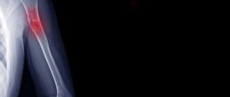
The decisive factor in making an unambiguous diagnosis of Osgood-Schlatter disease in children and adolescents was and remains radiography , which, in order to increase the information content of the pathology, is best carried out over time. To exclude other orthopedic diseases, such an examination of the affected knee joint must be carried out in two projections, namely lateral and direct.
In the initial phase of the development of the disease, X-ray images show a flattening of the tibial tuberosity in its soft part and a rise in the lower edge of the clearing, corresponding to the adipose tissue located in the anterior lobe of the knee joint. The last discrepancy with the norm is caused by an increase in the size of the infrapatellar bursa, which occurs as a result of its aseptic inflammation. There are most often no visible changes in the ossification nucleus itself at this stage of Schlatter’s disease.
X-ray of the knee joint in Osgood-Schlatter disease
As the pathology progresses, the x-ray picture changes for the worse. The photographs show a shift of the ossification nucleus by 2-5 mm upward and forward relative to the standard location of the tuberosity or its fragmentation. In some cases, there may be unevenness of the natural contours and unclear structure of the ossification nucleus, as well as signs of gradual resorption of its parts, but most often it fuses with the main body of the bone with the formation of a bone conglomerate in the form of a spiky protrusion. This “bump”, characteristic of Schlatter’s disease, in the later stages of the disease is especially clearly visible on a lateral radiograph and is clearly palpable during palpation in the area of the tuberosity.
In some atypical cases, it may be necessary to prescribe an MRI , CT and/or ultrasound of the problem knee and adjacent tissues to clarify the expected diagnosis. It is also possible to use a technique such as densitometry , which will provide comprehensive data on the structural state of the bones being studied. Other laboratory diagnostic methods, including PCR studies and blood tests for rheumatoid factor and C-reactive protein, are carried out in order to exclude the possible infectious nature of problems with the knee joint (mainly nonspecific and specific arthritis ).
Differential diagnosis of Osgood-Schlatter syndrome must be carried out with any fractures in the knee joint, bone tuberculosis , patellar tendinitis osteomyelitis , infrapatellar bursitis , Sinding-Larsen-Johanson disease and tumor neoplasms.
Symptoms (signs)
The disease has a gradual onset and is characterized by:
- cutting, burning pain in the upper half of the tibia, which intensifies with movement and decreases with rest;
- swelling in the growth area, without external skin changes;
- tightness of the thigh muscles;
- restriction of movements;
- the formation of a dense tumor under the knee, which does not interfere with walking.
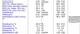
The disease is diagnosed using X-rays (in lateral projection), ultrasound, MRI, CT, radioisotope studies, CBC, blood biochemistry tests, tests for rheumatoid factor.
On palpation, the lump is painful, dense, swollen, with clear, uneven edges.
Treatment with folk remedies
With the permission of the attending physician and in addition to traditional methods of treating Schlatter's disease, the use of folk remedies is allowed, which mainly boil down to the use of various compresses and rubbing that relieve pain and inflammation. The following recipes have proven themselves well in this direction.
Honey compress
To make such a product, natural fresh honey should be mixed in equal proportions with medical alcohol and heated in a water bath until the honey is completely liquefied. Immediately after this, you need to moisten a clean piece of gauze in this mixture, apply it to the problem joint and wrap it first with cellophane and then with a warm cloth (preferably wool). Such procedures can be carried out twice a day for a month, keeping the compress on the knee for approximately 2 hours.
St. John's wort and yarrow
A kind of ointment is prepared from a crushed mixture of these herbs (in equal proportions), for which they are mixed with rendered pork fat, and then heated over low heat for 15 minutes. After cooling, the ointment is considered ready for use and can be rubbed into the skin around the injured knee 2-3 times a day.
Garlic
Two medium heads of garlic are peeled, passed through a garlic press and mixed with 400 ml of regular apple cider vinegar. Before use, this drug should be infused for a week in a dark glass container, where it can then be stored for six months. The method of application is to rub a small volume of this tincture into the damaged knee area 2-3 times a day.
Burdock
Finely chop a few fresh burdock leaves, place them on clean gauze and wrap it around the painful part of the leg for 3 hours. This dry compress is placed at night and applied once every 24 hours for one month (instead of burdock, you can take cabbage or plantain leaves).
Onion
Grate two small peeled onions on a fine grater and mix them with 1 tsp. granulated sugar. The resulting mixture is used for night compresses for about a month.
Healing oils
Camphor, clove, eucalyptus, menthol oil and aloe juice should be carefully mixed in equal proportions. This mixture should be rubbed into the skin over the damaged area several times a day, and then wrapped with a warm cloth.
Prevention
Prevention of the first occurrence or re-development of Schlatter's disease in general consists of controlling the intensity of physical activity performed by a child or adolescent on the lower extremities, especially if he is actively involved in sports, dancing, etc. This largely depends on the parents, since young people are rarely aware of the adequacy of their own training and can constantly overexert themselves. Also, an important role in the preservation of joints and the entire skeletal system during the period of its growth is played by nutritious nutrition, which should include the entire complex of minerals and vitamins . In addition, it is imperative to undergo full professional treatment for any injuries sustained by children, even if at first glance they seem insignificant.
Osgood-Schlatter disease in adults
The age group at increased risk of developing Schlatter's disease includes only children and adolescents, whose tibia in the area of their tuberosity are in the process of intensive growth. As it stops and the body naturally matures, the tuberosity zone becomes stronger and eventually completely ossifies, which in itself excludes the development of this disease in adults. The only thing that can connect adults with this osteochondropathy is its residual changes in the form of small tubercles under the knees.
Complications and consequences of Osgood-Schlatter
Most often, Osgood-Schlatter disease does not lead to any serious complications in the damaged knee joint and goes away over time with virtually no consequences. Sometimes, at first after treatment, local swelling or minor pain persists in the knee area, which usually occurs after excessive physical exertion.
Also, quite often, in the area of the previously affected lower leg, a formed bone growth remains noticeable, which, as a rule, does not affect the mobility of the knee joint and does not cause a feeling of discomfort both in everyday life and during sports. In rare cases, with severe cases and/or improper treatment of Schlatter's disease, such a bone growth can provoke deformation and displacement of the patella. osteoarthritis as adults and may experience pain when kneeling, as well as aching pain when weather conditions change.
Shock wave therapy: mechanisms of therapeutic action
A shock wave is essentially a sound pulse emitted by a generator located outside the human body, that is, extracorporeally, which is why the technique is called extracorporeal shock wave therapy (ESWT).
In a short period of time (about 1 microsecond), the wave has a powerful effect (up to 20 MPa) on tissues, due to which mechanisms are triggered in them that have a positive therapeutic effect:
- a new vascular network is formed at the site of exposure (neoangiogenesis);
- in the zone of action of shock waves, blood circulation improves, the outflow of decay products is restored;
- the permeability of cell membranes to drugs increases;
- the sound impulse irritates the nerve endings, which causes a local analgesic effect;
- endorphins are released, which is also accompanied by an analgesic effect;
- bone growths are loosened, fibrous and scar tissues are softened.
Thus, the use of shockwave therapy for Schlatter's disease leads to pain relief and improved mobility in the knee joint.
List of sources
- Abalmasova E.A. Osteochondropathies // Orthopedics and traumatology of childhood. - M., 1983. - P. 385-393.
- Gorodnik A.G., Lantsov V.P. The problem of Osgood Schlatter's disease // Vestn. X-ray Radiol. — 1963.- No. 38.-С14-17.
- Pozharsky V.F., Osteochondropathy of the tibial tuberosity (Osgood Schlatter disease) // Medical assistant Obstetrics.- 1982.- No. 47(9).- P.53.
- Pudovnikov S.P., Tarabykin A.N. “Method of surgical intervention for Osgood-Schlatter disease” // Military Medical Journal 1987. - No. 7. - P. 62.
- Esedov E.M. “Osgood-Schlatter syndrome” in the practice of a therapist // “Clinical Medicine”. - 1990, - No. 1. - P. 109-111.