Infarction-pneumonia is an inflammation that occurs after a local persistent disturbance of blood circulation in the lung tissue. The main cause of this phenomenon is considered to be thromboembolism of the branches of the pulmonary artery. When massively spread, it leads to instant death. When small branches are affected, only local infarctions of the pulmonary parenchyma are observed.
Pulmonary infarction - what is it?
According to statistics, pulmonary infarction accounts for approximately 10-25% of all cases of pulmonary embolism (PE).
Quite often the diagnosis is not made during the patient’s lifetime. The fatal outcome from the disease is recorded according to various sources in 5-30% of patients. The risk of death from pulmonary embolism increases if there is no proper treatment and there are concomitant background pathologies. It should be noted that infarction of the right lung is twice as common as that of the left. The lower lobes of the organ are affected four times more often than the upper ones.
Pulmonary infarction - classification of the disease
Depending on how strongly the lumen of the pulmonary artery is blocked by thromboembolism, the following types of pulmonary infarction are distinguished:
- Submassive thromboembolism (blockage at the level of segmental and lobar branches is diagnosed).
- Massive thromboembolism (thrombus blocks the main trunk or main branches of the pulmonary artery).
- Thromboembolism of small pulmonary arteries.
Pulmonary infarction can also be:
- primary (it is not known where the thromboembolus came from) and secondary (in addition to pulmonary embolism, the patient has venous thrombophlebitis);
- extensive (large area of damage) and limited (only subsegmental branches of the pulmonary artery are blocked);
- complicated (sepsis, hemoptysis, pleural empyema, abscess formation) and uncomplicated.
Hemorrhagic pulmonary infarction
Thromboembolism of the pulmonary arteries often leads to ischemia of the pulmonary parenchyma. As a result, the damaged lung tissue is filled with blood, which comes from areas with normal vascularization. A hemorrhagic pulmonary infarction develops.
At the site of inflammation, optimal conditions are created for the development of infection, which provokes the occurrence of symptoms of heart attack-pneumonia. If a branch of the pulmonary artery is blocked by an infected thrombus, destruction of the parenchyma is noted and a lung abscess is formed.
Treatment of heart attack-pneumonia
If the cause of pneumonia after a pulmonary infarction is thromboembolic complications, then therapy begins with fibrinolytic agents and anticoagulants.
Symptomatic treatment involves reducing pain by taking analgesics. Etiotropic treatment involves a course of antibiotics depending on the nature of the pathogen. The principle of therapy after myocardial infarction is based on the use of steroid hormones, which contribute to the rapid improvement of general well-being.
Only an experienced doctor can prescribe treatment, based on the results of the studies obtained, and taking into account the individual characteristics of each patient’s body. Pulmonologists, cardiologists, therapists, neurologists and other doctors at the Yusupov Hospital always prescribe a course of treatment for their patients on an individual basis. In this case, the general condition of the patient, age factor, the presence of concomitant diseases and much more are taken into account. Doctors at the Yusupov Hospital are putting back on their feet even those patients who were abandoned in other medical centers.
You can make an appointment with a doctor at the Yusupov Hospital by phone and by filling out the feedback form on the website.
Causes of pulmonary infarction
Most often, pulmonary infarction develops in people who suffer from diseases of the cardiovascular system:
- mitral stenosis;
- atrial fibrillation;
- coronary heart disease;
- myocardial infarction;
- infective endocarditis;
- cardiomyopathy;
- vasculitis;
- heart failure;
- atrial maxomas, etc.
In this case, blood clots form in the appendage of the right atrium and are carried with the blood flow into the arteries of the pulmonary circle.
Also, the causes of pulmonary infarction can be:
- thrombophlebitis of the deep pelvic veins;
- thrombosis of leg veins.
In these two diseases, floating thrombi that are attached to the distal part of the venous vessel are dangerous.
Other causes of pulmonary embolism include:
- fracture of tubular bones, due to which the patient is forced to remain in bed for a long time;
- caesarean section, natural childbirth;
- extensive thoracic, abdominal and gynecological operations;
- hemorrhoidectomy.
The risk of pulmonary infarction is high in people over 60 years of age, as well as in:
- recurrent venous thrombosis;
- hereditary burden of pulmonary embolism;
- taking hormonal contraceptives;
- the presence of a pancreatic tumor;
- obesity;
- pulmonary hypertension.
Dangerous background pathologies are considered:
- DIC syndrome;
- sickle cell anemia;
- polycythemia;
- heparin-induced thrombocytopenia.
1.General information
According to the definitions existing in modern medicine, a heart attack is a rapid, one-stage focal necrosis (death, death) of a large number of parenchymal, i.e. the main, functionally specialized cells of any tissue or organ. The cause is ischemia, a pathological state of oxygen and trophic starvation due to a lack of blood supply. Outside of medicine, the most famous example of such a scenario is myocardial infarction, while in clinical practice, infarctions of the spleen, brain (stroke), kidneys and other organs are also encountered.
Due to the peculiarities of the anatomical structure and vasculature (blood supply) of the organs of the human respiratory system, pulmonary infarction in most cases develops on the right side and in the lower lobes.
A must read! Help with treatment and hospitalization!
Signs of pulmonary infarction
The first symptoms of a pulmonary infarction appear two to three days after a branch of the pulmonary artery is blocked by a thrombus. The patient feels sudden pain in the chest, which intensifies when coughing, deep breathing, or bending over. Discomfort in the chest area is explained by reactive pleurisy in a necrotic area of the lung.
With a reaction of the diaphragmatic pleura, an “acute abdomen” is possible. In 30% of patients, hemoptysis is observed or “rusty” streaks appear in the sputum. Pulmonary hemorrhage occurs in 2-5%.
Also, pulmonary infarction can manifest itself:
- increase in body temperature to 38-39°C;
- severe shortness of breath;
- arrhythmia;
- cyanosis and pallor of the skin;
- arterial hypotension;
- hiccups;
- vomiting, nausea.
Half of the patients develop hemorrhagic or serous pleurisy. Occasionally, cerebral disorders occur. Their symptoms:
- jaundice;
- fainting;
- coma.
If you experience similar symptoms, consult your doctor
. It is easier to prevent a disease than to deal with the consequences.
Diagnosis of pulmonary infarction
Pulmonologists and cardiologists diagnose pulmonary embolism. During a physical examination, the following is revealed:
- small wheezing;
- decreased breathing;
- pleural friction noise;
- systolic murmur;
- shortening of percussion sound;
- gallop rhythm;
- splitting and emphasis of the second tone on the aorta.
While palpating the abdomen, the doctor notes an enlarged and painful liver.
Blood biochemistry, general blood test and blood gas analysis during pulmonary infarction show:
- moderate leukocytosis;
- arterial hypoxemia;
- total bilirubin with normal transaminase values;
- increased activity of lactate dehydrogenase.
Pulmonary infarction on the ECG is manifested by signs of overload of the right heart parts, incomplete blockade of the right bundle branch. EchoCG markers of pulmonary embolism may include:
- hypokinesia and dilatation of the right ventricle;
- increased pressure in the artery of the lung;
- detection of a blood clot in the right side of the heart.
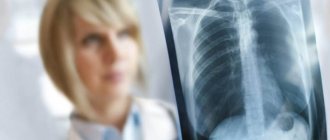
Ultrasound scanning of the veins of the lower extremities during pulmonary infarction diagnoses deep vein thrombosis. An x-ray of the lungs in lateral and frontal projections shows:
- deformation and expansion of the lung root;
- a wedge-shaped area of decreased transparency;
- effusion in the pleural cavity.
Pulmonary angiography for pulmonary embolism reveals narrowing of the lumen of the pulmonary artery due to intra-arterial filling defects. Scintigraphy is used to confirm the presence of areas of decreased pulmonary perfusion.
Pulmonary infarction must be differentiated from:
- atelectasis;
- lobar pneumonia;
- spontaneous pneumothorax;
- pericarditis;
- myocardial infarction;
- rib fracture;
- myocarditis.
Pulmonary embolism (PE) - symptoms and treatment
Pulmonary embolism (PE) is a blockage of the pulmonary arteries with blood clots of various natures, most often formed in the large veins of the lower extremities or pelvis.
Risk factors for pulmonary embolism include pathological conditions in which there is impaired venous blood return, endothelial damage or endothelial dysfunction, and hypercoagulability disorders.
As a result of thromboembolism of the pulmonary arteries, the blood supply to the lung tissue stops, necrosis (tissue death) develops, infarction-pneumonia, and respiratory failure occur. The load on the right parts of the heart increases, right ventricular circulatory failure develops: cyanosis (blue discoloration of the skin), edema in the lower extremities, ascites (fluid accumulation in the abdominal cavity). The disease may develop acutely or gradually over several hours or days. In severe cases, the development of pulmonary embolism occurs rapidly and can lead to a sharp deterioration of the patient’s condition and death.
Every year, 0.1% of the world's population dies from pulmonary embolism. In terms of the frequency of deaths, the disease is second only to IHD (coronary heart disease) and stroke. More patients with pulmonary embolism die than patients with AIDS, breast and prostate cancer, and victims of road accidents combined. The majority of patients (90%) who died from pulmonary embolism were not correctly diagnosed in time and did not receive the necessary treatment. PE often occurs where it is not expected - in patients with non-cardiological diseases (trauma, childbirth), complicating their course. The mortality rate for pulmonary embolism reaches 30%. With timely, optimal treatment, mortality can be reduced to 2-8%.[2]
Symptoms of pulmonary embolism depend on the size of the blood clots, the suddenness or gradual onset of symptoms, and the duration of the disease. The course can be very different - from asymptomatic to rapidly progressing, up to sudden death.
PE is a ghost disease that wears the mask of other heart or lung diseases. The clinic may be infarct-like, reminiscent of bronchial asthma, acute pneumonia. Sometimes the first manifestation of the disease is right ventricular circulatory failure. The main difference is the sudden onset in the absence of other visible reasons for the increase in shortness of breath.
Etiology
PE usually develops as a result of deep vein thrombosis, which usually develops 3-5 days before the onset of the disease, especially in the absence of anticoagulant therapy.
Risk factors for pulmonary embolism
When diagnosing, the presence of risk factors for thromboembolism is taken into account. The most significant of them are: fracture of the femoral neck or limb, replacement of the hip or knee joint, major surgery, trauma or brain damage.
Dangerous (but not so strong) factors include: knee arthroscopy, central venous catheter, chemotherapy, chronic heart failure, hormone replacement therapy, malignant tumors, oral contraceptives, stroke, pregnancy, childbirth, the postpartum period, thrombophilia. In malignant neoplasms, the incidence of venous thromboembolism is 15% and is the second leading cause of death in this group of patients. Chemotherapy treatment increases the risk of venous thromboembolism by 47%. Unprovoked venous thromboembolism may be an early manifestation of malignancy, which is diagnosed within a year in 10% of patients with an episode of PE.[2]
The safest, but still risky, factors include all conditions associated with prolonged immobilization (immobility) - prolonged (more than three days) bed rest, air travel, old age, varicose veins, laparoscopic interventions.[3]
Some risk factors are common to arterial thrombosis. These are the same risk factors for complications of atherosclerosis and hypertension: smoking, obesity, sedentary lifestyle, as well as diabetes, hypercholesterolemia, psychological stress, low consumption of vegetables, fruits, fish, low level of physical activity.
The older the patient, the more likely it is to develop the disease.
Finally, today the existence of a genetic predisposition to pulmonary embolism has been proven. The heterozygous form of factor V polymorphism increases the risk of initial venous thromboembolism by three times, and the homozygous form by 15-20 times.
The most significant risk factors contributing to the development of aggressive thrombophilia include antiphospholipid syndrome with an increase in anticardiolipin antibodies and a deficiency of natural anticoagulants: protein C, protein S and antithrombin III.
How to treat pulmonary infarction
Treatment of pulmonary infarction in children and adults involves the use of:
- non-narcotic or narcotic analgesics for pain relief;
- direct (Frakiparin, Heparin) and indirect coagulants to prevent further thrombus formation (coagulants are contraindicated in cases of bleeding, duodenal/gastric ulcers, hemorrhagic diathesis, malignant tumors);
- fibrinolytic therapy with Urokinase, Streptokinase or tissue plasminogen activator to dissolve existing blood clots.
If a pulmonary infarction is complicated by arterial hypertension, vasopressors (Dopamine, Norepinephrine), Reopoliglucin are administered intravenously. At the first symptoms of heart attack-pneumonia, antibiotics are prescribed. Also, patients with pulmonary embolism require oxygen inhalation through a special nasal catheter.
If conservative treatment does not provide improvement in well-being, thromboembolectomy is performed from the pulmonary artery - a vena cava filter is installed in the inferior vena cava system.
Treatment and prevention
To eliminate pain, the patient is prescribed non-narcotic (less often narcotic) analgesics. Coagulants are used to prevent further thrombosis. And to stimulate the dissolution of already formed blood clots - streptokinase and urokinase, tissue plasminogen activator.
If there is a suspicion of post-infarction pneumonia, antibiotics are prescribed. The drugs are prescribed orally and in the form of droppers. In cases where conservative treatment does not give the expected effect, surgical intervention is indicated - pulmonary embolectomy.
To avoid problems in the future, it is recommended not to start and treat thrombophlebitis in a timely manner. Since pulmonary infarction in the vast majority of cases develops precisely against their background. Therefore, for prevention it is necessary:
- wear compression stockings;
- do therapeutic exercises;
- Visit a phlebologist regularly if you are prone to varicose veins and thrombosis.
If you experience symptoms of a pulmonary infarction, consult your doctor immediately. At the medical center you will receive a consultation with a pulmonologist. If necessary, you will be examined by a surgeon and a cardiologist. At the clinic you will also be tested and undergo the necessary examinations. If therapy is started in a timely manner, the prognosis is favorable.
Prevention of pulmonary infarction
Prevention of pulmonary infarction is carried out taking into account the reasons that provoked the development of the disease. To avoid pulmonary embolism, cardiologists and pulmonologists recommend:
- promptly treat thrombophlebitis;
- wear compression stockings for pathologies of the veins of the lower extremities;
- do therapeutic exercises daily;
- observe the timing of the use of intravenous catheters during infusion therapy;
- follow the doctor’s recommendations after undergoing surgical treatment.
This article is posted for educational purposes only and does not constitute scientific material or professional medical advice.
4.Treatment
First aid for suspected pulmonary infarction consists of relieving intense pain and immediate hospitalization of the patient. Further, according to indications (and in the absence of contraindications), anticoagulants, antiplatelet agents, thrombolytics, hormonal vasopressors, and supportive oxygen therapy are used; if there is a reasonable or confirmed suspicion of post-infarction infection, broad-spectrum antibiotics are used in effective doses. In some severe cases, the method of choice, which does not have a reasonable life-saving alternative, becomes thoracic surgical intervention.
It should be emphasized that in case of pulmonary infarction, the time factor is critical. The prognosis is favorable if you seek help in a timely manner and apply an adequate protocol for its provision, when it is possible to quickly and accurately diagnose the cause and prevent possible complications.