Ehlers-Danlos syndrome (according to some sources, Ehlers-Danlos syndrome, EDS) is a rare hereditary collagen pathology that affects the skin, musculoskeletal system and other organs. Other names for the syndrome are “skin hyperelasticity”, cutis hyperelastica, Rusakov desmogenesis imperfecta and Chernogubov-Ehlers-Danlos syndrome. People with this syndrome could be seen in the circus in the past because of their ability to surprise ordinary people with their appearance and movements. Some famous people also suffered from this disease - for example, virtuoso violinist Niccolo Paganini.
History of the discovery of the syndrome
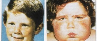
People with this disease were described by Hippocrates in 400 BC. e. in the essay “Airs, Waters and Places” (De aere, aquis, locis). He observed nomads and Scythians with weak joints and multiple scars around them. Hippocrates believed that the scars were traces of cauterization, with the help of which they tried to reduce joint hypermobility. Much later, in 1657, the Dutch surgeon J. Van Meekeren (1611–1666) described a small Spaniard with very elastic skin. A boy named George Albs could stretch the skin of his chin to his chest, and the skin of his knees to the middle of his shins, but this only affected the right half of his body. World-famous violinist and composer Niccolo Paganini (1782–1840) had hypermobile joints, a thin physique, and a deformed chest—all symptoms characteristic of a carrier of Ehlers-Danlos syndrome. At the end of the 19th century, some patients with EDS performed as people with unusual abilities in traveling shows, such as the “elastic lady” described by American doctors George M. Gould and Walter L. Pyle. For a long time, the diversity of clinical manifestations of the syndrome did not allow a detailed description of all forms of the disease.
Classification and name
The first detailed description of EDS was presented by the Russian doctor at the Myasnitskaya Hospital in Moscow, Andrei Chernogubov, at the Moscow Venereological and Dermatological Society in 1892. He described two patients with increased mobility of large joints. One of them was a 17-year-old boy with epilepsy who had fragile and hyperelastic skin that was unable to hold sutures.
Later, in 1901, Danish dermatologist Edvard Lauritz Ehlers published a description of a patient with weak joints and hyperelastic skin, with a predisposition to bruising. He demonstrated it at the Danish Dermatological Society in 1899. Seven years later, French physician Henri-Alexandre Danlos examined a patient with vascular skin lesions on the elbows and knees. Afterwards, separate descriptions of this syndrome appeared in both the USA and England, and by 1966 the total number of reports had increased to 300. In 1936, the English dermatologist Frederick Parkes Weber combined all cases with hyperelasticity and fragility of the skin, as well as hypermobility joints. He called the new disease “Ehlers-Danlos syndrome.”
In 1972, the first molecular defect in collagen was discovered in EDS. In 1986, at the International Congress on Hereditary Connective Tissue Diseases in Berlin, 9 types of Ehlers-Danlos syndrome were identified, but in 1997, in Villefranche-sur-Mer (France), experts developed and adopted a more precise classification. It already had 6 types of EDMS:
- classical;
- hypermobile;
- vascular;
- kyphoscoliotic;
- achondroplasyplastic;
- dermatosparaxis.
Ehlers-Danlos syndrome - symptoms and treatment
Ehlers-Danlos syndrome is a set of connective tissue dysplasias that differ in inheritance patterns and chemical abnormalities.
In most of the verified observations, an autosomal dominant mode of inheritance is noted.[7]
With the described syndrome, mutations are observed in the DNA structure of genes encoding the structure of fibrillar protein, which forms the basis of connective tissue - collagen.[2]
In types 1 and 2 of the disease, there is a decrease in fibroblast activity, increased production of proteoglycans, a defect in the activity or complete absence of proteins that contribute to normal collagen maturation. A molecular defect is found in Proalfa 1 (V) or Proalfa 2 (V)[3] collagen chains of the fifth type, an altered structure of collagen structures of the “broccoli” type is detected. Inherited according to AD type.[1]
The third type is caused by mutations in the genes for the synthesis of collagen III alpha1, tenascin X, and is manifested by insufficient collagen production, which is associated with damage to the ligamentous apparatus and frequent joint dislocations. Transmitted by AD type.[8]
The fourth type is determined by the insufficiency of production of collagen fibers of the third type, which are part of the structure of the vascular wall, which is manifested by pathology of the cardiovascular system, and the skin is damaged less than in other subtypes of the disease.[3] Ruptures of blood vessels of all sizes are common.
In the fifth variant, a recessive type of inheritance associated with the X chromosome is described, its pathogenesis has not been well studied.[1]
The sixth type is caused by a lack of lysyl hydroxylase protein, which ensures the addition of a hydroxyl group to lysine in the collagen structure, i.e., which is a collagen-modifying enzyme. Transmitted by the AR type. It manifests itself not only by damage to the ligamentous apparatus and dermis, but also by pathology of the visual and muscular apparatus.[5]
In type seven, the modification of the type 1 collagen precursor into collagen is impaired. AR and AD inheritance variants are described. Such patients are characterized by short stature and have severe joint pathology.
In type ten, there is a defect in plasma fibronectin, which takes part in the formation of the intercellular substance.[4] In addition to the classic manifestations of the disease, in type ten there is a blood clotting disorder associated with a violation of the aggregation properties of platelets, and strip-like scars on the skin.[3] Inherited according to the AR type.
Pathologically all types s. Ehlers-Danlos symptoms are manifested by a decrease in skin thickness, pathological orientation and decompactization of collagen structures, a predominance in the number of elastic fibers, hypervascularization, and an increase in the diameter of veins and arteries.
Diagnosis criteria
This classification not only systematizes the forms of the syndrome, but also identifies major and minor criteria for diagnosing Ehlers-Danlos syndrome, which is why it is often called the Villefranche diagnostic criteria.
Major diagnostic criteria:
- Thin translucent skin with a visible venous pattern.
- Predisposition to vascular, intestinal and uterine ruptures or weakness of these structures.
- Easy bruising and bleeding.
- Characteristic facial features: wide-set eyes, depressed middle part and epicanthus fold at the inner corner of the eye, covering the lacrimal tubercle
Some minor diagnostic criteria:
- Premature aging of the limbs (acrogeria - atrophy of the skin of the hands and feet).
- Hypermobility of small joints (interphalangeal and metacarpophalangeal joints of the hand).
- Rupture of tendons and muscles.
- Clubfoot.
- Pneumothorax/pneumothorax.
- Retraction (sagging) of the gums, their underdevelopment.
- Complicated family history, sudden death of close relatives.
Two or more major criteria allow one to suspect EDS; laboratory confirmation is required for clarification. If only small criteria exist, then this is not an EDS - there must be at least one large criterion.
Hypermobile EDMS
It is inherited in an autosomal dominant manner and is associated with the type 3 collagen gene COL3A1. Occurs in one person per 10,000–15,000 population. The dominant symptom is increased mobility of large and small joints, accompanied by pain. Dislocations and subluxations can occur spontaneously or due to minor injuries. Symptoms occur regardless of age, but in children with Ehlers-Danlos syndrome it is more difficult to diagnose due to the physiological weakness of the ligamentous apparatus of the joints. Patients with this type of EDS are also characterized by early development of osteoporosis (almost immediately after 30 years). This type of EDS is also characterized by functional disorders of the large intestine, hypermobility of the esophagus, gastroesophageal reflux and gastritis.
Vascular EDS
Develops with an autosomal dominant defect in the COL3A1 gene, which is involved in the synthesis of type III collagen. It is less common than the previous two types - one in 250,000 people. This type is characterized by a high risk of profuse bleeding from internal organs and ruptures of blood vessels. A patient with the vascular type of EDS has thin skin with vessels visible through it. It is precisely because of problems with blood vessels that such patients rarely live past 50 years. The appearance of a patient with the vascular type of EDS can be very characteristic - the face looks emaciated with prominent cheekbones and sunken cheeks, the nose and lips are thin. At the same time, there is no pronounced hyperelasticity of the skin.
The remaining types are much less common than the previous three - there are about 100 cases worldwide. The achondroplasisplastic type of EDS develops due to a defect in type 1 collagen. About 30 cases have been reported. Already at birth, children with Ehlers-Danlos syndrome are diagnosed with hip dislocation; patients suffer from early arthritis, frequent bruising of the skin and its increased elasticity, as well as atrophic scars. Dermatosparaxis - only 10 cases of EDS of this type have been registered in the world. Patients have extremely fragile skin with a soft, loose texture that is prone to bruising easily. Very early on, patients begin to experience chronic debilitating pain in the joints and muscles. Hyperelasticity of the skin manifests itself to varying degrees, but there is no atrophy of scars.
Ehlers-Danlos syndrome
Ehlers-Danlos syndrome (EDS) is a polymorphic group of diseases caused by hereditary connective tissue pathology (mesenchymal dysplasia). Synonyms: hyperelastic skin, “rubber man”.
The disease occurs in the practice of doctors of various specialties. Currently, 11 types of EDS manifestations have been identified, which differ in the type of inheritance, the prevalence of defects, clinical and morphological manifestations, and biochemical characteristics. Each type of EDS has its own characteristics of the distribution of type III collagen, as well as actin in the skin and in the walls of blood vessels. Abnormal collagen is found in the connective tissue of internal organs, in ligaments. In patients with EDS types I-III, thinning of the stratum corneum and loosening of the basement membrane of the epidermis, an abnormal structure of the reticular layer of the dermis, and a disorderly arrangement of collagen and elastic fibers are found. The walls of the vessels are thinned. These features explain the clinical manifestations of the disease and its extreme polymorphism. 8 types of the syndrome are better described and characterized than others.
Type I is characterized by generalized and severe hyperextension of the joints and hyperelastosis of the skin. The type of inheritance is autosomal dominant. The disease is more common in men. It usually starts in childhood. First of all, attention is drawn to the hyperelastic skin and its easy vulnerability, when even minor injuries lead to gaping wounds that heal with the formation of atrophic scars, similar to tissue paper, on the forehead, elbows, cheeks, and knees.
Bruises and other skin injuries can lead to the rapid formation of subcutaneous hematomas. In patients, postoperative wounds heal very poorly (frequent dehiscence and cutting of sutures, postoperative hernias, etc.); In women, premature rupture of the membranes occurs, which causes miscarriage.
Patients are characterized by excessive mobility and looseness of the joints. This leads to frequent subluxations and dislocations of the joints of the fingers and toes. But the paradox is that it is precisely these features that allow some individuals with EDS to achieve significant success when working in the circus and ballet.
Other malformations may also be detected: kyphoscoliosis, depression of the sternum, wide bridge of the nose, protruding frontal tubercles, chondrodystrophy, syndactyly, flat feet, eye defects, epicanthus. There are bronchiectasis, visceroptosis, diaphragmatic hernia, varicose veins, hyperpigmentation. Intellectual impairment is common. Disturbances in the shape of teeth and places where they are washed are described. In some patients, molluscum-like pseudotumors and subcutaneous balls are found.
In types I, II and V of EDS, signs of damage to the cardiovascular system are often found: mitral valve prolapse, congenital heart defects, aneurysm of the aorta and other vessels. Cardialgia, various conduction and rhythm disturbances are observed.
In patients with EDS, single dense subcutaneous nodules ranging in size from 2 to 5 mm, consisting of an accumulation of fat cells with a dense fibrous capsule, are often found on the anterior surface of the legs; calcifications may form in them. Less commonly, nodules and molluscum-like pseudotumors are found on the elbows and knee joints. Low-grade fever may occur periodically.
Type 2 EDS is characterized by less pronounced changes. Increased joint mobility can be found, for example, only in the fingers and toes. Skin changes are minimal, but bruising occurs and scars form poorly after injury.
Type 3 EDS, benign. Severe hypermobility and looseness of all joints, usually without musculoskeletal deformities. Skin changes are minimal.
Type 4 EDS, the so-called arterial or ecchymotic, is the most malignant. A pronounced tendency to spontaneous rupture of large and medium-sized arteries and to perforation of the intestine, mainly the sigmoid colon. The skin of these patients is very thin, but weakly extensible, with an accentuated pattern of superficial veins. In the genesis of this type of EDS, reduced synthesis of type III collagen is important. Autosomal recessive type of inheritance.
Type 5 EDS is manifested only by increased skin extensibility. Joint mobility is minimal. Moderate tendency to bruise. Unexpressed fragility of the skin. The primary defect in this type of EDS is lysyl oxidase deficiency. It occurs only in boys, since inheritance is recessive, linked to the X chromosome.
Type 6 EDS is ocular, characterized by “ocular fragility” and severe scoliosis in combination with minor changes in the skin and joints. With this type of EDS, ruptures of the sclera and cornea, and retinal detachment are possible even due to the slightest injury. The primary defect here is lysyl hydroxylase deficiency, with an autosomal recessive type of inheritance.
Type 7 EDS is represented by generalized joint mobility with short stature. Patients often have subluxations of the hip, knee joints, and feet. Moderate skin extensibility and tendency to hemorrhage. Hypertelorism and epicanthus are prominent on the face. The primary defect lies in the deficiency of procollagen peptidase. Autosomal recessive type of inheritance.
Type 8 EDS consists of necrobiosis fatty skin of diabetic origin. Periodontal disease is severe with premature tooth loss due to resorption of the alveolar bed. There are many small tissue paper-like scars on the knee joints. Joint mobility is variable, autosomal dominant type of inheritance.
Other types of EDS are less clinically defined. Differential diagnosis of EDS is primarily necessary with other variants of mesenchial dysplasia: with Marfan syndrome, with the so-called flaccid skin, when it hangs in folds and does not have extensibility. Joint hypermobility is known in Ashard's syndrome (see below).
In addition to the typical clinical picture corresponding to the specific type of EDS, a finger mobility test is performed. Usually, tests of extension of the little finger by 90° or more and active adduction of the 1st finger of the hand to the forearm are positive.
Diagnostics.
The morphological picture of EDS during skin biopsy usually reveals an increase in elastic fibers and a decrease in collagen structure, but electron microscopic changes in collagen fibrils are nonspecific. The types of EDS can be clarified with in-depth examination by a clinical geneticist. However, mild variants of EDS often go unnoticed by a doctor.
Treatment and prognosis
Patients with EDS are observed throughout their lives by various specialists - therapists, geneticists, orthopedists, physiotherapists, exercise therapy specialists, neurologists, cardiologists, etc., depending on the clinical symptoms. There is no specific treatment. Early diagnosis of Ehlers-Danlos syndrome in children allows one to make a prognosis, select the necessary lifestyle for the patient and reduce the number of complications associated with the underlying disease. Most people with this diagnosis can live relatively normal and long lives. The main condition is to reduce injuries while maintaining sufficient physical activity for the development of the muscular frame. In addition, regular preventive examinations and treatment by a dentist and ophthalmologist are necessary.
Sources
- Federal clinical guidelines for the diagnosis and treatment of Ehlers-Danlos syndrome. Rumyantsev A. G., Maschan A. A., Zhukovskaya E. V. - 2021
- Ehlers-Danlos syndrome—a historical review Liakat A. Parapia and Carolyn Jackson Department of Haematology, Bradford Teaching Hospitals NHS Foundation Trust, and University of Bradford, Bradford, UK.
- Vascular type of Ehlers-Danlos syndrome. M. V. Gubanova, L. A. Dobrynina, L. A. Kalashnikov. — Volume 10. — No. 4–2016. Annals of Neurology.
- Ehlers-Danlos syndrome in a 6-year-old child. E. F. Argunova, O. N. Ivanova, E. E. Gurinova, S. N. Alekseeva. Pacific Medical Journal. - 2014. - No. 2.
Ehlers-Danlos syndrome is a genetic disease
08.11.2021
Thanks to the advancement of medicine and science, various cutting-edge research is being carried out on many diseases every day. However, there are also diseases that are not well studied because they are extremely rare. One of these diseases, which is considered rare, is called Ehlers-Danlos syndrome. Although the concept of a rare disease is perceived and assessed differently around the world, the main feature of such diseases is that they are observed in a very small part of society. For example, this group includes diseases that affect 1 in every 2000 people in Europe. This figure corresponds to 0.05% of society.
Ehlers-Danlos syndrome is a group of genetic and hereditary diseases that occur in connective tissue and directly affect collagen material. This disease causes damage to the skin, joints and veins . Additionally, it is classified as a rare disease in Europe and many parts of the world. This syndrome most often occurs in people with sensitive skin and joints than normal. However, it should be emphasized that there are different species with different characteristics. However, the general features of the disease are repeated in many cases and manifest themselves in a similar way.
Types of Ehlers-Danlos syndrome
There are thirteen different types of Ehlers-Danlos syndrome. The most common types of this rare disease include classic, hypermobile, vascular and cardiovascular. The complete list of types is as follows:
- Classical
- Caused by Tenassin-X deficiency
- Valvular heart
- Hypermobility
- Vascular
- Kyphoscoliosis
- Dermatosparaxis
- Arises from the sensitive cornea
- Connective tissue dysplasia
- Periodontitis
- Myopathic
Symptoms
People with this syndrome begin to experience more changes and pain in their daily lives. Although each type of this syndrome has its own characteristic symptoms, it should be emphasized that, as we mentioned earlier, they all share a number of disorders that are very common. The occurrence of any of these symptoms does not necessarily indicate Ehlers-Danlos syndrome. Likewise, the absence of any symptoms does not mean that there is no discomfort.
- The most common symptom of the disease is back pain . Simply put, back is very tense. In this area, a state of contracture (continuous muscle contraction) is observed. People with this condition express the feeling of constantly carrying a bag of cement on their back .
- Another common symptom is connective tissue hypermobility . In other words, connective tissues can be said to be like “out-of-tune guitar strings” because humans are very elastic.
- Another common symptom in people with Ehlers-Danlos syndrome is that the skin is very soft, loose , and can be easily damaged. Even a simple blow to the skin of people with this syndrome can cause serious bruising or wounds.
- Another symptom of this syndrome is flat feet . This is why they often cramp their legs and walk unsteadily. The most important evidence of this is the sole of the shoe. One side of these people's shoes is always more worn than the other. This situation also indicates that visual .
- One of the most alarming consequences of the syndrome is increased joint mobility the joints disappears over time . In this situation, the bones are displaced from their normal positions and in some cases cause very severe displacement.
- When it comes to this syndrome, an important health issue to be aware of is calcification . Because arthritis can be seen in the early stages of the disease. On the other hand, people have very soft and sensitive skin. Sometimes the skin of these people is completely velvety.
Diagnosis and treatment
In many cases, people with this condition have to go through a long and stressful process. Because it takes an average of three years to diagnose the disease. The vast majority of patients are newly diagnosed with another condition, such as fibromyalgia.
The main reason conditions such as fibromyalgia and chronic fatigue syndrome or lupus are confused with each other is that the diagnosis is usually made by a rheumatologist . There are many diseases with overlapping symptoms. For this reason, mixing of various diseases with each other is a natural phenomenon.
The same situation is observed at the treatment stage. Because there is currently no specific treatment for this syndrome. In addition, after diagnosis, physical therapy and rehabilitation . Thus, it aims to stop a certain amount of joint .
Published in Articles without category Premium Clinic