In this article we will tell you:
- Causes of retinal detachment
- Types of retinal detachment
- Rhegmatogenous retinal detachment (RRD)
- Tractional retinal detachment (TRD)
- Exudative retinal detachment (ERD)
- Symptoms of retinal detachment
- Diagnosis and treatment of retinal detachment
- Prevention of retinal detachment
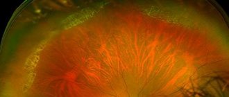
Retina
- the thinnest multilayer structure of the eye, covering 70% of the area of the inner surface of the eyeball. The retina of the eye is connected to the visual analyzers of the brain, into which it transmits information and is responsible for converting light impulses into nerve impulses.
The inner side of the retina is adjacent to the vitreous body, and the outer side is adjacent to the choroid: the choroid from which it receives nutrients and oxygen. Sometimes, under the influence of one reason or another, part of the retina moves away from the choroid. In this case, cells deprived of nutrition and oxygen quickly die. Without surgical intervention, the process leads to disturbances up to complete loss of vision. This condition is called retinal detachment.
Retinal disinsertion
–
a severe pathological process requiring emergency surgical care, in which the reticular layer of the eyeball is separated from the vascular layer.
The term itself was first proposed at the beginning of the 8th century, but its clinical confirmation was made only in 1851 after the invention of the ophthalmoscope. According to research, the incidence of the disease has increased significantly over the past decade: previously, 1 in 15,000 people were at risk of retinal detachment, now it is 1 in 10,000. Patients with myopia and aphakia often suffer from retinal detachment, especially if part of the vitreous lens was removed during surgery simultaneously with the lens. bodies. In the latter case, the risk of developing pathology increases to 10%.
Retinal detachment. What is important to know about it and how to recognize this disease
The retina is one of the membranes of the eyeball that lines it from the inside. This inner shell is a nervous tissue, the structure of which has more than 10 layers and provides image perception. Retinal detachment is a dangerous ophthalmological disease that requires immediate surgical intervention! It occurs due to a tear in the retina; through this tear, the vitreous body enters under the retina and detaches it from the choroid, which leads to complete or partial loss of vision.
At-risk groups:
- persons with moderate and high myopia
- elderly people with concomitant diabetes mellitus
- persons with hereditary dystrophic diseases of the retina (vitreoretinal degenerations)
- persons with inflammatory diseases of the posterior segment of the eyeball (retinitis, chorioretinitis)
- pregnant women
The connection between blunt trauma (contusion of the eyeball) and penetrating wounds of the eye with retinal detachment is beyond doubt. The causes of retinal detachment are the development of dense fibrous strands in the vitreous body, membranes on the surface of the retina and under it, their contraction and tension of the retina. As a result of hemorrhages in the retina and vitreous body, compaction of the retina develops, adhesions with the vitreous body are formed, which lead to the appearance of valve and perforated ruptures. Blunt trauma provokes retinal detachment within two years after injury in 80% of cases.
Symptoms of retinal detachment
The main symptoms characteristic of this disease are as follows:
- The appearance of a “veil” before the eyes, which causes a narrowing of the field of vision;
- “Flashes” before the eyes similar to lightning and sparks;
- Distortion of the shape of objects;
- Sharp, sudden loss of vision;
- Cloudiness before the eyes in the form of “rings” (Weiss symptom).
Symptoms of retinal detachment may depend on how far it has spread within the eye. For example, “dots and spots” before the eyes do not always indicate the occurrence of this disease; such problems often appear in older people. However, the patient must urgently contact an ophthalmologist for an initial examination to exclude the possibility of developing this pathology. Specialists at the ophthalmology department of the Federal Scientific and Clinical Center of the Federal Medical and Biological Agency recommend visiting a doctor at least once a year.
If you experience these symptoms, we recommend making an appointment with your doctor. Timely diagnosis will prevent negative consequences for your health. Phone for registration: 8 (499) 725-44-40
Symptoms
People who are not even aware of their pathology may experience the following symptoms in the eyes:
- sparks,
- flares,
- unclear vision of objects,
- flickering
- opacity of vision.
If you experience any such sensation, you should immediately consult an ophthalmologist. Only a specialized specialist can diagnose diseases. You won't be able to do this on your own. The study uses specialized instruments. As a result, it is possible to understand not only the degree of development, but also the reasons. This provides an opportunity to choose the most effective treatment.
Diagnostics
To correctly diagnose retinal detachment, an ophthalmologist will conduct a full eye examination at your initial appointment and prescribe the necessary treatment.
Diagnostic tests include:
- Visual field examination (perimetry);
- Visometry (visual acuity test);
- Ultrasound examination (A- and B-scan), which allows you to obtain the most accurate picture of the disease;
- Examination of the fundus using a special ophthalmological apparatus (ophthalmoscopy);
- Fundus examination using a 3-mirror Goldmann lens.
The diagnostic results will allow the ophthalmologist to quickly determine the optimal method of treating this pathology. It is extremely important to identify the development of retinal detachment as quickly as possible in order to prevent possible negative consequences for vision!
Information about the disease
Retinal degeneration, retinoschisis, is accompanied by cystic degeneration of its tissues, folding, tears, as well as detachment and lesions of the vitreous body. The name pathology was introduced into practice in 1935. The disease develops against the background of malformations of the photosensitive membrane or its dystrophic changes.
The incidence of retinoschisis in Russia ranges from 4 to 12% in the age group of people 40 years and older. In Europe, this figure is approximately 5%, while the disease occurs in the population after 30 years of age. In the case of a genetically determined form of pathology, clinical signs appear already in childhood - up to 10 years. Early diagnosis and timely treatment of retinoschisis are very important, since patients usually have no complaints during its development. Pathology is detected during an ophthalmological examination or after complications occur.[2]
Prevention
In order to prevent this disease, it is necessary to undergo an annual examination by an ophthalmologist. If there are risk factors (high degree of myopia, eye surgery and/or a history of penetrating eye trauma, diabetes mellitus in the stage of decompensation, the presence of peripheral retinal dystrophies), a more frequent examination by an ophthalmologist is required - at least once every 6 months.
Playing sports when the pathology is not diagnosed in time can lead to progression of the disease, and in the worst case, to complete loss of vision.
What forms of retinoschisis are there?
Russian ophthalmologists classify this disease into three types, depending on the cause of retinal separation:
- Hereditary. It develops as a consequence of genetic disorders and is transmitted as an autosomal dominant or recessive trait. Sometimes it is a symptom of genetic disorders such as Goldmann-Favre disease or Wagner syndrome.
- Degenerative. The acquired form usually occurs after the age of 40 due to degenerative changes in the retina.
- Secondary. It develops as a result of vascular diseases of the organs of vision (diabetic retinopathy, thrombosis of the central retinal vein), as well as inflammation of the eyes - uveitis, iridocyclitis in chronic stages, with oncology of the vascular system, after eye injuries.
With the development of retinoschisis, circulatory disturbances are observed in the macular and peripheral areas of the retina. In this case, a large number of compactions - cysts - appear. As they grow, they merge with each other, forming large cavities that stratify the retina along its entire length. This process leads to the development of retinal detachment and death of retinal structures.
Symptoms of retinal dissection include:
- decreased visual acuity;
- narrowing of peripheral fields;
- flashes in the eyes;
- distortion of the outlines of objects.
Usually these signs appear at a fairly advanced stage.
Treatment of retinal detachment
To identify a retinal tear, a complete ophthalmologic examination is necessary, including a detailed examination of the extreme periphery of the retina. Sometimes an ultrasound examination of the eye may be required. Retinal detachment does not respond to any conservative drug treatment. Surgical treatment of retinal detachment is the only effective treatment for this pathology.
The sooner treatment is performed, the greater the chance of saving vision!
The main methods of treatment of retinal detachment in the Federal Scientific Center FMBA:
- Laser coagulation of the retina. The essence of the laser coagulation method is the formation of point coagulates using a laser, which allows you to fix the retina to the choroid. Depending on the severity of retinal pathology, the number and location of coagulates may vary. The operation is performed under local anesthesia, does not require preoperative preparation and has a short recovery period. It often complements surgery in cases of widespread retinal detachment.
- Episcleral filling. The essence of the episcleral filling method is to form an indentation zone on the sclera. The scleral area is sealed with silicone material. As a result, the retina is “soldered” to the choroid.
- Cryopexy. The method is based on the local effect of low temperatures on the retinal tear area. This helps to avoid detachment due to the development of adhesions. Cryopexy often complements episcleral buckling. This method of treating retinal detachment can be supplemented with pneumoretinopexy - the introduction of special sterile gases into the vitreous body. This allows you to improve the fit of the eye shells to each other.
- Endovitreal operations. These surgical interventions are based on the removal of the vitreous humor from the eye cavity with the introduction of silicone oils or long-absorbing gases into the eye cavity.
Cost of retinal detachment surgery:
| № | Service name | Price in rubles | Make an appointment |
| 2011074 | Introduction of drugs of the 3rd degree of complexity into the vitreous cavity | 78000 | Sign up |
| 2011073 | Introduction of drugs of the 2nd degree of complexity into the vitreous cavity | 27000 | Sign up |
| 2011072 | Introduction of drugs of the 1st degree of complexity into the vitreous cavity | 22200 | Sign up |
| 2011071 | Peripheral vitrectomy 3rd category of complexity | 14400 | Sign up |
| 2011070 | Peripheral vitrectomy 2nd category of complexity | 12000 | Sign up |
| 2011069 | Peripheral vitrectomy of 1st category of complexity | 9600 | Sign up |
| 2011068 | Microinvasive revision of the anterior chamber | 14400 | Sign up |
| 2011075 | Intraoperative administration | 7200 | Sign up |
| 2011066 | Reconstruction of the anterior chamber | 7200 | Sign up |
| 2011065 | Removal of perfluoroorganic liquids from the vitreous cavity | 9000 | Sign up |
| 2011064 | Removing liquid silicone from the vitreous cavity | 12000 | Sign up |
| 2011063 | Circular retinotomy or retinectomy | 16800 | Sign up |
| 2011062 | Retinotomy and retinectomy | 9000 | Sign up |
| 2011061 | Injection of gas into the vitreous cavity | 12000 | Sign up |
| 2011060 | Injection of liquid silicone into the vitreous cavity | 18000 | Sign up |
| 2011059 | Introduction of perfluoroorganic liquids into the vitreous cavity | 15000 | Sign up |
| 2011058 | Circular peripheral endolaser coagulation of the retina | 16500 | Sign up |
| 2011057 | Endolaser coagulation of the retina, delimiting (one quadrant) | 7200 | Sign up |
| 2011056 | Endodiathermocoagulation | 9000 | Sign up |
| 2011055 | Removal of epiretinal membranes or posterior hyaloid membrane of the third category of complexity | 57600 | Sign up |
| 2011054 | Removal of epiretinal membranes or posterior hyaloid membrane of the second category of complexity | 47700 | Sign up |
| 2011053 | Removal of epiretinal membranes or posterior hyaloid membrane of the first category of complexity | 36600 | Sign up |
| 2011067 | Endodrainage of subretinal fluid | 7200 | Sign up |
| 2011052 | Unscheduled revision of the vitreal cavity | 42000 | Sign up |
| 2011051 | Revision of the vitreous cavity of the third category of complexity | 33000 | Sign up |
| 2011050 | Revision of the vitreous cavity of the second category of complexity | 28200 | Sign up |
| 2011049 | Revision of the vitreous cavity of the first category of complexity | 20400 | Sign up |
| 2011048 | Vitreoretinal surgery for complicated conditions (2-stage) of the highest category | 210060 | Sign up |
| 2011047 | Vitreoretinal surgery for complicated conditions (2-stage) of the third category | 162060 | Sign up |
| 2011046 | Vitreoretinal surgery for complicated conditions (2-stage) of the second category of complexity | 119940 | Sign up |
| 2011045 | Vitreoretinal surgery for complicated conditions (2-stage) of the first category of complexity | 84420 | Sign up |
| 2011042 | Vitrectomy for complicated conditions of the second category | 90000 | Sign up |
| 2011041 | Vitrectomy for complicated conditions of the first category of complexity | 83400 | Sign up |
| 2011039 | Vitrectomy for uncomplicated hemophthalmos or vitreous opacities of the second category | 64500 | Sign up |
| 2011038 | Vitrectomy for uncomplicated hemophthalmia or vitreous opacities of the first category | 54000 | Sign up |
| 2011037 | Removal of a silicone filling implanted in another medical institution | 24900 | Sign up |
| 2011036 | Removal of a silicone filling within a period of more than 6 months. after the first operation | 18660 | Sign up |
| 2011035 | Pneumoretinopexy for retinal detachment | 22200 | Sign up |
| 2011034 | Additional extrascleral filling in case of detachment | 28860 | Sign up |
| 2011033 | Combined extrascleral filling for detachment | 64800 | Sign up |
| 2011032 | Circular extrascleral filling for detachment | 48420 | Sign up |
| 2011031 | Local extrascleral filling for retinal detachment | 37800 | Sign up |
| 2011030 | Extrascleral ballooning for retinal detachment | 31800 | Sign up |
| 2011028 | Set of disposable consumables for vitreoretinal surgery | 57600 | Sign up |
| 2011027 | Introduction of drugs into the vitreous cavity (Eylea, Lucentis, Ozurdex) | 55200 | Sign up |
| 2011040 | Vitrectomy for uncomplicated hemophthalmia or vitreous opacities of the third category | 78600 | Sign up |
| 2011044 | Vitrectomy for complicated conditions of the third category of complexity | 144900 | Sign up |
Making an appointment Today registered: 21
Reasons why you should contact the Federal Scientific Clinical Center for treatment of this disease
- The Department of Ophthalmology of the Federal Scientific and Clinical Center of the Federal Medical and Biological Agency has the most advanced technologies and extensive experience in the treatment of diseases of the retina;
- Modern surgical equipment: a DORC (Eva) combine is used during operations on the retina, has high accuracy, provides optimal clinical results, and a quick recovery period;
- The extensive clinical experience of ophthalmologists of the Federal Scientific and Clinical Center of the Federal Medical and Biological Agency and first-class technical equipment make it possible to successfully treat various eye diseases even in advanced stages of the disease;
- At the Federal Scientific and Clinical Center of the Federal Medical and Biological Agency, all necessary preoperative preparation is carried out in just one day.
- Taking into account the presence of a multidisciplinary hospital, resuscitation and anesthesiology service, we perform operations on somatically severe patients with a risk of developing cardiovascular complications.
Classification of pathology
Clinical classification T.A. Bagdasarova, distinguishes three stages of retinoschisis:
- First. The retinal split is limited to a separate area, there is no vascular elevation, and there is no intraretinal fluid.
- Second. Retinal splitting progresses without a definite boundary between the affected area and the healthy retina. The formation of microcysts is detected in its layers.
- Third. The lesion with extensive cysts is localized in several quadrants of the retina. Their cavities rupture under the retina. There is a risk of transition of the pathological process to a limited detachment.
Separately, it should be said about central (macular) retinoschisis , which is less common, but has more severe consequences due to damage to the center of the visual field and possible complications - the formation of a macular hole.[4]
Fig.2 Picture of maculoschisis (OCT image - optical coherence tomography)
Symptoms and signs
What signs indicate the development of the disease? Retinal detachment has the following symptoms:
- sudden loss of visual fields;
- lack of coordination;
- distortion of the contours of objects;
- presence of opacities;
- flashes, sparks, lightning - these symptoms occur when there is bright light or pressure on the eyeball.
Characteristic signs of retinal detachment: sudden onset of symptoms. Such manifestations intensify when a person is in an upright position, and after sleep a noticeable improvement may occur, since in this case there is a temporary restoration of contact between the separated layers.
The ambiguity of retinal detachment lies in its painlessness (there are no sensations of pain and pain in the eye if the pathology is not associated with eye injury), and in the irreversibility of the process. Therefore, the earlier the disease is detected, the greater the chance of complete restoration of visual functions.
Clinical manifestations
The course of the pathology is not accompanied by severe pain symptoms. You can guess the problem by the presence of the following signs:
- “flash” or “lightning” effect;
- floating “spots” or dark spots, blurred vision;
- blurred vision that takes the shape of a ring;
- distortion of the shape and size of observed objects;
- blurred vision;
- loss of peripheral vision.
The “flash” effect is usually observed at the initial stage of development of rhegmatogenous detachment. Other types of pathology have a less pronounced clinical picture. For example, decreased vision occurs only after damage to the macula, the central part of the retina.