03 Jun 2021 at 09:50 MRI of the heart in Tushino 14126
Left ventricular hypertrophy is a fairly common heart disorder. The disease in most cases begins to develop in patients suffering from hypertension. Hypertrophy provokes an increase in the size of the wall of the left ventricle. The disease can provoke a change in the size of the septum, which is located between the left and right ventricles. The development of hypertrophy in most cases occurs over several years.
Left ventricular hypertrophy: causes
The myocardium can increase in size under the influence of certain factors that force it to work harder. In this way, the heart cells try to compensate for the increased load. These factors include:
- High blood pressure. One of the most common causes of left ventricular hypertrophy.
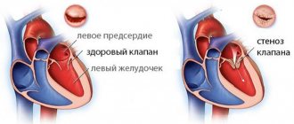
- Aortic valve stenosis. It is located between the left muscular chamber and the aorta. If it narrows, the muscle has to work harder, causing it to increase in size.
- Intense physical exercise. During prolonged cardio and strength exercises, thickening of the myocardium is considered as an adaptation of the body to increased loads.
Sometimes left ventricular hypertrophy of the heart develops due to a genetic disease that causes structural changes in heart cells - hypertrophic cardiomyopathy. Another possible cause may be amyloidosis, which is accompanied by abnormal protein deposits in all organs, including the heart.
Diagnosis of cardiomyopathy
Diagnosis of left ventricular hypertrophy occurs in several ways: by identifying signs of the disease on an ECG, examining the heart using ultrasound or using a magnetic resonance imaging scanner. If you experience any heart problems or symptoms of illness, you should contact a cardiologist, and if you have already suffered some kind of defect and suspect complications, you need a cardiac surgeon and, possibly, a treatment system.
Left ventricular hypertrophy on ECG
ECG is a common diagnostic method that helps to find out the thickness of the heart muscle and voltage characteristics. However, it can be difficult to identify LVH on an ECG without the participation of other methods: an erroneous diagnosis of hypertrophy may be made, since on the ECG the signs that are characteristic of it can be observed in a healthy person. Therefore, if they are found in you, this may be due to increased body weight or its special constitution. Then it is worth conducting another echocardiographic examination.
LVH on ultrasound
Ultrasound examination helps to more likely judge individual factors and causes of hypertrophy. The advantage of ultrasound is that this method allows not only to diagnose, but also to determine the characteristics of the course of hypertrophy and the general condition of the heart muscle. Indicators of cardiac echocardiography reveal changes in the left ventricle such as:
- ventricular wall thickness;
- ratio of myocardial mass to body mass;
- coefficient of asymmetry of seals;
- direction and speed of blood flow.
MRI of the heart
Magnetic resonance imaging helps to clearly calculate the area and degree of enlargement of the ventricle, atrium or other compartment of the heart, and to understand how strong the degenerative changes are. MRI of the myocardium shows all the anatomical features and configuration of the heart, as if “stratifying” it, which gives the doctor complete visualization of the organ and detailed information about the condition of each department.
Diagnosis of the disease
At the first stage, the doctor will listen to the patient's complaints, take a medical history and conduct a thorough physical examination, including measuring blood pressure and a preliminary assessment of heart function (auscultation or auscultation). Further additional studies will be prescribed:
- Electrocardiogram (ECG). Thickening of the wall of the left heart chamber can lead to disruption of the electrical activity of the heart, which is reflected in the results of the ECG.
- Echocardiogram. Allows you to evaluate blood flow, detect myocardial hypertrophy and identify the causes that caused it, for example, aortic valve stenosis.
- MRI. It has a high discriminative ability when assessing soft tissues, including the heart muscle.
Laboratory tests and, in some cases, invasive tests such as coronary angiography and myocardial biopsy may also be performed.
Drugs for left ventricular hypertrophy
Medicines for high blood pressure help prevent further enlargement of the myocardium and, in some cases, reverse it. For this purpose, the following may be prescribed:
- ACE inhibitors (angiotensin-converting enzyme). They lower blood pressure and reduce the load on the heart by dilating blood vessels and improving blood flow. Examples: Captopril, Enalapril, Lisinopril, Ramipril.
- Angiotensin II receptor blockers (ARBs). Drugs such as Lozap, Closart, Sentor also relax blood vessels, reducing blood pressure.
- Calcium channel blockers. These drugs inhibit the penetration of calcium ions from the intercellular space into the muscle cells of the heart and blood vessels. The blood vessels relax, blood pressure decreases. Examples: Amlodipine, Diltiazem, Verapamil, Lercanidipine.
- Diuretics. Diuretics used for left ventricular hypertrophy are called thiazide diuretics. They work by decreasing the kidneys' ability to reabsorb salt and water from urine, thereby increasing urine production and output (diuresis). Also, diuretics of this type directly help relax the smooth muscles of blood vessels. Examples: Tonorma, Hypothiazide, Hydrochlorothiazide.
- Beta blockers. Drugs such as Metoprolol, Atenolol, Lokren, Betak help reduce heart rate and blood pressure, as well as prevent the negative effects of stress hormones. Beta blockers are generally not prescribed as the primary treatment for hypertension. Your doctor may recommend them if therapy with other drugs has not been successful.
In cases where the disease is caused by aortic valve stenosis, surgery may be required. The decision on the advisability of the operation is made by the doctor after a thorough diagnostic examination of the patient.
Reasons for the development of the anomaly
Enlargement of the left ventricle can occur if some unfavorable factor causes the heart to work harder than usual. This means that the heart muscle will need to make several times more contractions in order to pump blood around the body.
Model of the heart with left ventricular hypertrophy
Reasons that can provoke a significant deterioration in heart function:
- High blood pressure (hypertension) is considered the most common cause of thickening of the ventricular wall. More than one third of all patients learn about hypertrophy at the time of diagnosis of arterial hypertension.
- Aortic valve stenosis is a disease that is a narrowing of the flap of muscle tissue that separates the left ventricle from the aorta. Narrowing of the aortic valve causes the heart to contract several times more often in order to pump blood into the aorta.
- Hypertrophic cardiomyopathy is a genetic disease that occurs when the heart muscle becomes abnormally thick and stiff.
- Professional sports. Intense, long-term strength training, as well as irregular performance of endurance exercises, can lead to the fact that the heart is not able to quickly adapt and cope with the additional load. As a result, the left ventricle may swell (enlarge).
Is left ventricular hypertrophy dangerous?
Without effective treatment, pathological thickening of the wall of the heart chamber can lead to serious complications such as:
- weakening of the blood supply to the heart;
- pumping dysfunction (heart failure);
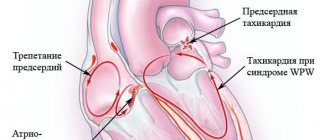
- heart rhythm disturbance (arrhythmia);
- insufficient oxygen supply to the myocardium (ischemic disease);
- stroke and heart attack.
Timely consultation with a doctor and properly prescribed therapy can avoid serious consequences of the disease.
What can hypertrophy lead to?
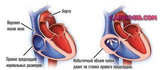
The disease cannot be ignored, because a significant enlargement of the ventricle can greatly change the structure and function of the heart. An enlarged ventricle may weaken and lose elasticity, increasing pressure in the heart. Hypertrophied tissue can also compress blood vessels and restrict blood flow directly to the heart muscle.
On the left is a normal heart, on the right is an enlarged ventricle
As a result of these changes, the following complications may occur:
- complete interruption of blood supply to the heart;
- inability of the heart to pump enough blood around the body (heart failure);
- abnormal heart rhythm (arrhythmia);
- irregular, fast heartbeat (atrial fibrillation);
- insufficient oxygen supply to the heart (coronary heart disease);
- enlargement of the aorta (dilatation of the aortic root);
- stroke;
- unexpected deterioration in cardiac function (sudden cardiac arrest);
- sudden loss of consciousness.
Hypertrophy is fraught with a significant deterioration in heart function
The consequences of hypertrophy can be called catastrophic for health, so if the patient has identified the reasons for the development of the disease, it is necessary to consult a cardiologist.
Popular questions about left ventricular hypertrophy
What medications should I take for left ventricular hypertrophy?
The choice of medications primarily depends on the causes of LVH. Therefore, drug therapy should be prescribed by a doctor. Most often, it includes taking ACE inhibitors, angiotensin II receptor blockers, calcium channel blockers, thiazide diuretics and beta blockers.
Why is left ventricular hypertrophy dangerous?
If the patient does not receive proper treatment for severe LVH, this can lead to the development of complications such as acute heart failure, arrhythmia, coronary artery disease, heart attack and stroke.
Is it possible to cure left ventricular hypertrophy of the heart?
You can achieve good results by eliminating the underlying cause of the disease, namely high blood pressure. Correctly selected antihypertensive therapy in many cases makes it possible to stop the progression of the pathology, and sometimes leads to a reduction in the hypertrophied heart wall.
What is concentric left ventricular hypertrophy?
It is a disease that occurs as a result of stressors on the heart, such as hypertension, congenital heart defects (such as tetralogy of Fallot), valve defects (narrowing of the aorta or stenosis), and primary myocardial defects that directly cause hypertrophy (hypertrophic cardiomyopathy). It is characterized by thickening of the myocardium without a corresponding increase in the size of the ventricles, and is often accompanied by symptoms such as chest pain and shortness of breath on exertion, general fatigue, syncope and palpitations.
Symptoms of the development of an abnormal condition
Dilatation of the left ventricle in most cases develops very slowly. The patient may not experience any unpleasant signs or symptoms, especially in the early stages of the disease. But as hypertrophy develops, the following may occur:
- shortness of breath;
- unexplained fatigue;
- chest pain, especially after exercise;
- sensation of fast, fluttering heartbeats;
- dizziness or fainting.
You should seek medical help if:
- there is a feeling of pain in the chest that lasts longer than a few minutes;
- there are serious breathing difficulties that interfere with daily activities;
- have severe, recurring memory problems;
- there are loss of consciousness;
- shortness of breath combined with rapid heartbeat is troubling.