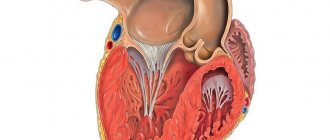
Right atrium hypertrophy is an expansion, thickening of the myocardial wall at the level of this chamber. At the initial stages it does not manifest itself in any way, except for rare exceptions. Further, the symptoms are represented not only by cardiac, but also by respiratory signs. The clinical picture is bright, the patient’s quality of life drops significantly.
Without competent, timely assistance, there is a high probability of death from cardiac arrest or pulmonary edema. Therapy is conservative or surgical, if the root cause is eliminated. Depends on the diagnosis.
In most cases, the process has a closed, paradoxical character: hypertrophy is provoked by problems with the bronchopulmonary system and aggravates them in the future.
In extremely rare cases, it can be a variant of the physiological conditional norm (in athletes).
Development mechanism
Enlargement of the right atrium is the result of an increase in pressure in the pulmonary artery, other vessels of the small circle, as well as overload of the chamber with blood as a result of the influence of one or another factor.
Detection plays a diagnostic role, because hypertrophy has several types.
An increase in pressure in the pulmonary and other arteries develops as a result of hemodynamic disturbances, vascular defects, and hypertension.
The normal outflow of blood from the right atrium weakens, the remains of liquid connective tissue press on the walls, provoking adaptive mechanisms.
The load on the atrium increases, the organ grows. Builds muscle mass to improve contractility and prevent tissue tearing.
Another mechanism is associated with dysfunction of the tricuspid valve. It covers the passage between the right atrium and the ventricle.
Malfunction leads to incomplete ejection of blood, increased pressure in the chambers and their hypertrophy for the same reasons.
Based on the origin of the process, the mechanism of formation, three forms are distinguished:
- Cardiosclerotic variety. It occurs as a result of inflammation, heart attack and other phenomena of the same kind (coronary insufficiency, coronary artery disease). The affected area becomes scarred, cells are roughly arranged around the area, and tissue grows artificially. Hence the increase in volume, disruption of normal activity.
- Compensatory type. It develops as a result of the influence of a negative factor: an increase in pressure in the pulmonary artery, pulmonary vessels, and other processes.
- Working variety. It develops in patients who are professionally involved in sports. Less likely to engage in similar physical activity as part of work (loaders, others).
Determining the mechanism plays an important role in diagnosis. This is the basis for prescribing competent therapy.
Causes
Factors may be cardiac or non-cardiac. Some lead to the development of the defect faster. Others for a long period (from 3 years and more).
Sample list:
Arterial hypertension
Represents a stable increase in pressure. The result of a combination of a group of moments that lead to a change in the tonometer readings.
As it progresses, blood pressure levels stabilize at high levels, and increased atrial output is noted. The tension on the walls of the chambers increases, the first blow falls on the right structure. Hence the thickening and the beginning of a compensatory mechanism.
At the 3rd stage of hypertension, it is no longer possible to radically help. There is a high risk of developing resistance to therapy, in which case the rate will decrease slightly or not completely.
The load is still there. Therefore, regular hospitalizations are indicated to correct the treatment regimen. Otherwise, there is a high risk of death within 5-10 years from diagnosis. Perhaps less.
Change in body weight towards increase
Obesity is not recognized by everyone as a disease. This is a pathological process, but it is not to blame for hypertrophy.
An enlarged right atrium is the result of ongoing atherosclerosis, blockage of the lumen primarily of the pulmonary and coronary arteries with cholesterol deposits - plaques.
Hence the increase in pressure, overstrain of the chambers, ischemia of myocardial tissue and the dual mechanism of development of the disorder.
Metabolic conditions are treated with statins. Treatment is prescribed in the early stages, before generalized cellular disorders begin.
Long-term stress
Accompanied by the release of corticosteroids and catecholamines. Cortisol and adrenaline as the main ones. They cause constriction of blood vessels, including coronary ones. An increase in blood pressure, then cardiac dysfunction occurs.
Recovery is very difficult. Psychotherapeutic assistance, mastering relaxation methods, changing professional activities, and other activities are indicated. Depends on the case.
The body cannot exist in this state for a long time.
Congenital and acquired vascular malformations
An example is stenosis of the pulmonary artery or aortic valve.
It ends with a disturbance in the movement of blood in the small (pulmonary) circle and an increase in pressure in the vessels. The stress on the chamber walls also increases.
Such processes are difficult to correct. Medicines only treat the symptoms, while the root cause remains untouched.
Tricuspid valve defects
The tricuspid structure bridges the gap between the right atrium and the ventricle. If there is a malfunction, an incomplete release of blood into the next chamber occurs, or its reverse flow occurs - regurgitation.
Some of the liquid connective tissue remains in the atrium and presses on the walls. Thickening occurs as a compensatory mechanism.
On the one hand, this prevents rupture, on the other hand, it allows blood to be pumped more actively. Read more about tricuspid regurgitation in this article.
Treatment is prescribed immediately after detection. Advanced or dangerous cases are corrected surgically. The operation significantly increases the chances of survival and health.
Previous heart attack
Necrosis of cardiac tissues, myocardium. It is accompanied by severe symptoms, so it is difficult to confuse the pathological process with others. Requires urgent hospitalization and first aid.
Even in case of success, which is recognized as saving the patient’s life, coronary disease is observed. It imposes a lot of restrictions on everyday and professional conditions.
Constant treatment under the supervision of a cardiologist is also required. Protectors, antiarrhythmics, antihypertensives and others are used.
The cause of right atrium hypertrophy is tissue scarring and rough epithelization of damaged areas.
Angina pectoris
A variant of coronary insufficiency on a par with a heart attack. A process that is so dangerous gives a chance for a cure and time for diagnosis.
It occurs in fits and starts. Each such episode leads to the death of a small part of the muscle layer, necrosis. Further, just as during a heart attack, the dead tissue is replaced by thick, scar tissue.
Read more about the symptoms of an angina attack and methods of treating it here.
Pneumonia or pneumonia
Septic lesion of the parenchyma. Accompanied by long-term disturbances in gas exchange and an increase in pressure in the small circle.
Damage to cardiac structures is possible, especially with a long-term or chronic process.
Treatment in a hospital, with elimination of signs of respiratory failure, antimicrobial therapy and restoration of heart activity.
Bronchial asthma
Most cases are of the allergic variety. It cannot be completely cured because it is immune in nature. There are chances to achieve a stable long-term remission.
A long course is accompanied by the process getting out of control, an increase in the phenomena of respiratory failure and disruption of normal gas exchange, and an increase in pressure in the small circle.
The result is hypertrophy not only of the atrium, but also of the right ventricle (pulmonary heart).
Systematic use of glucocorticoids and bronchodilators is required (with caution, the latter wash out potassium and impair myocardial contractility).
Chronic obstructive pulmonary disease
The scourge of smokers and workers of hazardous industrial enterprises.
The violations are irreversible. You shouldn't expect a total cure. It is also not easy to transfer the pathological process into remission. Long periods of therapy with powerful hormone-based drugs are required. Quitting smoking will no longer help.
Extended sports activities
Lead to artificial growth of the chambers of the heart. This is the result of excessive physical activity, a response to the need to normalize blood flow.
The organic defect is associated with bradycardia (decreased heart rate) and a drop in blood pressure. This is part of the overall clinical picture.
“The heart of an athlete” is considered a variant of the physiological norm, but individuals with this type of professional activity must be carefully monitored.
Thickening of the right atrium is not the only change. The remaining chambers are also expanding.
Inflammation of the myocardium, pericardial sac
Infectious or autoimmune. They lead to consequences similar to those of a heart attack.
The causes of RA hypertrophy are the result of cardiac and bronchopulmonary pathologies.
Treatment of myocardial hypertrophy
To carry out qualified treatment, it is necessary to diagnose the disease, as well as determine its nature and course features. Based on these data, the optimal treatment method is selected. In most cases, treatment of myocardial hypertrophy consists of taking the drug verampil with beta blockers. The complex use of medications can reduce the symptoms of the disease, as well as normalize the general condition of the patient. As an additional therapy, it is necessary to follow the prescribed diet, and also stop smoking and drinking alcohol. In addition, it is necessary to reduce the amount of salt and consumed foods with the highest fat content. During treatment, physical activity should be moderate.
In particularly severe cases, surgical intervention is required. In this case, the specialist removes the hypertrophied part of the muscle. During treatment, the patient's condition is monitored using an ECG.
Rehabilitation
As you know, after treatment or surgical intervention, full rehabilitation is required. Therapeutic diet is an integral part of the rehabilitation process. In this case, you need to eat up to 6 times a day in small portions. Flour products should be limited; fatty and smoked foods should also be excluded from the diet. The level of physical activity needs to be reduced. Full rehabilitation allows the patient to recover much more quickly after treatment.
Make an appointment Make an appointment and get a professional examination at our center
Symptoms
Signs of the process depend on the degree and severity of the disorder. In the early stages, the following points arise:
- A slight burning sensation in the chest, possibly tingling. The pain is weak, not pronounced. Duration is minimal. Treated with Nitroglycerin.
- Cough. Intense, non-productive, no sputum. It intensifies at night and may be accompanied by hemoptysis.
- Paleness of the skin. Mucous membranes.
- Cyanosis of the nasolabial triangle. Blue discoloration of the area around the mouth.
- Dyspnea. First associated with intense physical activity. Deviations can only be detected by athletes; a person without daily training does not reach the threshold when the symptom manifests itself. Then at rest. The manifestation accompanies the patient on an ongoing basis and is not controlled by bronchodilators.
- Excessive fatigue without physical activity. Weakness, drowsiness, apathy. Decreased ability to work.
Similar symptoms are observed at a later time, but are aggravated and supplemented by a number of signs.
Among the alarming points:
- Hemoptysis. It has nothing to do with tuberculosis, but differential diagnosis is necessary. At the very least, an X-ray, or better yet a computed tomography scan to differentiate.
- Swelling of the lower extremities. At first only the feet are affected, then the ankles. Next, the process covers the entire leg or both at once.
- Enlarged liver. Painful sensations in the right hypochondrium; palpation reveals an organ protruding beyond the edge. In the absence of treatment, the phenomena of the inflammatory, destructive process increase. Possible accumulation of fluid in the abdominal cavity or ascites, jaundice, and other phenomena.
- Swelling of the cervical vessels. Indicates an increase in venous pressure. This is a consequence of blood stagnation.
- Heart rhythm disturbances. According to the type of bradycardia, ventricular fibrillation. Subjectively it is felt as freezing in the chest, interruptions, heaviness and other moments
- Headache. In the occipital region. Top of the head.
- Vertigo. To the point of being unable to stand on your feet.
- Nausea. Extremely rarely - vomiting. Signs of neurological deficit.
Fainting is possible. Indicate circulatory disorders in cerebral structures. Evaluation in the system.
Heart defects
Rubella
Cold
Stroke
Atherosclerosis
Diabetes
4985 19 October
IMPORTANT!
The information in this section cannot be used for self-diagnosis and self-treatment.
In case of pain or other exacerbation of the disease, diagnostic tests should be prescribed only by the attending physician. To make a diagnosis and properly prescribe treatment, you should contact your doctor. Heart defects: causes, symptoms, diagnosis and treatment methods.
Definition
Heart disease is defined as an atypical or abnormal structure of its structures (chambers, valves, large vessels), which is a consequence of disturbances in the formation and development (congenital heart disease) or various pathological changes (acquired heart disease). As a result of the formation of this pathology, the functioning of the heart is disrupted and oxygen deficiency of organs and tissues of the body is formed, which can ultimately lead to heart failure.
Causes of heart defects
In humans, the blood returning to the heart from all organs and tissues (oxygen-poor blood) passes through the right atrium and then through the right ventricle into the pulmonary artery, and from there it enters the lungs. In the lungs, the blood is enriched with oxygen, releases carbon dioxide and enters the left atrium and left ventricle, and from there it is pumped to all organs and tissues through the aorta, and then through smaller arteries. The performance of the heart muscle depends on the functioning of the valves, which, when contracting, freely allow blood to flow into the next section, and when relaxing, do not allow blood to flow back.
If the function of the valves is impaired, the function of the heart is also impaired.
Congenital heart defects are very diverse. More than one and a half hundred different variants of atypical heart structure have been described. About one in 100 babies is born with a heart defect. Common heart pathologies diagnosed in infancy and childhood are atrial and ventricular septal defects (the openings between the chambers of the heart). Often the defects are combined with anomalies of the valvular apparatus of the heart or large vessels. The most common congenital heart defect is the bicuspid aortic valve, which opens with every heartbeat, allowing blood to flow from the heart to all organs. A normal aortic valve has three leaflets.
Bicuspid aortic valve does not usually cause problems during infancy or childhood, so it is often not diagnosed until adulthood.
The causes of congenital heart defects include genetic, environmental and infectious. In addition, their formation can be caused by certain diseases (gestational diabetes mellitus, rubella and systemic lupus erythematosus) in the mother, taking certain medications, drugs and alcohol during pregnancy, and other factors.
Acquired defects are anomalies and defects of the heart valves, its openings or partitions between the chambers, and the vessels extending from it, which appeared during life under the influence of morphological and functional changes in the functioning of the heart. The mitral valve is affected more often than the aortic valve. Pathologies of the tricuspid (tricuspid) valve and pulmonary valve are less common. Diseases can manifest at any age under the influence of atherosclerosis, cardiosclerosis, ischemic or hypertension, rheumatism, systemic pathology, trauma, syphilis and some other reasons. Also, degenerative changes in the valves lead to valvular heart defects - as the disease develops, their structure and function are disrupted, which causes a restructuring of hemodynamics, overload of the corresponding parts of the heart occurs, hypertrophy of the heart muscle, and impaired circulation in the heart and in the body as a whole.
Classification of the disease
It is customary to highlight:
- defects of the “white” type, when there is no tendency to mix arterial and venous blood;
- “blue” type defects - venous blood enters the arterial bed, resulting in oxygen deficiency.
Type of functional pathology:
- stenosis - as a result of the pathological process, deformation of the valve tissue occurs and the hole through which blood flows to the next part of the heart narrows;
- valve insufficiency - failure of the heart valves to close due to changes in shape, their shortening as a result of scarring of the affected tissues;
- combined and concomitant heart defects:
- combined – in the presence of both stenosis and insufficiency of one valve;
- combined – when several valves are affected at once.
Based on the reasons for their formation, acquired defects are classified as follows:
- degenerative, or atherosclerotic (occur in 5-6% of cases) - more often these processes develop after 40-50 years, when calcium is deposited on the leaflets of the affected valves, which leads to the progression of the defect;
- rheumatic, developing against the background of rheumatic diseases (80% of cases);
- defects that arise as a result of inflammation of the inner lining of the heart (endocarditis);
- syphilitic (in 5% of cases).
Based on the general hemodynamic condition, compensated, subcompensated and decompensated heart defects are distinguished.
Symptoms of heart defects
Clinical symptoms of heart defects include shortness of breath, weakness, fatigue, swelling of the lower extremities, sleep disturbances, interruptions in heart function, arrhythmia (usually tachycardia), change in skin color (blueness or pallor), swelling of the veins of the neck and head, causeless anxiety, pressing pain in the heart area (especially during physical activity) or between the shoulder blades, in rare cases - loss of consciousness.
Specific symptoms of congenital heart defects depend on the patient's age. Because normal circulation of oxygen-rich blood is essential for normal growth and development, infants may experience difficulty or rapid breathing, poor appetite, sweating or increased breathing during feeding, cyanosis of the lips and/or skin, unusual irritability, or failure to gain weight. .
Children and adolescents may experience decreased exercise tolerance, dizziness, and fainting.
Most serious heart defects in children are identified based on symptoms noticed by parents and abnormalities found when examined by a doctor. Impaired blood flow through the heart usually manifests as a heart murmur that can be heard with a stethoscope. Abnormal heart murmurs are often loud or harsh. However, in the vast majority of cases, heart murmurs noted in childhood are functional and not caused by heart defects.
The severity and nature of symptoms depend on the location of the affected valve. With valve defects of the left half of the heart (mitral and aortic), the lungs are primarily affected, since blood stagnates in their vessels, which is manifested by shortness of breath and cough. In addition, there are signs of insufficient blood supply to the brain and the heart itself, dizziness, fainting, and angina. In the presence of cyanosis, one of the most common pathologies is tetralogy of Fallot (restriction of blood flow to the lungs).
When the valves of the right half of the heart (the tricuspid and pulmonary valves) malfunction, blood stagnates in the vessels of the systemic circulation, so all organs except the lungs suffer. Swelling of the legs and feet, ascites (accumulation of fluid in the abdominal cavity), enlarged liver, and other lags in weight gain develop.
Signs of acquired defects are often combined with other heart diseases, in particular coronary artery disease, which makes their clinical differentiation difficult.
Diagnosis of heart defects
Diagnostic measures carried out to detect heart defects require an integrated approach, but always begin with collecting an anamnesis: the doctor finds out the patient’s complaints, the time and circumstances of their manifestation, intensity, and hereditary factors. Next, a physical examination is carried out, which includes visual inspection, palpation, percussion (tapping), auscultation (listening).
Laboratory tests that are prescribed for suspected heart disease:
- clinical blood test;
Diagnostics
It takes place on an outpatient basis. In most cases, there is time for a quality examination; the main thing is to contact before the development of threatening phenomena.
Approximate list of events:
- Oral questioning of the patient, collection of anamnestic data.
- Auscultation. Listening to heart sounds. Noises of various types are noted. But not always.
- Measurement of blood pressure, contraction frequency. Both indicators may be normal or abnormal. Based on the nature of the disorder, one or another probable diagnosis is indicated.
- Daily monitoring. Record vital signs within 24 hours. Used to assess the state over time.
- Electrocardiography. Detection of functional disorders of the muscular organ.
- Echocardiography. Visualization technique. Used to determine defects, their location and severity.
- MRI or CT scan of the chest as needed.
- Assessment of neurological status.
- Ultrasound of the liver and abdominal organs.
General blood test, biochemical, also not hormones (if endocrine disorders are suspected).
Signs on ECG
{banner_banstat9}
- A characteristic sign on the cardiogram is deformation of the atrial P wave (pointed, in the normal state it is round, without notches) and its widening.
- Change in contraction frequency. First towards tachycardia, in the later stages the heart rate drops to 60 and below.
The assessment is carried out by a diagnostician, then again by a cardiologist at the appointment.
Hypertrophy of the right atrium on the ECG is manifested by an increase and sharpening of the P-wave. The remaining points play a secondary role and allow us to assess the very nature of the accompanying violations.
Right ventricular hypertrophy
N.B. Familiarize yourself with the lesson content and get a general idea of the topic. Don't try to remember all the details right away. It will be more convenient to do this when further performing step-by-step exercises for the lesson. The link to the exercises is at the bottom of the page.
Right ventricular hypertrophy is accompanied by increased electrical activity when the right ventricle is excited. This is reflected on the ECG by an increase in the amplitude of the R waves in the right leads (III, aVF, V1-V3) and an increase in the depth of S in the left leads (I, aVL and V4-V6).
The main criteria for right ventricular hypertrophy on the ECG are the dominance of the R wave in lead V1 and the dominance of the S wave in leads V5 and V6, as well as deviation of the EOS to the right.
In this case, the following parameters must be met:
- RV1 must be greater than 6 mm and/or greater than SV1;
- S in lead V6 (or V5) should be greater than 7 mm and/or greater than RV6 (or RV5, respectively).
Look at the cardiograms below.
ECG 1. Cardiogram without deviations from the norm
ECG source.
ECG 1 shows no right ventricular hypertrophy. RV1 < 6 mm and RV1 < SV1. In the left leads: S in V5 and V6 is less than 7 mm and less than R in these same leads.
ECG 2. Right ventricular hypertrophy
ECG source.
ECG 2 shows right ventricular hypertrophy. RV1 = 12 mm, i.e. RV1 > 6 mm and at the same time RV1 > SV1. In the left leads: SV5 = 9 mm, SV6 = 7 mm and SV6 > RV6. The EOS is deviated to the right: (RIII - SIII) > (RII - SII) > (RI - SI).
Often, with right ventricular hypertrophy, the ECG shows ST segment depression and T wave inversion in the right precordial leads V1-V3, as well as in II, III, aVF, which is also recorded on ECG 2.
Look at another cardiogram with an incomplete set of parameters considered.
ECG 3. Right ventricular hypertrophy
ECG source.
On ECG 3, RV1 < 6 mm, in leads V5 and V6 R > S. At the same time, RV1 > SV1 (there is dominance of R in V1), the EOS is deviated to the right and the TV1 wave is negative. Therefore, in this case there is right ventricular hypertrophy. Thus, not all signs of right ventricular hypertrophy are always present. In such cases, the dominance of the R wave in lead V1 and the deviation of the EOS to the right is important.
The main signs of right ventricular hypertrophy:
- EOS is deviated to the right.
- R wave dominance in lead V1: RV1 > 6 mm and/or RV1 > SV1.
- ST segment depression and negative T wave in leads V1-V3, as well as in leads II, III, aVF.
- Dominance of the S wave in leads V5, V6: SV5,V6 > 7 mm and/or SV5,V6 > RV5,V6.
Go to exercises
Treatment
{banner_banstat10}
Carried out under the supervision of a cardiologist. In the early stages, if there are no catastrophic defects yet, dynamic monitoring is indicated. Every 3-6 months, cardiography and ECHO-CG, also changes in pressure, heart rate.
Deterioration and progression are grounds for urgent prescription of medications. According to indications, surgical treatment of pathology is used.
List of medications for symptomatic and etiotropic, cardiac action:
- Cardioprotectors. Normalize metabolism in cells.
- Antiarrhythmic. With extrasystole, fibrillation, paroxysmal tachycardia.
- Beta blockers. To correct blood pressure levels and relieve sinus arrhythmia.
- Cardiac glycosides. Stabilize myocardial contractility. But they are used in small courses, with caution. They are not prescribed after a heart attack.
- Diuretics. To remove excess fluid and reduce the load on the heart.
- Hepatoprotectors. To prevent damage to liver cells.
Other names are indicated for non-cardiac causes.
Surgery is a last resort. Prescribed for vascular defects, the heart itself and valves. Tumors, aneurysms, etc.
Attention:
You cannot resort to traditional methods; at best they are useless, then they are not worth the time and money spent, at worst they are dangerous.
As part of rehabilitation and further treatment, smoking and alcohol cessation is indicated. Normalization of the diet (table No. 10).
The working or physiological variety does not require therapy at all. The patient must be constantly monitored. Cardioprotectors are used if necessary.
Progression of pathology is associated with poor outcome. Only organ transplantation can help.
Cardiac hypertrophy
Cardiac hypertrophy
Cardiac hypertrophy is an enlargement of the heart muscle, which occurs mainly due to an increase in the number of cardiomyocytes - specialized muscle cells of the heart. This condition occurs in children, adolescents, young adults and the elderly.
Cardiac hypertrophy is a manifestation of a special state of the body: physiological or pathological. That is, it is not a disease, but a symptom.
Physiological
Physiological hypertrophy of the heart is observed in athletes and people who lead an active lifestyle. For regular physical activity, the body requires large amounts of oxygen. Oxygen is delivered through the blood. And to meet the increased oxygen needs, the heart increases the frequency and strength of contractions. And this requires greater metabolism in the heart muscle itself. This gradually increases the volume and mass of cells (cardiomyocytes). More often in athletes, cardiac hypertrophy begins from the left ventricle. Sports that can lead to cardiac hypertrophy are rowing, hockey, football, cross-country skiing, cycling, long-distance running, etc. When you stop training, this condition reverses. That is, the hypertrophied heart again becomes of normal size with normal wall thickness.
Pathological
Pathological hypertrophy of the heart occurs due to various diseases of the body. The human heart consists of four sections: two atria and two ventricles. The atria are reservoirs where blood enters from the body's circulation (blue vessels). The ventricles are a buoyant force that pushes blood through the vessels (red vessels). So each department has its own reasons for increasing.
Causes:
- Left ventricle – enlarges due to arterial hypertension, aortic valve stenosis, aortic atherosclerosis, general obesity, diabetes mellitus
- Right ventricle – due to congestive heart failure, chronic pulmonary failure
- Left atrium – with arterial hypertension, general obesity, aortic and mitral valve defects
- Right atrium - due to pulmonary diseases (when there is stagnation in the pulmonary circulation).
Development
The above reasons force us to maintain normal blood flow by increasing the mass of the heart. It must be taken into account that an increase in one part of the heart leads to hypertrophy of another. In addition to cardiomyocytes, the heart also contains connective tissue. With cardiac hypertrophy, it also grows, and this leads to a decrease in the elasticity of the walls and disruption of the heart.
If the load on the heart does not decrease, then the myocardium is gradually depleted because the blood flow cannot cope with the nutrition of the enlarged heart. This can lead to disruption of nerve impulses (arrhythmia), sclerosis and atrophy of the heart muscle.
Symptoms
- An asymptomatic course of cardiac hypertrophy is possible.
- If the left half of the heart is affected: pain in the heart area (increases after physical activity), arrhythmia, loss of consciousness, shortness of breath, dizziness.
- If the right half of the heart is affected: cough, shortness of breath, cyanosis (cyanosis) or pallor of the skin, swelling, arrhythmia.
Diagnostics
- Ultrasound examination of the heart
- ECG (electrocardiography)
- X-ray of the chest organs.
Treatment It is necessary to eliminate the cause of cardiac hypertrophy.
If it is arterial hypertension, it is necessary to take antihypertensive and diuretic drugs. Severe heart valve defects require surgical treatment and prosthetics. Respiratory diseases require anti-inflammatory and bronchodilator therapy. In any case, the approach is always individual. To monitor blood pressure and early detect arrhythmia, I recommend using automatic tonometers from the manufacturer Microlife, presented in our online store.
The author of the article is a practicing neurologist Maxim Nikolaevich Starshinin.
Forecast
Depends on the underlying pathological process. Early forms provide 100% survival rate. The way of daily living is almost unaffected, the patient can continue the same activities with minor restrictions.
When defects are added, progression is many times faster. Death occurs in 30% of cases.
Arterial hypertension, increased pulmonary pressure and other incurable pathologies do not guarantee early death. Depends on the start of supervision.
The possibility of radical intervention is characterized by a sharp improvement in prognosis.
Questions about the likely outcome should be directed to the leading physician. Therefore, it is recommended to contact the same cardiologist. He will take into account all factors and give an approximate answer.