Photo: UGC Many women are aware that during pregnancy the placenta protects the baby in the womb. However, not many people know about its functions in detail. The term “degree of maturity of the placenta” also causes confusion. Let's try to consider the questions in detail and answer them in layman's language.
Attention! The material is for informational purposes only. You should not resort to the treatment methods described herein without first consulting your doctor.
Placenta - what is it?
In Latin, the word “placenta” means “pie, flatbread.” It's all about how the placenta looks: the organ received this name due to its flattened shape, because in appearance it resembles a flat disk.
When does the placenta form during pregnancy?
Its formation begins only when conception occurs, and when the child is born, it is excreted along with the membranes.
The placenta performs the following functions:
- respiratory - through it oxygen enters the fetus and carbon dioxide comes out;
- barrier – protects the fetus from those harmful substances that are in the mother’s blood;
- nutritious - nutrients come from mother to fetus;
- hormonal - produces a number of hormones that determine the normal development of pregnancy;
- excretory - waste products of the fetus are excreted through it.
Development of the placenta
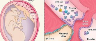
The development of this organ does not occur immediately after conception. In the fourth week of pregnancy, the fertilized egg is surrounded by the chorion - this is a special villous tissue. The formation of the early placenta occurs around the ninth week. It is formed by chorionic villi that penetrate the upper layer of the uterus and connect with the blood vessels there.
By the end of gestation, the placenta already weighs about half a kilogram, and its diameter is 15-20 cm. The permeability of the placenta membrane increases until the 32nd week of pregnancy. After all, every week the size of the fetus increases, and it requires more and more oxygen and nutrients.
Accordingly, in order to provide such nutrition, the number of vessels in the child's place increases, and the membrane becomes thinner. After the thirty-second week, the development of the placenta stops and it begins to age. If this happens earlier, then early aging of the placenta .
Degrees of placenta maturity
During the development of pregnancy, the doctor looks for all internal changes during an ultrasound examination. And the degree of maturity is also determined during the ultrasound process, taking into account certain parameters.
Based on the duration of pregnancy, the degrees of maturity are determined as follows:
- degree of maturity 0 – up to 30 weeks;
- degree of maturity of the placenta 1 – 27-36 weeks;
- degree of maturity of the placenta 2 – 34-39 weeks;
- degree of maturity of the placenta 3 – after 36 weeks.
To determine what degree of aging - first, second or third - the doctor determines its thickness, looks for calcium deposits and cysts.
Not so long ago, the maturity of a child's place was viewed differently than it is now. Thus, it was believed that with premature aging, miscarriages , there is a high risk of antenatal fetal death, and low-weight children are born. However, after scientists conducted a series of studies, such interdependence was refuted.
If a woman has a third degree of maturity before 35 weeks (for example, at 32 weeks), she is considered to be at increased risk.
A gynecologist will explain in more detail about the characteristics of maturity, for example, the degree of placenta maturity 2, what this means.
In the table below you can clearly see the degrees of ripeness by week.
Table of degrees of placenta maturity by week of pregnancy
| Chorionic part (the one that is adjacent to the fetus) | Structure | Are there calcium deposits? | |
| 0 tbsp. (up to 30 weeks) | Completely smooth | Homogeneous | Virtually none |
| I Art. (27-36 weeks) | Wavy | A little seal | Microscopic |
| II stage (34-39 weeks) | There are indentations | There are seals | Visible |
| III Art. (after 36 weeks) | Recesses to the basement membrane | Cysts | A large number of |
Ultrasound standards for first screening
The very first screening should be done from the 11th week of pregnancy to the 13th. It is during this period that the doctor prescribes an examination. Some expectant mothers can’t wait to go for an ultrasound before the 11th week. But there is no urgent need for this. After all, many indicators simply cannot be determined before this date. Therefore, the information content of the study will be small.
At the first screening, the doctor:
- reveals the exact duration of pregnancy;
- calculates the size of the fetus;
- looks at the uterus.
There are standards for each value. Let's look at them.
- The CTE or, more simply put, the length of the fetus is assessed. The norm is from 43 to 65 mm. When the deviation increases, it is believed that the baby will be born large. If it is to a lesser extent, then this indicates a slowdown in development, illness, or even the death of the fetus.
- The next indicator they look at is called BPR. The distance from one temple to another temple is measured. Normally it should be from 17 to 24 mm. Low scores may indicate developmental delays. High - about a large fetus (if other criteria also indicate this), or about hydrocephalus.
- Thickness of the collar space. The norm is 1.6-1.7 mm. If other numbers are revealed, then this is a bad sign, and we may be talking about Down syndrome.
- The length of the nasal bone should be from 2 to 4.2 mm.
- Heart rate. The norm is from 140 to 160 beats.
- Amnion. The norm of amniotic fluid is 50-100 ml.
- Cervical length. The norm is 35-40 mm.
Placenta thickness
This parameter is a very important point in the diagnosis, since deviations from the norm can be evidence of pathologies. There is a special table of placental thickness by week, which indicates the normal limits.
The thickness of the placenta by week is determined by ultrasound, which is performed after 20 weeks. If the process of bearing a baby proceeds normally, then the greatest thickness of the placental tissue is observed at 34 weeks, and at 36 weeks. its growth stops, and its thickness may even decrease slightly.
If the placenta is too thin, placental hypoplasia . However, as a rule, this is not a very threatening phenomenon, unless a significant decrease in size is noted. Very often, such deviations are associated with genetic disposition, the influence of unfavorable factors, and diseases of the woman. If this is due to illness, treatment is carried out; in all other cases, supportive therapy is practiced.
This indicator is also influenced by the physique of the expectant mother. Short, thin women have smaller baby seats than tall, curvy women. If thickening of the placental tissue is diagnosed, this condition can threaten the termination of pregnancy. But with the right approach to treatment, pregnancy can be saved.
Thickening can occur due to Rh conflict , diabetes mellitus , gestosis , iron deficiency anemia , or previous infections . In case of serious deviations, careful monitoring of the expectant mother is important. The fact is that with the rapid growth of a child's place, its active aging occurs. When thickened, hormonal function is disrupted, which can negatively affect pregnancy.
However, if after a series of additional examinations the doctor concludes that the fetus is developing normally, only careful observation will be needed.
Should I worry if an ultrasound reveals abnormalities of the placenta?
The placenta develops from the cells of the fetal egg, and not from the maternal cells, so the organ cannot be ideal. Violations are mild, moderate and significant. If abnormalities are detected, a diagnosis of placental insufficiency is made.
Gynecologists believe that only significant changes are dangerous for the fetus, since thanks to the extensive vascular network, the organ can perform all functions even with partial damage or detachment.
To understand how dangerous the changes are, it is not enough to assess the degree of problems in the placenta; it is important to take into account the quality of fetal development. If there is a discrepancy between the parameters of growth and development and the norms for the period, you need to sound the alarm and take action. If the baby develops normally, you can limit yourself to observation. For this purpose, non-screening (unscheduled) ultrasounds are performed.
Why does the placenta age too early?
If an old placenta is diagnosed during pregnancy, this may be due to various factors.
Hypertension
Gestational hypertension , that is, increased blood pressure during pregnancy, is very often associated with the function of the placenta. Due to various reasons, defective blood vessels are formed in the child's place, and this affects both the condition of the fetus and the health of the woman. As a result, the expectant mother develops edema , high blood pressure, and sometimes, in severe cases, preeclampsia . A child, developing in the womb, does not receive enough oxygen due to defective blood vessels. As a result, the placenta has to “work” at full capacity, which is why it ages prematurely.
Infections
If the expectant mother falls ill with any infectious disease, then the placental tissue works more actively. It filters the mother's blood from viruses, allows more oxygen and antibodies to pass through to the baby in order to intensify the fight against the disease. As a result, its maturation and, accordingly, aging accelerates.
Eating Excessive Calcium
Calcium deposits are one of the main signs of aging in a child's place. The closer to the end of pregnancy, the more calcifications are detected in the placenta. And if a woman’s body constantly receives a large amount of this microelement, then gradually calcium replaces the placental tissue, which provokes its active premature aging. This often happens if a pregnant woman takes vitamin supplements uncontrollably.
37th week of pregnancy for baby
At the 37th week of pregnancy, the baby’s height is approximately 48 cm, and his weight is 2,600 g. Externally, the fetus is almost no different from a newborn; all facial features are developed, and cartilaginous tissue is clearly visible. The accumulation of subcutaneous fat at this stage of pregnancy makes the body contours softer and rounder. The baby’s skin gradually smoothes out, it is no longer as pink as in the previous weeks of intrauterine development, and the integument gradually becomes lighter. The baby's body is still abundantly covered with lubricant, but the amount of vellus hair decreases noticeably, vellus hair remains only on the shoulders and back, and in some babies it disappears almost completely.
This week the accumulation of fatty tissue continues. It reaches a maximum of 15% of the child’s total body weight. It is difficult to overestimate the importance of adipose tissue for newborns; it is this tissue that protects the child from overheating or hypothermia, since the baby’s thermoregulation system after birth is not yet sufficiently formed and continues to develop in the first months of the little person’s life.
At this stage, not only the volume of subcutaneous fat increases, but also the muscles and skeleton intensively develop. The child constantly moves his arms and legs. These unique workouts help increase muscle mass. The baby also makes rhythmic breathing movements that strengthen the intercostal muscles and diaphragm, preparing the respiratory organs for childbirth.
What does early maturation of the placenta lead to?
For every expectant mother who is interested in the dangers of early maturation of the placenta during pregnancy, it is important to realize that this phenomenon in itself is not a threat to the health of the mother and baby. Only if other signs of fetal aging are observed during aging can a health threat be noted. Such signs are the following manifestations:
- Severe intrauterine developmental delay.
- Violation of the fetal-placental and uteroplacental blood supply.
- Presence of signs of Rh conflict in the fetus.
- Severe hypertension in the mother.
- Diabetes mellitus in a pregnant woman (decompensated).
It is important to understand that even if there is no premature maturation of the placenta at 32 weeks of pregnancy or at another period, the conditions listed above are dangerous in themselves. In such conditions, special therapy is required, and in some cases, urgent delivery is necessary.
Possible pathologies
The most common violations:
- discrepancy between the pace of development of the placenta and the baby;
- the appearance of blood clots that obstruct blood flow;
- incorrect location of the organ;
- excessive thickening;
- heart attack - cessation of blood flow through the arteries;
- various inflammations, tumors.
The reasons for these phenomena may be:
- toxicosis;
- diabetes;
- atherosclerosis;
- infectious phenomena in the mother's body;
- Rhesus conflict;
- excessive or insufficient weight of a woman;
- emotional stress;
- bad habits;
- woman's age over 35 years.
It is important that the expectant mother monitors her own well-being throughout the entire period of carrying the child. The appearance of any unusual symptoms - vaginal discharge, pain, cramps, increased swelling should be a signal for urgent contact to our center.
What is an immature placenta and why is it dangerous?
If the placenta does not reach 2-3 degrees of maturity by the end of pregnancy, it is considered immature. This is a fairly rare occurrence; as a rule, it is associated with errors in the diagnostic process.
For example, if there is a Rh conflict between mother and fetus, then the baby’s place may “swell.” When an ultrasound examination is performed, due to the edematous smoothness, the placenta looks like it is at 0 degree of maturity. Therefore, an immature placenta is not a dangerous phenomenon, but such signs can often mask dangerous complications for the mother and child.
What additional research methods are practiced?
To determine the degree of maturity of the fetus and placenta and to diagnose or exclude dangerous conditions, additional research methods are practiced.
Ultrasound with Doppler
It is impossible to assess the child’s condition based on information about the degree of maturity of the placenta. Therefore, it is the data obtained from Doppler ultrasound that is considered to be indicators of a normal pregnancy.
Data is obtained due to the reflection of ultrasonic waves from a variety of biological media. They make it possible to assess blood circulation through the placenta. Provided that everything goes well, after 20 weeks the blood resistance in the vessels that connect the uterus, fetus and baby's place decreases. Thanks to stable resistance, oxygen and nutrients are constantly supplied to the fetus. If a good result was obtained during an ultrasound with Doppler, then there is no need to be afraid, even if during an ultrasound examination the placental tissue looks older than it should be at this moment.
It is also important to take into account that the opposite situation is possible: placental tissue may be of normal maturity, but it may not perform its functions as needed. Accordingly, this will negatively affect the child’s condition. Therefore, it is important to carry out regular examinations.
Cardiotocography CTG
This method is good because it makes it possible to assess the baby’s current condition. With the help of special sensors, it is possible to detect the fetal heartbeat, count the number of its movements, and register how the uterus contracts. The totality of the data obtained allows us to determine even the most minor disturbances in the functions of the placenta.
If an ultrasound examination reveals premature aging of the placental tissue, then it is necessary to perform CTG and Dopplerography to determine the condition of the child.
How to stop the aging process?
If the doctor, based on research results, has concluded that the placental tissue is aging prematurely, then pregnant women, of course, worry about this and try to find out how to “rejuvenate” it. If it is diagnosed that aging of the placenta is stage 3 during pregnancy, or aging of a lesser degree, any attempts at “rejuvenation” are pointless.
Every expectant mother needs to be aware of the following:
- Immediately early maturation of the child's place is not a threatening condition for either the mother or the baby.
- If ultrasound reveals aging of the placental tissue, additional studies must be performed - CTG and Dopplerography. However, this fact is not a reason to worry.
- In the process of determining the maturity of a child's place, diagnostic errors occur quite often.
- If, in the course of additional studies, normal indicators of the fetal heartbeat and placental blood flow are determined, then the expectant mother has no reason to worry about the aging of the placenta.
- Provided that severe fetal hypoxia , then the condition of the pregnant woman and child must be monitored. The doctor makes a decision on treatment or emergency delivery.
- There are no drugs or methods to slow down the aging process of placental tissue.
- Currently, there is no evidence base regarding the use of Curantil , Actovegin , Pentoxifylline , multivitamin complexes, etc. for this purpose.
Pregnant woman at 37 weeks
As the due date approaches, pregnant women begin to notice the appearance of their precursors, that is, certain signs and changes that occur under the influence of hormones. A woman’s body is preparing to give birth to a child, progesterone gives way to the dominant role of the birth hormone estrogen, and the pregnant woman’s well-being changes.
From the 37th week, expectant mothers may observe the following changes:
- slight loss of body weight;
- reduction in abdominal volume;
- the appearance of training or “false” contractions and an increase in their intensity;
- discharge of mucus from the cervix.
The nature of the stool changes, it becomes weaker, aching pain in the lower back of varying intensity may appear, and the fundus of the uterus descends. The woman notes some signs on her own, others are observed by the gynecologist during a routine examination.
Precursors do not appear in all women. Some expectant mothers notice only some of the symptoms listed above, while others observe signs of impending labor not two or three weeks before the due date, but just a few hours. Both the appearance of precursors at the 37th week and their absence are normal and depend on the individual characteristics of the woman’s body.
This week, the female body is intensively preparing for the birth of a child. If the fetus is positioned correctly, head down, it gradually lowers, goes to the lower part of the uterus, presses it to the body and bends its limbs, intuitively taking the most comfortable position for passing the birth canal. The consequence of fetal movement is prolapse of the uterine fundus. The abdomen lowers, the pressure on the diaphragm decreases significantly, the pregnant woman can breathe easily, and the shortness of breath that plagued her in the previous weeks disappears. The pressure on the stomach also decreases, heartburn, a feeling of heaviness after eating and other unpleasant sensations disappear. Moving your baby can put pressure on your bowels and bladder. A pregnant woman at this stage often experiences the urge to urinate and may suffer from frequent loose stools. The reason for frequent bowel movements is not only the mechanical effect of the uterus on it, but also an increase in the content of estrogen in the body, hormones that promote the excretion of fluid. At the 37th week, the expectant mother can have bowel movements up to 3-4 times a day and at the same time observe significant dilution of the stool.
How to prevent early aging of the placenta?
To prevent such manifestations, a woman must be very careful in planning her pregnancy and in her lifestyle while carrying a baby. It is important to practice the following preventive measures:
- plan pregnancy;
- do not drink alcohol or smoke;
- practice moderate physical activity;
- to walk outside;
- carry out screenings, CTG, Doppler ultrasound in a timely manner;
- take Folic acid ;
- for anemia, take iron supplements;
- try to avoid crowds of people to prevent infection with infectious diseases.
If a woman is worried about certain indicators obtained during the examination, she should definitely seek additional advice from a doctor. Perhaps he will order additional examinations.
Pregnant woman
The long wait to meet your unborn child is coming to an end; the 40th week of pregnancy is the last for most women. Every day the expectant mother’s anxiety increases, and the long wait affects her mood and well-being. Women strive to give birth to a child as quickly as possible, so that pregnancy and painful contractions become a thing of the past. Every pregnant woman dreams of meeting her baby, wants to hold him to her breast and stroke his tender head.
Many women, especially first-time mothers, worry that labor will begin unnoticed, but such cases are extremely rare. A woman feels the onset of labor, feels regular contractions that repeat at certain intervals and gradually increase, the time interval between them shortens.
Contractions may be preceded by prenatal rupture of amniotic fluid, which is observed in a certain percentage of women in labor. After the water breaks, contractions may be quite weak or completely absent. Regardless of the intensity of contractions, the release of water is one of the signs of the onset of labor and requires immediate contact with specialists and the woman’s hospitalization in a maternity hospital or hospital, since when the water breaks, the integrity of the bladder is disrupted and the risk of microorganisms dangerous to the baby’s health entering the uterine cavity increases. It is important that after the water breaks, the baby is born within a maximum of 10-12 hours.
A pregnant woman must properly tune in to childbirth, concentrate on the desired result and believe in her own strength, and complete the task intended for her by nature. The right psychological attitude and theoretical knowledge will help a woman become a mother, successfully go through all stages of childbirth and hold the long-awaited child to her heart.