Aplastic anemia is a serious disorder of the formation, development and maturation of blood cells. It is characterized by inhibition of the hematopoietic function of the bone marrow, which is manifested by a deficiency in the formation of white and red blood cells, as well as platelets. Sometimes there is a lack of formation of only red blood cells. The disease is considered one of the most severe disorders of hematopoiesis and, in the absence of adequate treatment, can cause death within several months. It affects male and female patients equally between the ages of ten and twenty-five or over fifty. According to medical statistics, two cases of pathology per one million people are diagnosed every year.
The Department of Hematology at CELT offers diagnosis and treatment of aplastic anemia in Moscow. Our multidisciplinary clinic was one of the first in the Russian Federation to begin operating in the market of paid medical services and has been successfully continuing it for the third decade. The hematology department receives consultations from leading domestic specialists, who have a modern diagnostic and treatment facility that allows them to accurately diagnose and carry out treatment in accordance with international standards. The cost of services is available in our price list, which we update regularly. To avoid misunderstandings, we ask you to check the numbers with our information line operators.
Aplastic anemia: etiology
According to origin, congenital and acquired anemia are distinguished. The first develops as a result of chromosomal mutations, the second - under the influence of chemicals, radiation, and infections. Experts believe that suppression of bone marrow hematopoiesis can be initiated by the appearance of cytotoxic T-lymphocytes in it and in the blood. They produce TNF (extracellular protein) and interferon “y”, which have a suppressive effect on hematopoietic germs. The reason for starting the mechanism may lie in:
- Exposure to ionizing radiation or chemicals in the form of aromatic compounds, arsenic, pesticides;
- Entering the body of infectious agents (causative agents of hepatitis “D”, “B”, cytomegalovirus, DNA-containing Epstein-Barr virus);
- Taking myelotoxic drugs while undergoing treatment with tranquilizers, anticonvulsants, antithyroid and antitumor drugs;
- Development of a number of autoimmune processes (systemic lupus erythematosus, connective tissue damage - Sjögren's syndrome).
In 50% of cases, the cause of the development of the pathology cannot be determined. This form of aplastic anemia is called idiopathic.
And a few more words about the forecast...
{banner_banstat10}
Although the prognosis of aplastic anemia was briefly touched upon during the description of the disease, I would like to add a few more words.
The main causes of death in patients with AA are infections and bleeding. Some authors believe that very bad prognostic signs are: taking chloramphenicol (even in short courses) and the development of the disease after infectious hepatitis (severe bone marrow aplasia is considered an indication for bone marrow transplantation at the very beginning of the disease).
However, you should not immediately despair and perceive AA as a death sentence.
The use of modern treatment methods, in general, improves the situation and allows many patients to significantly increase their life expectancy, or even get rid of a serious illness altogether.
More than half of AA patients live 10 years or longer after using immunosuppressive drugs. The chances increase markedly in people (up to 75%) who have had a successful donor bone marrow transplant.
In general, the prognosis is based primarily on the form of the disease.
For example, idiopathic AA, if it
does not
occur in an acute, super-severe form, gives more hope for long-term survival or recovery. But only the attending physician can predict something in advance (and even then with great caution); our task is only to provide general information about such a pathological condition as aplastic anemia.
Classification of aplastic anemia
| Form of pathology | What is the difference? |
| By duration | |
| Acute | No more than one month |
| Subacute | From one month to six months |
| Chronic | More than six months |
| According to the severity of selective aplasia | |
| Moderate | Granulocytes less than 0.0x10⁹/l, platelets less than 20.0x10⁹/l. |
| Heavy | Granulocytes less than 0.5x10⁹/l, platelets less than 20.0x10⁹/l. According to diagnostic results, bone marrow cellularity is less than a third of normal. |
| Very heavy | Granulocytes more than 0.5x10⁹/l, platelets more than 20.0x10⁹/l. |
Aplastic anemia: symptoms
The disease begins acutely, it is accompanied by a feeling of severe weakness and fatigue. The patient's skin and visible mucous membranes look pale, and he himself suffers from the following clinical manifestations:
- noise in ears;
- the appearance of shortness of breath even with insignificant efforts;
- unpleasant tingling in the chest area.
When the number of platelets per unit volume of blood decreases, hemorrhagic syndrome appears:
- even after slight compression or shock to the skin, bruises and hemorrhages appear on it;
- a rash in the form of small dots can be seen on the body, arms and legs;
- bleeding gums are observed;
- spontaneous nosebleeds;
- heavy menstruation (in women).
A decrease in the number of leukocytes per unit volume of blood is characterized by the regular frequent development of infectious diseases of the skin and structures of the urinary system, inflammatory processes of the oral mucosa, and pneumonia.
The congenital form of anemia develops in children under ten years of age and is accompanied by a number of other disorders:
- underdevelopment of the skull and brain;
- reduction in the size and weight of the kidneys (hypoplasia);
- intense coloring of certain areas of the skin - hyperpigmentation;
- severe hearing loss and speech impairment due to it.
Symptoms
Suppression of bone marrow hematopoiesis will certainly change the situation in the periphery. A blood test will show pancytopenia (a sharp decrease in all populations of formed elements), which will undoubtedly sooner or later affect the patient’s well-being. Idiopathic aplastic anemia sometimes occurs in an acute form: the disease begins suddenly, progresses rapidly and practically does not respond to treatment. But more often it comes little by little, slowly changing the blood picture. The patient does not notice anything for a long time, since the process proceeds smoothly and adapts to the decrease in blood cells. But for the time being, because sometimes a critical moment comes that forces you to seek help.
When pancytopenia reaches a great depth, the remaining amount of formed elements, which still continues to circulate in the blood, cannot cope with the tasks of a normal community, depression of hematopoiesis begins to manifest itself with symptoms:
- Anemia, which is caused not only by impaired formation of red blood cells, but also by bleeding that occurs due to a decrease in blood platelets - platelets. Anemia is manifested by hypoxia, pale skin, headache, dizziness, shortness of breath, general weakness and malaise, inflammatory reactions of the oral mucosa;
- Thrombocytopenia with clinical signs of hemorrhagic syndrome (skin rashes, bleeding from the gums, nose, uterine cavity);
- A decrease in white blood cells - neutrophil leukocytes, which makes the body unable to resist infections. Neutropenia can be quite severe and cause various inflammatory diseases (otitis media, pneumonia, suppuration of hematomas, sepsis and other infectious complications accompanied by fever);
- Impaired cardiac activity during anemia will manifest itself as a systolic murmur, which the doctor will note during auscultation;
- Concomitant hemolysis - it occurs in some patients (hypoplastic anemia with a hemolytic component) due to the early death of red blood cells, which indicates not only quantitative abnormalities of red cells in the periphery, but also indicates their qualitative defects (the cause of hemolysis is a special syndrome of a combination of AA and paroxysmal nocturnal hemoglobinuria - PNH). Due to an increase in bilirubin in the blood, the patient’s skin becomes jaundiced;
- signs of the initial stage of AA
The change in the size of the spleen and liver is imperceptible in the first stages, but this happens somewhat later, for example, massive transfusions of red blood cells will cause hemosiderosis and splenomegaly, and circulatory failure will affect the liver (it will increase).
Acute anemia is usually an acquired variant of the disease. It should be noted that the acute super-severe form progresses quickly, it is difficult to fight it; within a few weeks, regardless of the intensive measures taken, the patient dies. Super-severe anemia often (10 times more often than in others) develops in people who were treated with chloramphenicol, known as chloramphenicol.
In congenital and hereditary forms, the disease has a predominantly chronic course.
Chronic anemia continues for a long time - sometimes subsiding, sometimes worsening, leaving the patient a chance to live, since sometimes it leads to complete relief from the disease, that is, recovery.
Aplastic anemia: diagnosis
Before starting treatment of the disease, CELT hematologists carry out a comprehensive diagnosis aimed at accurately making a diagnosis and identifying the etiological factor. It includes:
- examination by a hematologist;
- general and biochemical blood tests;
- collection of a bone marrow sample and its examination - sternal puncture.
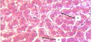
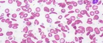
If the disease is present, the patient is diagnosed with a serious decrease in hemoglobin, up to a critical level of 20-30 g/l, and agranulocytosis is observed - a decrease in granular leukocytes and monocytes. The number of lymphocytes may be normal or reduced, platelets are always reduced, sometimes they are not detected at all. Erythrocyte sedimentation rate – increases to 4-60 mm/h. A study of a bone marrow sample reveals an increased content of adipose tissue - 90%, which includes elements of stroma and lymph, but hematogenous cells are present in very small quantities.
Diagnostic measures
Diagnosing aplastic anemia is not that difficult. Most cases are found during routine examinations. One general blood test is enough to suspect the presence of aplastic anemia.
Then the doctor begins a detailed collection of the patient’s complaints, anamnesis, and notes symptoms characteristic of aplastic anemia.
And then the doctor prescribes more detailed studies to make an accurate diagnosis and determine the degree of complexity.
So, if AA is suspected, the doctor prescribes a bone marrow aspiration. Next, the bone marrow is examined under a microscope, the presence and condition of blood cells are assessed at the stage of their development. In aplastic anemia, the bone marrow is depleted of all cell lineages. The main diagnostic method for AA is bone marrow biopsy, which allows the most accurate assessment of the state of hematopoiesis.
The Doctor also prescribes:
— Serological tests for hepatitis and other viral formations;
— Laboratory tests to study kidney and liver function;
— Ultrasound diagnostics. Enlarged lymph nodes can be specifically examined.
— Test for antibodies to infections;
- number of reticulocytes
— presence of a PNG clone;
Sometimes it is necessary to undergo other tests to clarify the diagnosis and establish the cause of the development of aplastic anemia.
Although, quite often, the cause remains unknown, due to the large number of factors and triggers that can provoke it.
https://www.cinj.org/sites/cinj/files/documents/December%20National%20Aplastic%20Anemia%20Awareness%20Month%202018.pdf
Treatment of aplastic anemia
Treatment of idiopathic and other types of aplastic anemia is a very complex task that requires a comprehensive individual approach. When developing tactics, CELT specialists take into account the diagnostic results and the patient’s indications. The patient is placed in an isolation room with aseptic conditions, which eliminates the risk of developing infections and their complications. Drug therapy consists of taking:
- Glucocorticoids – in identifying autoimmune mechanisms and the formation of antibodies against one’s own blood cells;
- Cytostatics – in the absence of effect from treatment with glucocorticoids for autoimmune anemia;
- Cyclosporine “A” – to suppress the production of TNF and interferon “y”;
- Anabolic steroids – to stimulate hematopoietic function;
- Androgens – to stimulate the formation of red blood cells.
All patients with aplastic anemia receive transfusions of red blood cells and/or platelets, in volumes determined based on the clinical picture and peripheral blood parameters. In addition, the patient may be prescribed a splenectomy, a surgical procedure aimed at removing the spleen. Bone marrow transplantation can provide the most favorable prognosis. It consists of transplanting donor or own hematopoietic stem cells, previously removed from the iliac bones by puncture. Unfortunately, the procedure is not widely available due to the difficulty of selecting a compatible donor. If this is not possible, the patient is prescribed palliative therapy with cyclosporine A.
The hematology department of our clinic welcomes candidates, doctors and professors of medical sciences with over twenty-five years of practical and scientific experience. You can make an appointment with them online or by contacting our operators. Highly qualified specialists also work in the urology department. You can make an appointment with them for cystoscopy of the bladder.
At CELT you can consult a hematologist.
- Initial consultation – 3,500
- Repeated consultation – 2,300
Make an appointment
By making an appointment with a hematologist, you can get a comprehensive consultation. The doctor is competent to treat various blood diseases, most of which can be identified in the early stages and prescribe timely treatment to cope with the disease quickly and easily.
Publications in the media
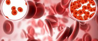
Aplastic anemia is a group of pathological conditions characterized by pancytopenia in the peripheral blood due to inhibition of the hematopoietic function of the bone marrow.
Classification • Congenital (Fanconi, Diamond-Blackfan anemia) • Acquired (the result of exposure to chemical, medicinal, infectious agents and ionizing radiation) • Idiopathic (the cause of the disease is unclear).
Etiology • Acquired anemia •• Infections (viral hepatitis, infectious mononucleosis, CMV, parvovirus, influenza) •• Lymphoproliferative disorders (thymoma, lymphoma, chronic lymphoblastic leukemia) •• Ionizing radiation •• Toxins and chemicals (benzene, insecticides, lead compounds etc.) •• Drugs, for example cytostatics, gold preparations, NSAIDs, anticonvulsants, antibiotics • Congenital anemia (Fanconi anemia). There are 4 complementation groups, i.e. at least 4 genes, mutation of any of which leads to the development of aplastic pancytopenia of 4 types (all r) •• Group A (type 1 Fanconi anemia, *227650, 20q13.2–q13.3, defects in the genes FA1, FA, F, r ) •• Group B (type 2, *227660, FA2 gene defect, r) •• Group C (type 3, *227645, 9q22.3, FACC gene defect, r) •• Group D (type 4, *227646, FA4, r gene defect).
Pathogenesis of aplastic anemia • Intrinsic defect of hematopoietic stem cells • Immune reaction to hematopoietic tissue • Impairment of the supportive function of the stromal environment in the bone marrow
Clinical picture
• Characteristic features of aplastic anemia •• General signs (shortness of breath, pallor of the skin and mucous membranes, tachycardia, weakness, systolic murmur at the apex of the heart, loss of body weight) •• Frequent addition of infectious-inflammatory and purulent-necrotic processes of various localization due to leukopenia ( neutropenia) •• Hemorrhagic syndrome: large and small hemorrhages (including in the retina), bleeding of various locations (menorrhagia, melena, nosebleeds) caused by thrombocytopenia. Features in children • The predominant age is 14–16 years • Anemia is usually preceded by an infectious disease (chicken pox, rubella, viral hepatitis) • The disease develops very quickly • In 70% of cases, treatment with cyclosporine can achieve complete clinical and hematological remission.
• Fanconi anemia. There are 2 types •• Classic (type I) ••• Short stature ••• Microcephaly ••• Skeletal deformities (absence or hypoplasia of the first metacarpal or radius) ••• Kidney and heart abnormalities ••• Hypospadias ••• Skin hyperpigmentation ••• Deafness •• Type 2 is characterized by the presence of minor developmental anomalies.
• Congenital aplastic anemia (Diamond–Blackfan syndrome) is an allelic variant of Fanconi anemia (*205900, r). It is distinguished from Fanconi anemia by the early onset of anemia (usually in the first months of life), the absence of neutropenia and thrombocytopenia.
According to severity, they distinguish between non-severe (granulocytes less than 1.5´109/l, platelets 100´109/l, megakaryocytes less than 80´109/l) and severe (in peripheral blood granulocytes less than 0.5´109/l, platelets less than 20´ 109/l; in bone tissue, the predominance of fibrous tissue is more than 60%).
LABORATORY RESEARCH
• Acquired anemia •• Pancytopenia •• Normochromic erythrocytes •• Prolonged bleeding time •• Marked decrease in reticulocyte content (regenerative anemia) •• Increased iron concentration in the blood serum (due to blood transfusions) •• Normal indicators of total serum iron-binding capacity (TIBC) •• Serum erythropoietin concentrations are usually increased in proportion to the degree of anemia •• Impaired liver function tests.
• Fanconi anemia •• Macrocytosis (as opposed to acquired aplastic anemia) •• Increased HbF levels •• Absence of pronounced pancytopenia until 3–8 years of age •• Chromosome fragility, repair defects, increased sensitivity of chromosomes to diepoxybutane, mitomycin and ultraviolet radiation are characteristic.
• Diamond–Blackfan anemia: macrocytosis, reticulocytopenia, elevated HbF.
Bone marrow puncture • Increased iron stores • Reduced number of cells •• Decreased number of megakaryocytes •• Decreased number of myelocytes •• Decreased number of erythroid precursors •• Replacement of normal hematopoietic tissue with adipose and fibrous tissue • Fanconi anemia - bone marrow puncture often shows no changes.
Instrumental studies • CT of the thymus region if thymoma is suspected • X-ray examination of the bones of the forearm (possibly detecting abnormalities in the development of the radius) and thumbs (constitutional anemia) • Ultrasound of the kidneys.
Differential diagnosis • Transient erythroblastopenia in children • Myelodysplastic syndrome • Paroxysmal nocturnal hemoglobinuria • Acute leukemia • Hairy cell leukemia • SLE • Disseminated infection • Hypersplenism • Anemia in hypothyroidism, hypopituitarism and liver diseases.
TREATMENT
Management tactics • Inpatient treatment in the hematology department. In case of neutropenia, isolation is indicated to prevent possible infection • Exclusion of causative factors • Study of the HLA antigen system of patients and their family members.
Bone marrow transplantation in patients with severe aplastic anemia, especially under the age of 30 years, is the method of choice (if an HLA-identical donor is available) • Transplantation from unrelated donors in case of ineffective treatment.
Conservative treatment
• Carried out in the absence of a donor for bone marrow transplantation •• Antithymocyte globulin. To identify hypersensitivity, a skin test is first necessary ••• Drug of choice for the treatment of elderly patients or in the absence of a donor for bone marrow transplantation ••• Course dose of 160 mg/kg, divided into 4 doses, during the first 4 days of treatment •• • The required dose of the drug is dissolved in 500 ml of 0.9% sodium chloride solution and administered intravenously over 4–6 hours ••• The development of an anaphylactic reaction is an absolute contraindication to further administration of the drug •• Cyclosporine ••• Initial dose - 5 mg/kg/day orally or 3 mg/kg intravenously. Next, doses are selected based on the concentration of cyclosporine in the blood, determined daily ••• If there is no effect within 120 days, the drug is discontinued •• Methylprednisolone - 2 mg/kg/day IV from days 1 to 14 of treatment, 1 mg/day kg - from 15 to 21 days •• Granulocyte colony-stimulating factor (for example, molgramostim) - if thymoglobulin or cyclosporine is ineffective ••• Initial dose - 5 mcg/kg/day subcutaneously until the granulocyte count increases to more than 1,000/μl ••• If there is no effect within 14 days, the dose is doubled •• Androgens are effective in some types of Fanconi anemia, acquired aplastic anemia, although cases of successful treatment are extremely rare. If there is no effect within 4–6 months, the drug is discontinued gradually.
• Maintenance therapy •• Oxygen therapy •• Sanitation of the oral cavity and other mucous membranes •• Transfusion of washed red blood cells through leukocyte filters; platelet mass •• Antibacterial therapy •• Hemostatics
• Complications of therapy • Hemorrhagic syndrome • Infectious complications • Hemosiderosis during blood transfusion • Heart failure • Renal failure • Toxic hepatitis • Anaphylactic shock and serum sickness (when using antithymocyte globulin) • Development of acute leukemia.
Surgical treatment • Thymectomy for thymoma.
Treatment outcomes • Complete clinical and hematological remission: Hb concentration above 110 g/l, platelets more than 100´109/l, granulocytes more than 1.5´109/l • Partial clinical and hematological remission: Hb concentration 90–110 g/l, platelets 30–100´109/l, granulocytes 0.5–1.5´109/l • Minimum hematological response: Hb level 80–90 g/l, platelets more than 10–30´109/l (without transfusions).
Course and prognosis • Acquired anemia. The course and prognosis are directly proportional to the patient's age. In the absence of timely and adequate therapy, 80% of patients die within 3 months from the manifestation of the disease. With the introduction of cyclosporine into clinical practice, the chances of prolonging the patient's life have increased. In case of proposed bone marrow transplantation, in order to avoid sensitization in the preoperative period, it is advisable to avoid transfusions of blood products (especially from relatives) • Fanconi anemia. Survival does not exceed 4 years with isolated replacement therapy. The prognosis is much better with the effectiveness of androgens or bone marrow transplantation.
Synonyms: For Fanconi anemia - Fanconi syndrome.
ICD-10 • D60 Acquired pure red cell aplasia [erythroblastopenia] • D61 Other aplastic anemias
Notes • Bone marrow aplasia is a congenital or acquired severe condition manifested by pancytopenia. The congenital form is combined with phenotypic and cytogenetic abnormalities, which facilitates diagnosis • Pancytopenia - a decrease in the number of red blood cells, leukocytes and platelets. Characterized by pallor and drowsiness, infections caused by neutropenia; hemorrhagic diathesis due to thrombocytopenia. For differential diagnosis, a bone marrow examination is necessary (aplasia [usually hypoplasia] of the bone marrow, displacement of bone marrow sprouts by blast cells, autoimmune destruction of blood cells - Evans syndrome) • Congenital and sometimes idiopathic types of anemia are called constitutional.