Muscle atrophy leads to loss of volume and number of fibers in striated muscles. This pathology is caused by many different reasons. It develops from prolonged restriction of physical activity. Muscle atrophy is caused by various diseases or injuries.
Some types of atrophy are caused by genetic problems. If these disorders are not consulted and treated, the patient may lose the ability to move. Therefore, if you feel weakness and soreness in your muscles, you should visit a doctor as soon as possible.
Description
Muscular atrophy is a disorder of muscle tissue trophism. It is accompanied by gradual thinning and then degeneration of the fibers. Over time, their contractility decreases. Muscles decrease in volume. Their fibers become thinner until they disappear completely.
Muscle tissue is replaced by connective tissue fibers. They are unable to contract. The patient's motor function suffers. His muscle strength and tone decrease. This leads to loss of motor activity or its sharp decrease. There are simple and degenerative types of the disorder.
Degenerative muscle atrophy is a leading sign of a group of neuromuscular hereditary pathologies. Simple muscle atrophy develops due to the increased sensitivity of muscle fibers to damaging factors of various types.
This disorder can be a syndrome associated with various diseases, intoxications, and injuries. Muscle atrophy occurs due to exhaustion, tissue hypoxia, nerve damage, microcirculation disorders, intoxication, compression by tumors, and metabolic disorders.
1.General information
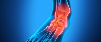
The quadriceps muscle (quadriceps) is located on the front side of the thigh and partially covers it from the side. It received its name due to its anatomically complex structure: the muscle is formed by four structures (heads) with a separate origin and a common tendon, which is attached to the knee joint. The functional task of the quadriceps femoris muscle is to flex the lower leg and raise the knee upward; in addition, the quadriceps muscle is one of the extensors (extensors) of the hip joint. Accordingly, any pathology of this quadricomponent muscle inevitably affects the gait and, in general, a person’s mobility.
Atrophic is the process of gradual death of cells in any tissue, which entails a reduction in its volume and a progressive (at varying speed) loss of functional viability. Thus, quadriceps atrophy is a serious threat to motor function; Left without attention and medical care, this process in the later stages can completely disable the limb and result in disability.
A must read! Help with treatment and hospitalization!
Causes
Muscle atrophy develops for the following reasons:
- hereditary predisposition;
- mechanical injuries;
- hard physical labor;
- infections;
- myopathy;
- diabetes;
- malignant neoplasms;
- aging of the body;
- paralysis due to damage to peripheral nerves;
- starvation;
- intoxication;
- metabolic disorder;
- pathological process in the superior parietal lobe;
- prolonged immobility;
- spinal cord damage;
- thrombosis of large vessels;
- birth injury;
- neurological disorders;
- polio;
- myositis;
- pathology of the pancreas.
In case of a back injury with damage to the spinal cord, muscle dystrophy is observed in the area of innervation of the damaged segments.
Sources[edit | edit code]
- ↑ 1,01,11,2 Kots Ya.M. Sports physiology Textbook for physical education institutes. - M.: Physical culture and sport, 1998.
- Solodkov A.S., Sologub E.B. Human physiology. General. Sports. Age: Textbook. - M.: Terra-Sport, Olympia press, 2001.
- Solodkov A.S., Sologub E.B. Human physiology. General. Sports. Age: Textbook. - M.: Terra-Sport, Olympia press, 2001.
- Kraemer, William J.; Zatsiorsky, Vladimir M. (2006). Science and practice of strength training. Champaign, IL: Human Kinetics. p. 50. ISBN 0-7360-5628-9.
- MacDougall JD, Sale DG, Alway SE, Sutton JR Muscle fiber number in biceps brachii in bodybuilders and control subjects // Journal Applied Physiology, 1984. V 57. No. 5. P. 1399-1403.
- Samsonova A.V. Hypertrophy of human skeletal muscles: Textbook. — 3rd ed. - St. Petersburg: Politekhnika, 215.
- MacDougall JD Hypertrophy and Hyperplasia // In: The Encyclopedia of Sport Medicine. Strength and Power in Sport. - Bodmin, Cornwall: Blackwell Publishing, 2003.
- Gibala, MJ, MacDougall JD, Tarnopolsky MA, Stauber WT, Elorriaga A. Changes in human skeletal muscle ultrastructure and force production after acute resistance exercise // Journal of Applied Physiology. - 1995. - P. 702-708.
- MacDougall JD, Elder GCB, Sale DG, Moroz JR & Sutton, JR Effects of strength training and immobilization on human muscle fibers // European Journal of Applied Physiology. - 1980. - No. 43. - P. 25–34.
- Yazvikov V.V. The influence of sports training on the composition of muscle fibers of mixed human skeletal muscles // Theory and practice of physical culture. - 1988. - No. 2. - P. 48-50.
- Yazvikov V.V., Morozov S.A., Nekrasov A.N. Correlation between the content of slow fibers in the vastus externus muscle and athletic performance // Human Physiology. - 1990. - T. 16, No. 4. - P. 167-169.
- Goldbeig et al., 1975
- Wong and Booth, 1990; Chcsley et al., 1992; Biolo ct al., 1995; Philips et al., 1997
- Laurent et al., 1978; Wong, Booth, 1990
- Frederickson, Sonebcig, 1993; Wada et al., 1996
- Biolo et al., 1997
- Cheek, 1985; Hall, Ralston, 1989; Allen et al., 1999
- Goldberg et al., 1975; Cabric, James, 1983; Winchester, Gonyea, 1992; Allen et al., 1995; Kadi, 2000
- Kadi et al., 1999a; Kadi, 2000
- Hikida et al., 1998; Kadi, Tomcll, 2000
- Hawke, T. J., and D. J. Garry. Myogenic satellite cells: physiology to molecular biology. Journal of Applied Physiology. 91:534-551, 2001.
- Egginton, 1987; Salmons, 1992
- Basin et al., 1996
- Powers, Florini, 1975; Rogozkin, 1979
- Kadi et al., 1999b
- Kadi et al., 1999b
- Galavazi, Szirmai, 1971; Sassoon, Kelley, 1986; Joubcrt, Tobin, 1989; Joubert, Tobin, 1995
- Kadi et al., 1999b
- Doumit et al., 1996
- Douglas R. Bolster, Leonard S. Jefferson and Scot R. Kimball. Regulation of protein synthesis associated with skeletal muscle hypertrophy by insulin-, amino acid- and exercise-induced signaling. Department of Cellular and Molecular Physiology, The Pennsylvania State University College of Medicine, PO Box 850, Hershey, PA 17033, USA
Symptoms
With the initial manifestations of muscle atrophy, the patient quickly gets tired. There is a significant decrease in muscle tone and twitching of the limbs is noticeable. With atrophy of infectious or traumatic origin, damage to motor neurons occurs.
It leads to movement disorders that vary in severity from slight limitation of movements in the upper and lower extremities to paralysis. The patient develops weakness in the legs.
It is difficult for him to move on if he is forced to stop. The patient notes a feeling of heaviness in the legs. His movements are difficult, numbness appears, and his gait is disturbed. Then atrophy of the muscles of the gluteal region occurs.
The patient develops a waddling gait. Numbness in the buttock area is detected. Pallor of the skin is characteristic. Muscle atrophy in the upper extremities is expressed in limited movement.
The patient is bothered by tingling and numbness in the hands, decreased tactile sensitivity. Mechanical impact on tissue causes severe discomfort. The patient's tissue trophism is disrupted, which leads to blue discoloration of the skin.
Republican Children's Clinical Hospital
Malnutrition in children is a chronic nutritional disorder. Hypotrophy is a type of dystrophy. If dystrophy in a child occurs with a deficiency of body weight, then it is called malnutrition, but if dystrophy occurs with excess body weight, then it is paratrophy.
What could be the reasons?
Hypotrophy is distinguished between prenatal (congenital) and postnatal (acquired). Congenital malnutrition develops during a pathological course of pregnancy, various diseases of the mother, the presence of occupational hazards, smoking, drinking alcohol during pregnancy, etc.
The causes of acquired malnutrition are divided into internal and external.
Internal - caused by pathologies of the body that interfere with food intake and digestion, absorption of nutrients and metabolism: congenital malformations, central nervous system lesions, immunodeficiency, endocrine diseases, metabolic disorders.
In the group of endogenous factors, it is worth highlighting food allergies and three hereditary diseases that occur with malabsorption syndrome - one of the common causes of malnutrition in children:
- — cystic fibrosis — disruption of the exocrine glands, affecting the gastrointestinal tract and respiratory system.
- — celiac disease — gluten intolerance, changes in a child’s intestinal function begin from the moment gluten-containing foods are introduced into the diet — barley, semolina, wheat porridge, rye, oatmeal.
- - lactase deficiency - the digestibility of milk is impaired (lactase deficiency).
External - caused by incorrect actions of parents and unfavorable environment:
- nutritional factor: insufficient supply of breast milk, incorrectly selected formula for artificial feeding, late introduction of complementary foods, low amount of calories in complementary foods, imbalance in proteins, fats and carbohydrates;
- infectious factor: intestinal infections, chronic infections, frequent acute respiratory viral infections and other diseases;
- toxic factor: unfavorable environmental conditions, medication, poisoning, excessive intake of vitamins, especially A and D;
- social factor: inattention of parents to the child, lack of walks in the fresh air, lack of gymnastics and massage.
How does malnutrition manifest itself?
Hypotrophy of the first degree - characterized by thinning of the subcutaneous fat layer on the trunk, limbs, body weight deficiency compared to the norm is 10-20%. Tissue turgor is reduced. The child's height is appropriate for his age. The child does not lag behind in psychomotor development, but is characterized by unstable emotional tone and increased excitability.
Hypotrophy of the second degree - the subcutaneous fat layer is sharply thinned on the trunk, limbs, absent on the abdomen, the weight deficit compared to the norm is 20-30%. There is a delay in growth and psychomotor development. The child is restless, irritable, or has a sluggish response to his surroundings. The skin is pale, dry, “marbled”; tissue turgor is flabby, muscle tone is reduced; appetite is reduced, there may be vomiting, regurgitation, unstable stool, sometimes “hungry” - scanty, dry, discolored, with an unpleasant putrid odor, or dyspeptic.
Hypotrophy III degree. The subcutaneous fat layer is absent on the trunk and limbs, facial features are pointed. Body weight deficiency compared to the norm is more than 30%. The skin is pale gray, dry, flaky, the skin fold almost does not straighten out (the elasticity of the skin is sharply impaired). The mucous membrane of the lips is bright, dry, and there are “seals” in the corners of the mouth. Dry cornea. Hypertonicity of the muscles of the arms and legs is often observed. Shallow breathing, muffled heart sounds, arterial hypotension. The stomach is swollen. The stool is unstable with a tendency to constipation. Urination is reduced. Children are lethargic and indifferent to their surroundings. As a result of a sharp violation of immunoreactivity, complications develop (otitis media, pneumonia, pyelonephritis, meningitis), which are sluggish and asymptomatic.
What are the principles of treatment?
In a polyclinic, stage I malnutrition is treated in young children, and stage II and III malnutrition is treated inpatient settings.
The main activities are aimed at: normalizing nutrition; diet therapy (gradual increase in calorie content and volume of food consumed by the child + fractional, frequent feeding); compliance with the daily routine; organizing proper child care; correction of metabolic disorders; drug therapy (enzymes, vitamins, anabolic hormones); in the presence of a severe form of the disease, intravenous administration of glucose, protein hydrolysates, vitamins, and saline solutions is prescribed; Ural Federal District; massage with elements of exercise therapy.
With timely treatment of stage I and II disease, the prognosis is favorable, but with grade III malnutrition, up to 50% of cases are fatal!
What is prevention?
Prevention of malnutrition in children involves regular examination by a pediatrician, constant anthropometry and nutritional correction. During pregnancy, the mother must: maintain a daily routine; eat in a timely manner; promptly treat existing diseases; eliminate all possible unfavorable factors. maintain a daily routine; eat in a timely manner; promptly treat existing diseases; eliminate all possible unfavorable factors. After the birth of a child, an important role is played by: high-quality and balanced nutrition of the nursing mother; timely and correct introduction of complementary foods; body weight control; rational, competent care for a newborn; treatment of any, even spontaneously occurring, concomitant diseases.
Having heard a diagnosis such as malnutrition, parents should understand that the child needs the help of a qualified doctor, and not postpone examination and treatment for the future. If you provide your child with normal conditions of routine, care and nutrition in a timely manner, quick and effective treatment of possible infections and concomitant pathologies - severe forms can be avoided!
head Pediatric Department, pediatrician of the highest category G.D. Levchenko
Diagnostics
If signs of muscle atrophy are detected, you should consult a therapist, traumatologist, neurologist or orthopedist. To clarify the diagnosis, the following studies are performed:
- general clinical blood test;
- electroneuromyography
- electromyography;
- muscle tissue biopsy;
- blood biochemistry;
- genetic examination.
To identify the causes of muscle atrophy, it is necessary to diagnose the condition of the pancreas, thyroid gland, and liver.
Treatment
Therapy for muscle atrophy is determined by the degree of loss of this tissue, as well as the nature of the underlying disease. This slows down the progression of muscle fiber breakdown. Exercise therapy is prescribed as additional measures.
The complex consists of stretches, as well as exercises to prevent the development of limb immobility. They increase muscle strength, improve blood circulation, and reduce spasticity.
The method of functional electrical stimulation is actively used in treatment. Here, electrical impulses are used to stimulate muscles. Electrodes are applied to the affected areas. The current, passing through the tissues, causes muscle contraction.
Specific drug therapy will depend on the underlying disease. To do this, identify the cause of muscle atrophy as early as possible and begin treatment.
Methodology for assessing the degree of hypertrophy[edit | edit code]
In order to assess the degree of skeletal muscle hypertrophy, it is necessary to measure the change in its volume or mass. Modern research methods (computer or magnetic resonance imaging) make it possible to assess changes in the volume of skeletal muscles in humans and animals. For this purpose, multiple “slices” of the cross-section of the muscle are performed, which makes it possible to calculate its volume. However, until now, the degree of skeletal muscle hypertrophy is often judged by changes in the maximum cross-sectional value of the muscle obtained by computed tomography or magnetic resonance imaging.
In bodybuilding, muscle hypertrophy is assessed by measuring the coverage of the arms (at the level of the forearm and biceps), thighs, legs, and chest using a meter tape.