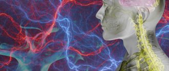
Causes of esophageal varicose veins
The disease develops against the background of increased pressure in the portal vein (also known as the portal vein), but other pathologies (hypertension, congenital anomalies, and so on) can contribute to its occurrence. Esophageal varicose veins, the causes of which lie in pathological changes in the portal vein, often begin to progress with previously diagnosed severe diseases of the liver or pancreas - cirrhosis, tumors, insufficiency. The disease often develops against the background of severe diseases of the thyroid gland, in which case we will be talking about pathological disorders of the vena cava.
According to statistics, esophageal varicose veins are more often diagnosed in men aged 50 years and older.
External factors
In addition to pathological causes, there are a number of extraneous causes that can affect the appearance and development of the disease, these include:
In addition to pathological causes, there are a number of extraneous causes
- alcohol abuse;
- medications that negatively affect the condition of the liver;
- some diseases transmitted by inheritance;
- viral diseases affecting the liver (hepatitis).
Important! Pregnant women should pay increased attention to their health, since any viral infections suffered during this period can be transmitted to the fetus, increasing its risk of developing esophageal varices.
Esophageal varicose veins: symptoms
Depending on the degree of development, the following signs are distinguished:
- First degree. The diameter of the veins is increased by no more than 3 mm, the formation of single nodes is possible. Diagnosis of the disease is possible only by the endoscopic method; there are no clinical signs.
- Second degree. A rather dangerous period of varicose veins of the esophagus - there are no symptoms (even the mucous membrane of the organ still remains unchanged), but at the same time the contour of the veins already becomes unclear, and X-ray diagnostics reveal thickenings on the veins (harbingers of nodules). Already at this stage, bleeding is possible, which in the absence of immediate medical attention often leads to death.
- The third and fourth degrees of varicose veins of the esophagus are characterized by severe symptoms - the patient is often bothered by heartburn, pain occurs in the sternum, shortness of breath is possible, not only during physical activity, but also at rest. Often the disease is diagnosed only in the later stages of its development, when the first bleeding appears; blood is released directly from the esophagus or found in the vomit.
Course of the disease
The course of the disease can be divided into 4 stages:
Stage I - initial
The disease has already appeared, but cannot yet be diagnosed. It is asymptomatic, or patients feel heaviness in the right hypochondrium and mild malaise.
Stage II - moderate
Pronounced clinical symptoms of the disease appear. Pain in the upper abdomen, flatulence, digestive disorders (belching, heartburn, heaviness in the abdomen, etc.), splenomegaly and hepatomegaly (enlarged liver).
Stage III - pronounced
All the signs and symptoms of portal hypertension are present in a pronounced form.
Stage IV - stage of complications
Intense ascites develops, difficult to treat, repeated bleeding, as well as possible complications.
Treatment
Treatment of varicose veins of the esophagus begins with finding out the true causes of the development of the disease. If a pathology of the liver, thyroid or pancreas is diagnosed, then it must be treated - the provoking factor must be eliminated or its influence must be weakened. Medications include vitamin complexes, antacids and astringents. If bleeding is diagnosed, there is always a risk of its reoccurrence. In this case, the following is prescribed:
- blood transfusion;
- hemostatic drugs;
- introduction of a special probe that can compress the vessels.
Varicose veins of the esophagus, bleeding in which occurs too often, require surgical treatment.
Surgery is also performed if there is no effect from therapeutic treatment. Can esophageal varicose veins be stopped in this case? The prognosis is more favorable - the survival rate of patients increases 3 times. After any treatment, you must follow the dietary recommendations of doctors, undergo regular examinations and avoid excessive physical activity.
You can find out more about what esophageal varicose veins are and which doctor you should contact on the pages of our website Dobrobut.com.
2.What are the symptoms of bleeding from varicose veins?
As we have already said, varicose veins of the esophagus themselves do not cause any discomfort. But if a vein ruptures and starts bleeding, it is dangerous and requires emergency medical attention. Symptoms of varicose veins of the esophagus or stomach and the onset of bleeding from them may include:
- Vomiting blood;
- Black or bloody stools;
- Low blood pressure;
- Increased heart rate;
- Shock (in particularly serious cases).
Visit our Gastroenterology page
Diagnostic methods
A competent therapist is quite capable of suspecting the presence of problems with the esophagus during a routine examination, in which case he is obliged to give a direction for additional examination. Three research options are possible:
Laboratory test - complete blood count test for platelet count
- Anamnesis of the disease - the examination data and complaints of the patient are recorded by the attending physician, the medical record reveals previous illnesses, and if among them diseases are found that provoke damage to the venous system of the esophagus, then the specialist, after tapping and pressing the abdominal area, can draw some conclusions.
- Laboratory test - a general blood test is taken to determine the number of platelets. A biochemical examination is also prescribed to check the amount of liver enzymes, protein and other important indicators in the body. Additional liver testing is prescribed if the initial results do not reveal a problem. It is even possible to prescribe an in-depth blood test to determine its group and coagulability.
- Instrumental examination - for each individual organ, a fluoroscopic examination, esophagoscopy or ultrasound is prescribed.
When formulating a diagnosis of esophageal varicose veins, the cause of its appearance is initially indicated. The conclusion also indicates the degree of damage and possible complications associated with this disease.
When formulating a diagnosis of esophageal varicose veins, the cause of its appearance is initially indicated.
References
- Rubin, Rafael; Strayer, David S.; Rubin, Emanuel, ed. (2012). Rubin's Pathology: Clinicopathological Fundamentals of Medicine
(6th ed.). Lippincott Williams and Wilkins. item 612. - Cushman, James (2018-01-01), Harken, Alden H.; Moore, Ernest E. (ed.), "Chapter 44—Portal Hypertension and Esophageal Varices," Abernathy's Surgical Confidential (Seventh Edition)
, Elsevier, pp. 195–199, doi:10.1016/b978-0-323-47873-1.00044-9, ISBN 978-0-323-47873-1, received 2020-11-23 - ^ a b
Awad, Joseph;
Wattaheril, Julia (01/01/2012), Jarnagin, William R.; Blumgart, Leslie H. (ed.), "Chapter 75B—Esophageal Varices: Emergency Management of Portal Hypertension," Blumgart's Liver, Pancreatic, and Biliary Surgery (fifth edition)
, Philadelphia: W. B. Saunders, pp. 1135–1138.e1, doi:10.1016/b978-1-4377-1454-8.00120's, ISBN 978-1-4377-1454-8, received 2020-11-23 - Bikker E, Schepke M, Sauerbruch T (2005). "The role of endoscopy in portal hypertension." Dig Dis
.
23
(1): 11–7. Doi:10.1159/000084721. PMID 15920321. - Arguedas M (2003). "Patient liver in critical condition: variceal bleeding." Semin Gastrointest Dis
.
14
(1):34–8. PMID 12610853. - Lebrecq, D., Poynard, T., Hillon, P., & Benhamou, J. P. (1981). "Propranolol for the prevention of recurrent gastrointestinal bleeding in patients with cirrhosis: a controlled study." N Engl J Med
.
305
(23):1371–1374. Doi:10.1056/NEJM198112033052302. PMID 7029276. - Talwalkar J. A., Kamath P. S. (2004). "An evidence-based approach to beta-blocker therapy in patients with liver cirrhosis." Am J Med
.
116
(11):759–766. doi:10.1016/j.amjmed.2004.03.006. PMID 15144913. - Grosmann, R. J., Garcia-Cao, G., Bosch, J. et al. (2005). "Beta-blockers for the prevention of gastroesophageal varices in patients with cirrhosis" (PDF). N Engl J Med
.
353
(21):2254–2261. doi:10.1056/NEJMoa044456. PMID 16306522. - Garcia-Cao G., Sanyal A. J., Grace N. D., Carey W. (2007). "Prevention and treatment of varicose veins of the esophagus and varicose veins in cirrhosis." Hepatology
.
46
(3):922–938. Doi:10.1002/hep.21907. PMID 17879356.CS1 maint: several names: list of authors (link to site) - de Ledingen, W., Bo, P., Mannant, P. R., et al (1997). “Early feeding or enteral nutrition in patients with cirrhosis after bleeding from esophageal varices? Randomized controlled trial." Dig the ground.
Dis. The science .
42
(3):536–41. Doi:10.1023/A: 1018838808396. PMID 9073135. - Abid S, Jafri W, Hamid S, et al (March 2009). "Terlipressin versus octreotide for bleeding esophageal varices as adjuvant therapy with endoscopic tape ligation: a randomized, double-blind, placebo-controlled trial." Am.
J. Gastroenterol .
104
(3):617–23. doi:10.1038/ajg.2008.147. PMID 19223890.
OVERBIG ZAVROYUVANNYA
Sculo-stravochoidal varicose veins (PVVs) are the most clinically important portosystemic collaterals, so their ruptures lead to varicose bleeding, the most common lethal form of cirrhosis - the first This episode is associated with a mortality rate of 30–50%. Varicose veins and varicose bleeding are aggravated by cirrhosis, which is directly caused by portal hypertension. Patients with cirrhosis and gastroesophageal varix show HPVT readings of less than 10–12 mmHg. Art.
Schlunkovo-stravochodnye varicose veins are detected in about 50% of patients with cirrhosis. Their presence correlates with the importance of liver disease (Table 2): while in class A for Child-Pugh, only 40% of patients have varicose veins, then in class C veins, 85% of patients have varicose veins. In patients with primary biliary cirrosis, varicose veins and varicose bleeding may develop at an early stage of illness, leading to the diagnosis of cirrhosis. It was also found that 16% of patients with hepatitis C and pontic fibrosis have varicose veins.
In patients without varicose veins, the remaining varicose veins develop with a frequency of 8% per river; the strongest predictor for the development of varicose veins in patients who are not visible during the initial endoscopic examination is an HPVT reading >10 mm Hg. Art. In patients with small varices, large varices develop with a frequency of 8% per population. Decompensated cirrhosis (Child B/C), alcoholic cirrhosis and the presence of “worm signs” during the first endoscopy are the head signs associated with the transition from small varixes to large ones (Merli M. et al., 2003). Japanese authors, in their classification, give a substantive meaning to the bluish bark of the VRV.
Variceal bleeding occurs with a frequency of 5–15% per river, and the most important predictor of bleeding is the size of the variceal vein, with the highest risk of first bleeding (15% per river) in patients with large variceal veins (The North Italian Endoscopic Club for the Study and Treatment of Es. ophageal Varices , 1988). Other predictors of bleeding are decompensated cirrhosis (Child B/C), bluish coloration of the veins, and the appearance of red signs during endoscopy. While bleeding from varicose veins usually disappears on its own in up to 40% of patients, regardless of treatment in the hospital for the remaining 10 years, stench is associated with a mortality rate of at least 20% between 6 years iv. Patients with HPVT >20 mm Hg. Art. (over a period of 24 years after varicose bleeding) it was found that there is a high risk of early recurrence of bleeding (re-bleeding in the first life after surgery) or distant spots of bleeding (83% versus 29%) and I see a single mortality rate (64% per against 20%) was equal to patients with lower pressure. Later, relapses of bleeding occur in about 60% of serious patients, mostly in 1-2 cases after the initial bleeding.
The portal pressure, the size of the vein and the thickness of its wall are related to each other by Laplace’s law - the stress of the vein wall (T) is directly proportional to the pressure in the middle of the vessel (P) and its radius (R) and is proportional to the thickness of the vessel Inki (W): T = P × R/W.
Apparently, dilatation of the node from the thinning of its wall is the main factor that causes stress on the wall and rupture of the varix. Under pressure, however, a vessel with a larger diameter will rupture, but a vessel with a smaller diameter will not. Around the diameter of the vessel, another source of stretching of the varicose wall is the pressure in the middle of the VRV, which is directly connected with the HPVT. Thus, a decrease in HPVT is likely to reflect a change in the stretching of the wall of the varicose vein and, apparently, a change in the risk of rupture. In fact, varicose veins do not bleed when HPVT decreases to <12 mmHg. Art. It has also been shown that the risk of re-bleeding significantly changes when GPVT is reduced by more than 20% of the cob level. Diseases in which HPVT decreases to <12 mmHg. However, at least 20% of the cob level (“HVPG responders”) may reduce the risk of recurrent variceal bleeding, as well as a lower risk of ascites, spontaneous bacterial peritonitis, and death. Table 2. Classification of the importance of cirrhosis according to Child-Pugh
| Items* | |||
| 1 | 2 | 3 | |
| Encephalopathy | No | Riven 1–2 (induced) | Riven 3–4 (chronic) |
| Ascites | No | Minor/moderate (responds to diuretics) | Tension (resistant to diuretics) |
| Bilirubin (mg/dl) | <2 | 2–3 | >3 |
| Albumin (g/dl) | >3,5 | 2,3–3,5 | <2,8 |
| Prothrombin hour (PT), daytime (s) or international normalized ratio (INR) | <4 <1,7 | 4–6 1,7–2,3 | >6 >2,3 |
*5–6 points: Child A. 7–9 points: Child B. 10–15 points: Child C.
CLINICAL DIAGNOSIS OF PORTAL HYPERTENSION
With physical examination, it is easiest to detect ascites, redness, spider-like angiomas on the skin, erythema pubis, testicular atrophy, gynecomastia, Dupuytren's contracture and muscle atrophy. Portosystemic encephalopathy may also manifest itself - in some cases, asterixis (blurring or “purrying” tremor of the hands) and lethargy, in the lungs - restlessness, sleep disturbance. Most patients with PG also exhibit splenomegaly, although the size of the spleen correlates poorly with portal venous pressure and its increase may be associated with other illnesses. A hyperdynamic state can be indicated by a high (jumping) pulse, warm, well-perfused endings in conjunction with arterial hypotension.
Signs of portosystemic collaterals are dilated veins on the anterior surface of the abdomen (umbilical-epigastric shunts), the “jellyfish head” becomes a sinuous colateral vein near the navel. Internal hemorrhoids are not a specific finding, but may also manifest collateralization. The weakness of the visible collateral vessels on the back does not indicate portal hypertension, but obstruction in the level of the inferior venous vein. A rare cause may be compression of the lower empty vein by regenerative nodes in liver cirrhosis.
Ascites is easy to detect when there is a lot of swelling in the stomach - in such cases the life is tight, and swelling of the spine can be detected. However, minor ascites can be diagnosed instrumentally, for example, with the help of ultrasonography. Paracentesis helps to establish the etiology of ascites: the portal (non-peritoneal) etiology indicates a serum ascitic albumin gradient (SAAG) of over 1.1 g/dL (11 g/L). However, this method cannot distinguish between hepatic and suprahepatic PH, for example, in cases of persistent heart failure. Recent studies indicate a decrease in SAAG following the placement of transjugular portosystemic shunts (TIPS).
Laboratory testing of gas is not specific for the detection of PG. Indicates an increase in prothrombin hour and thrombocytopenia. In patients with cirrhosis of the liver, these indicators are considered predictors of the presence of varicose veins (see below). Hypoalbuminemia indicates the importance of liver function and, indirectly, PG. Similarly, hyponatremia and low sodium concentrations in the tissue may contribute to water and sodium retention in portal hypertensive syndrome.
ASSESSMENT OF PORTAL HYPERTENSION
The shortest, albeit indirect, method of assessing portal pressure involves wedged hepatic venous pressure (WHVP), which is determined by inserting a balloon catheter (from the jugular, brachial or stegnosus access) under the fluid. oroscopic guidance of the hepatic vein and “jamming” it in the small hilt (introduced all the way) or (which is still sticky) - by inflating the balloon and occluding the larger hilt of the liver vein. The correction of WHVP will ultimately involve the improvement of the internal hepatic vein pressure (for example, with ascites) by releasing the pressure in the hepatic vein - VTPV (free hepatic vein pressure - FHVP) or the internal hepatic vein pressure pressure in the empty vein, which is respected by the internal zero level. They survive when the balloon is deflated. The hepatic venous pressure gradient (HVPG) is determined by the pressure gradient in the hepatic vein. The indicator, as a matter of course, dies three times; If the values are technically correct, they are also well-designed and reliable. Although there is a gradient of pressure between the sinusoids (and not the portal vein veins) and the hepatic vein, HPVT will be advanced in case of intrahepatic causes of portal hypertension, such as cirrhosis, however normal for prehepatic causes, such as portal vein thrombosis. Normal HPVT readings are 3–5 mmHg. Art. It was found that WHVP closely correlates with portal pressure in both alcoholic and non-alcoholic cirrhosis. HPVT and changes in its symptoms, which are observed over time, may have a transferable value to the development of varicose veins in the vein, the risk of varicose bleeding, the development of non-varicose folds of portal hypertension And death. Single extinction is useful for the prognosis of both compensated and decompensated cirrhosis, while repeated extinction is useful for monitoring the effectiveness of drug therapy and the reversal of liver disease. The main considerations for the widespread viability of HPVT include the lack of evidence from practicing physicians and their continued recommendations on how to eliminate reliable and effective indicators, as well as other the simplicity of the procedure.
Duplex Doppler ultrasonography
Ultrasonography is a safe, inexpensive and effective method for screening for PG. The knobby structure of the liver, splenomegaly and the presence of colateral circulation give rise to suspicion of liver cirrhosis and PG. The diameter of the splanchnic vessels, directly and the fluidity of the bloodstream, changes in the diameter of the vessels during breathing, the index of portal venous stagnation, the pulsativity index and the resistance index of the liver, upper pars and glands also depend. Hepatic artery and hepatic vessel index. Normally, the diameter of the portal vein does not exceed 13 mm during quiet breathing, but during deep breathing it can increase by 50%. Expand a vein with a diameter of >15 mm while breathing calmly. When the pressure of the hemorrhage at the portal vein is increased and becomes monotonous (without breathing sounds) and resolves completely in the portal vein, straightened at the non-functioning portocaval shunts.
However, the creation of many ultrasonic parameters, their accuracy is insufficient, lies both in the tracking technology and in the form of additional rhythms, the development of the sympathetic nervous system, medicine, etc. So, for example, the diameter of the portal vein may increase in case of severe congestive heart failure (water from the inferior empty vein). Therefore, duplex Doppler ultrasonography itself is not recommended for establishing an accurate diagnosis of PG; it is unclear that its diagnostic value for the early stages of PG is lost (in most of these studies, patients were monitored with severe cirrosis and severe PG). Nevertheless, this method is valuable for primary closure, and also helps to identify the etiology of PG, for example, obstruction of blood vessels at different levels, highly sensitive to the identification of functional portocaval anastomoses. After placement of TIPS or portocaval anastomoses, Doppler ultrasonography is of great importance in monitoring shunt patency.
Computed tomography is a test with clear (not sharp) values, whereas ultrasonography did not give clear results. Apart from ultrasonography, CT results do not indicate the presence of gas in the intestine. Reconstruction of the vascular tree with the help of 3-D technology makes it possible to accurately create the actual portal venous system and collaterals. A common finding in portal hypertension during CT scanning is dilatation of the inferior vein. However, CT does not allow assessing the flow of blood through the venous and arterial vessels. MAR makes it possible, in addition to clear information, to obtain detailed information, for example, to assess venous flow in the portal and azygos veins. However, this does not exclude the need for invasive surveillance.
In cases of doubt about the genesis of liver disease, puncture biopsy may be necessary: necrosis of the third zone, for example, indicates portal hypertension, secondary to cardiac failure, and normal liver parenchyma - about subhepatic cause of PG.
Contraindications for liver biopsy include coagulopathy and ascites. Table 1. Causes of portal hypertension and pressure gradient in the hepatic vein
| GTPV | |||
| Rhubarb occlusion | Normal | Normal or movement | Advances |
| Subhepatic syndromes of PG | Portal vein thrombosis Thrombosis of the splenic vein Splanchnic arteriovenous fistula | ||
| Intrinsic hepatic syndromes of PG More important than the level of presinusoidal level | Schistosomiasis, primary biliary cirrosis, idiopathic PG (early stage)* Vuzlova regenerative hyperplasia* | Myeloproliferative diseases*† Polycystic disease*† Liver metastases*† Granulomatous disease † (sarcoidosis, tuberculosis)*† | |
| More important is the level on the sinusoidal or postsinusoidal level | Budd-Chiari syndrome‡ | Cirrosis of the liver Schistosomiasis, primary biliary cirrosis, idiopathic PG (late stage) Hospital and fulminant hepatitis Gostria alcoholic hepatitis Wilson's illness Veno-occlusive disease Budd-Chiari syndrome‡ | |
| Post-hepatic syndromes of PG | Obstruction of the inferior venous vein Right heart failure Clutching pericarditis Tristucular valve insufficiency | ||
* Rare causes of PG. † For the mind of the sinusoids. ‡ Sinusoidal symptoms are weak.