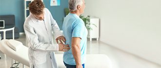
Traumatologist-orthopedist (adults and children)
Bogatov
Victor Borisovich
21 years of experience
Highest qualification category. Doctor of Medical Sciences, Professor of the Department of Traumatology and Orthopedics, First Moscow Medical University. I.M. Sechenov.
Make an appointment
Arthrosis is a disease that affects human joints. First, the disease affects the articular cartilage, and then becomes chronic and spreads to nerves, ligaments, muscles and bone.
What is arthrosis
Arthrosis is a disease of the joints of a degenerative-dystrophic nature with the gradual destruction of cartilage and proliferation of bone tissue. The process is accompanied by deformation, impaired joint function and pain. Recently, the term osteoarthritis (OA osteoarthritis) is more often used - a group of diseases that are based not on purely dystrophic, but on dystrophic-inflammatory processes leading to gradual destruction of the joint. More and more experts believe that the causes of arthrosis and osteoarthrosis, the mechanisms of their development are the same, that is, in fact, they are the same disease.
According to statistics, from 10 to 20% of the population suffers from arthrosis in different countries. By the age of 80, almost everyone has age-related disorders in the musculoskeletal system. At the same time, patients do not always consult a doctor on time and take a long time to treat themselves, which leads to disability. Whereas the right treatment can relieve suffering and stop the progression of the disease. Arthrosis codes according to ICD 10: M15-M19.
Prevalence
Arthrosis is the most common joint disease.
According to Russian experts, 6.43% of Russians suffer from it. Men and women suffer from arthrosis equally often, but there is a slight predominance of men among young patients, and women among older patients. An exception to the general picture is arthrosis of the interphalangeal joints, which develops in women 10 times more often than in men. With age, the incidence increases sharply. Thus, according to studies, arthrosis is detected in 2% of people under 45 years of age, in 30% of people from 45 to 64 years of age and in 65-85% of people aged 65 years and older. Arthrosis of the knee, hip, shoulder and ankle joints has the greatest clinical significance due to its negative impact on the standard of living and ability to work of patients.
Causes and mechanism of development of arthrosis
The reasons for the development of the articular degenerative-dystrophic process are diverse. Arthrosis is a disease that begins gradually against the background of congenital structural features of connective tissue, as well as prolonged microtrauma, acute joint injuries and diseases. Almost all chronic inflammatory processes (arthritis) eventually turn into degenerative-dystrophic ones with periodic relapses of inflammation. Finally, the largest group of arthrosis is age-related.
Factors predisposing to the development and progression of this pathological process are:
- sedentary lifestyle;
- heavy professional or sports physical activity;
- professions that involve standing for long periods of time without moving;
- excess weight;
- endocrine diseases and hormonal disorders leading to circulatory and metabolic disorders: diabetes mellitus, atherosclerosis, thyroid disease, obesity;
- venous insufficiency;
- poor irregular diet, bad habits (smoking, alcohol abuse) - increase metabolic disorders;
- hereditary predisposition to diseases of the musculoskeletal system.
Under the influence of all these reasons, the cartilage cells covering the articular surfaces of the bones begin to gradually deteriorate. The cartilage first thins, loses its elasticity, and then cracks. The subchondral bone tissue begins to rub against the same dilapidated bone surface on the other side of the joint and collapse. The reaction of bone tissue to this process is its growth, especially at the edges of the joint, which leads to limited mobility and joint deformation.
Joint deformity due to arthrosis
A feature of articular tissue cells is the ability to respond to any, even minor, injuries. Destroyed cells produce pro-inflammatory (causing and supporting the inflammatory process) cytokines. Therefore, the destruction of cartilage and bones is accompanied by an aseptic inflammatory process in the synovium (synovitis) and the effusion of inflammatory fluid into the joint cavity. Periodically developing inflammation contributes to even more active death of hard tissue cells and progression of the disease.
The process is long, at first it does not manifest itself in any way, since the cartilage tissue does not contain nerve endings, so patients do not experience pain in the early stages. They appear when the periosteum located under the cartilage is injured and during exacerbation of inflammatory processes (the periosteum and synovial membrane are very well innervated).
Arthrosis that develops against the background of chronic inflammatory processes is more severe. Complete destruction of articular tissues occurs with the formation of ankylosis (immobility) and disability. Age-related disorders are not so aggressive and rarely lead to severe disorders.
At risk are women over 50 years of age, men over 40 years of age, persons suffering from endocrine pathology and chronic arthritis, as well as persons engaged in heavy physical labor, professions involving prolonged standing, athletes (weightlifters and those involved in traumatic sports) . For these individuals, prevention of arthrosis is of particular importance.
Prevention
Arthrosis is easier to prevent than to treat. To maintain healthy joints for many years, it is recommended:
- to live an active lifestyle;
- do exercises regularly and visit the pool;
- eat right, consume enough omega-3 and collagen;
- do not exceed the BMI norm;
- wear comfortable shoes.
If the disease is diagnosed at an early stage, it is recommended to undergo regular spa treatment, as well as to exclude occupational risk factors: prolonged standing, heavy lifting, vibration.
Symptoms of arthrosis
Signs of arthrosis do not appear immediately, but several years after the onset of the degenerative-dystrophic process or when inflammation occurs.
First signs
It begins imperceptibly with pain in the limbs during physical activity. In the morning, after a long stay at rest, so-called “starting pains” appear in the joints, combined with mild stiffness. All this goes away after the start of active movements. The pain is aching, dull, not too intense. All this leads to the fact that patients in the early stages rarely go to the doctor, preferring to be treated with folk remedies. Then at this stage it is easiest to stop the progression of the disease.
Obvious symptoms
The pain intensifies, becomes constant, keeps you awake at night, and increases with weather changes. Often the entire limb hurts. Elderly people report aching bones, aching pain in muscles and joints. Arthrosis of the joints of the lower extremities – the knee and hip – develops especially often. The patient quickly gets tired when walking, joints with arthrosis have difficulty bending, and stiffness develops.
Patients complain of limb instability and the appearance of an unsteady gait. When bending, a rough crunch appears in the knees due to the fact that the articular surfaces, devoid of cartilage, rub against each other. It differs from a slight crunching sound during inflammation - synovitis. The knee is deformed due to marginal growth of bone tissue. As patients try to move less, muscle atrophy (decrease in volume) develops, causing the gait to become even more unstable.
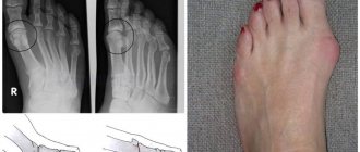
In the upper limb, arthrosis often develops after injuries or against the background of chronic arthritis. The deformation is clearly visible in the interphalangeal joints of the hand. Bone growths form on the fingers - Heberden's and Bouchard's nodes, and the hand itself takes on a square shape.
Heberden's and Bouchard's nodes on the interphalangeal joints with arthrosis
Dangerous signs of arthrosis
Since degenerative-dystrophic processes develop slowly, even dangerous symptoms of arthrosis are not always noticed on time.
Dangerous symptoms include:
- the appearance of swelling and pain in a constantly aching limb is a sign of inflammation that requires treatment;
- development of joint deformation;
- pain in the muscles and bones of the limb;
- unsteady gait, impaired mobility when flexing and extending the limb;
- constant pain that spreads to the entire limb.
Types of disease
In modern orthopedics, the diagnosis of arthrosis begins with determining its type. Thus, according to localization, diseases of the ankle, elbow, shoulder and other joints are distinguished.
At the appointment, the stage of spread of the disease is determined:
- the first implies the absence of pronounced changes. The cartilage tissue lacks nutrients, and the cartilage itself becomes less resilient. Primary inflammation leads to unpleasant pain;
- the second stage of arthrosis involves the destruction of the cartilage structure and the spread of bone growths. The patient suffers from regular pain, which either subsides or reappears;
- in the third degree of arthrosis, the cartilage becomes especially thin and deformed, and joint mobility is sharply limited due to shortening of the ligaments.
Are you experiencing symptoms of arthrosis?
Only a doctor can accurately diagnose the disease. Don't delay your consultation - call
Why is arthrosis dangerous?
Arthrosis is a disease that develops slowly and rarely causes severe disability. Periodically developing inflammatory processes pose a danger.
Therefore, in recent years, they have begun to distinguish a disease such as osteoarthritis or osteoarthritis (OA), depending on which process predominates in the joint - degenerative-dystrophic or inflammatory. It is OA that causes severe impairment of limb function.
Stages of arthrosis
Clinical and radiological stages of arthrosis according to the Kellgren-Lawrence classification:
- Zero. The patient feels discomfort, sometimes pain when walking. There are no changes on the x-ray.
- Initial (doubtful). The patient is bothered by moderate dull pain when walking for a long time, sometimes a slight crunch when bending a limb. X-ray: slight narrowing of the joint space, small areas of marginal bone defects.
- Easy. The pain intensifies, appears in the morning along with short-term stiffness, and intensifies with movement. X-rays of grade 2 show: a clear narrowing of the joint space and isolated bone growths (osteophytes) along the edges of the articular surfaces.
- Moderate (degenerative). Night pain syndrome appears. Bones and muscles hurt. Sometimes the joint swells slightly and the pain intensifies (a sign of inflammation). On X-ray: even greater narrowing of the joint space and proliferation of osteophytes; bone density increases (osteosclerosis).
- Heavy (deforming). The pain is constant, aching, intensifies with movement, a rough crunching sound when bending the limb, muscle atrophy and deformation. On the x-ray: the joint space is sharply narrowed, the edges of the articular surface have grown significantly, which has led to a change in the structure and deformation of the joint. Read more about deforming arthrosis in this article.
Stages of arthrosis
Possible complications
Arthrosis is a disease complicated by:
- chronic pain syndrome that increases from physical activity;
- joint deformity;
- dysfunction of the limb - stiffness, alternating with complete or partial immobility;
- loss of ability to work and disability.
Exacerbations of arthrosis
Degenerative diseases are characterized by a slow progressive course. Increased pain occurs in damp, cold and windy weather, as well as when inflammation occurs. Inflammatory processes occur with slight swelling and moderate pain. As a rule, the inflammatory process is aseptic in nature, but in the presence of foci of infection and chronic diseases, infection is possible. Therefore, if symptoms of inflammation appear during arthrosis, it is better to consult a doctor. You can do the following yourself:
- take any painkiller - Analgin, Nise, Paracetamol;
- apply anesthetic ointment or gel to the skin over the affected joint - Voltaren, Bystrumgel, Pentalgin, etc.;
- provide rest to the sore limb.
Stages of the disease
Arthrosis of the joints develops gradually and in the process goes through three successive stages that determine the severity of the disease:
- Stage 1: the pathology is not detected on X-ray or ultrasound, but the destruction processes have already started; the composition of the joint fluid changes, as a result of which the tissues receive fewer nutrients and become more sensitive; increased stress on the affected area causes inflammation (arthritis) and pain;
- Stage 2 is characterized by active destruction of cartilage tissue, and bone spines and growths appear along the edges of the articular platform (the area of contact of surfaces); at this time, the pain becomes habitual, and the inflammatory processes become stronger and weaker; spasms of the muscles associated with the joint are periodically observed;
- Stage 3: areas of destruction affect almost the entire surface of the cartilage, the articular area is deformed, the affected limb deviates from its axis; range of motion decreases and ligaments weaken and become short.
Some experts also distinguish stage IV of the development of arthrosis. It is characterized by almost complete immobility of the joint.
Localizations and clinical forms
Any localization and form of arthrosis has serious complications, so you should not delay treatment.
See how easily the disease can be cured in 10-12 sessions.
Arthrosis develops mainly in the most loaded joints – the knee and hip. But after an injury or against the background of arthritis, degradation-dystrophic processes can progress in any joint.
Classification of osteoarthritis
There are several classifications. The most famous are:
- Classification by etiology (reasons for development):
- primary – the causes of development have not been established;
- secondary – develop against the background of injuries and diseases.
- Classification by clinical forms:
- polyosteoarthrosis – multiple joint damage; divided into nodular (Heberden's and Bouchard's nodes) and nodular;
- oligoosteoarthrosis – the number of affected joints is no more than two;
- monoosteoarthrosis – damage to one joint;
- in combination with osteochondrosis or osteoarthritis of the spine.
- Classification by localization:
- interphalangeal;
- hip;
- knee;
- other.
Arthrosis of the lower extremities
Due to the high load, the legs suffer first of all:
- Arthrosis of the hip joint (coxarthrosis) is the most severe. The structural features of the hip joint (deep joint cavity, narrow joint space) contribute to the rapid development of degenerative disorders involving muscles and ligaments. If inflammation occurs, the effect of partial or complete immobility develops. Often develops against the background of congenital dysplasia, dislocations, subluxations of the hip, osteochondropathy (aseptic necrosis of the femoral head - Perthes disease). Symptoms of arthrosis: pain initially appears only towards the end of the day, but gradually increases, worries all day, radiates to the groin and buttock area. To reduce pain during coxarthrosis, the patient holds the leg in a forced position, which makes it seem shorter than the healthy one. The pain syndrome is very strong, so patients often agree to endoprosthetics.
- Arthrosis of the knee joint (gonarthrosis) - the most common. The knee withstands the highest loads and is injured, so gonarthrosis develops most often. Arthrosis of two joints is distinguished:
- patellofemoral - develops after injuries to the patellofemoral joint and is initially characterized by an inconspicuous course, since the joint has a lot of shock absorber cartilage that does not allow injury to the bone for a long time; but pain gradually appears after physical exertion, long walking or standing, when going up or down stairs; Over time, they become constant, dull, aching, and intensify when the weather changes; inflammation of the synovial membrane (synovitis) often develops, causing the pain to become acute;
- tibiofemoral arthrosis (femoral-tibial joint) - develops less frequently and is easier. The pain radiates down to the area of the lower leg and foot; Complete immobility with gonarthrosis rarely occurs. Prevention and timely treatment of arthrosis of the knee joint are very important - this will allow a person to live without pain. But even with an advanced disease, it is quite possible to relieve the patient from pain.
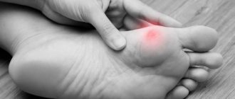
- Ankle – a large load also falls on the ankle, which is why arthrosis often develops in it. He is also often injured and the degenerative process is post-traumatic in nature. It is also affected in reactive arthritis. Symptoms: the disease is asymptomatic for a long time, but then pain appears. First, during physical activity, and then constant, aching pain. Stiffness of movement after a long rest is also characteristic, which goes away within half an hour. Complete immobility of the ankle is rare and only if the underlying cause of the disease is a long-term inflammatory process.
- Heel - arthrosis can develop in the area of the subtalar or talocalcaneal-navicular joints after injuries and diseases. They do not manifest themselves for a long time, then painful sensations in the heel begin to appear, gradually acquiring a constant painful character. Disability is rare.
Arthrosis of the upper extremities
Arthrosis in the joints of the hands develops less frequently. Main features of localizations in individual joints:
- Arthrosis of the shoulder joint. It usually develops after injuries and against the background of microtrauma in weightlifters, as well as in people engaged in heavy physical work. Acromial (acromio-clavicular) arthrosis is a consequence of trauma and inflammatory processes. At first it goes unnoticed, but then pain appears in the upper part of the shoulder, radiating to the elbow and neck, stiffness of movements and crunching when moving. The pain can be permanent and debilitating. Sometimes accompanied by inflammation, which contributes to the progression of the disease. If left untreated, partial ankylosis develops. Read more about shoulder arthrosis here.
- Elbow arthrosis - occurs rarely, mainly in miners, blacksmiths and workers of some other professions who deal with vibrating tools. Symptoms: pain in the elbow when bending and straightening the arm, stiffness after a long rest. If left untreated, there will be persistent dysfunction.
- Arthrosis of the joints of the hand. Most often, the degenerative process develops in the carpometacarpal joint of the first finger, since it is usually subject to trauma during household work. It manifests itself as a periodically appearing dull soreness in the outer side of the palm, radiating to the thumb.
- Arthrosis of the finger joints. Develops when performing small tasks (knitting, embroidery, sewing). In the distal (uppermost) interphalangeal joints, the pathological process manifests itself in the form of growths of bone tissue - Heberden's nodes; joint pain usually does not occur or they appear only occasionally, for example, when the weather changes. In the proximal interphalangeal joints, the disease manifests itself in the form of the same bone growths on the joints of the fingers located below - Bouchard's nodes.
Crunching in joints - when to worry
Intra-articular injections of hyaluronic acid
Arthrosis of the spine (vertebral)
In different parts of the spinal column, arthrosis manifests itself in the form of different symptoms:
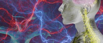
- Cervical uncovertebral arthrosis (cervical facet spondyloarthrosis) . Degenerative-dystrophic changes in small facet joints of the cervical vertebrae. Arthrosis of the joints of the cervical spine develops mainly in the second half of life in people who work for a long time in a stationary state with their heads bowed. It can also develop after injuries and against the background of chronic arthritis. It manifests itself in the form of headaches, dizziness, pain in the neck, radiating to the shoulder and crunching when moving. Decreased vision, hearing, and the appearance of high blood pressure (BP) are also possible. Since overgrown bone tissue can compress the vessels supplying the brain, the disease sometimes threatens the patient’s life. Requires long-term rehabilitation treatment.
- Thoracic spondyloarthrosis (arthrosis of the joints of the thoracic spine). It is much less common than the cervical one. First, moderate and then quite severe pain in the spine appears, intensifying with coughing, sneezing and deep breathing. Sometimes the symptoms are similar to manifestations of diseases of the cardiovascular and respiratory systems. To correctly establish the diagnosis, additional examination is required. In the thoracic region there are also costovertebral joints, 2 of them on each rib (rib head and costotransverse). They can also develop degenerative-dystrophic processes, mainly in older women. The disease manifests itself as pain in the chest. If it lasts for a long time, it can cause severe complications from the cardiovascular and respiratory systems.
- Lumbar spondyloarthrosis. It is a consequence of heavy physical labor and spinal injuries. Arthrosis of the joints of the lumbar spine manifests itself in the form of aching pain, aggravated by bending the body. Characterized by stiffness of movement after a long rest, a crunch in the back when bending over.
- Sacrococcygeal spondyloarthrosis. Most often develops after injuries, for example, after a fall and injury to the tailbone. It manifests itself as pain, aggravated by sitting and prolonged walking. Requires long-term rehabilitation treatment.
Tissue changes in spondyloarthrosis
Arthrosis of the temporomandibular joint (TMJ)
The disease develops with chronic arthritis of the TMJ, malocclusion, absence of lateral teeth, and problems with prosthetics. There is a violation of blood circulation and metabolism in the TMJ area with the development of degenerative-dystrophic processes in it. Symptoms of arthrosis of this joint: aching pain in the lower jaw, stiffness and crunching when opening the mouth and chewing. The pain increases with changes in weather, as well as with the development of synovitis. The long course of the disease leads to the appearance of asymmetry: displacement of the jaw tissue to the affected side.
Read more about the disease here.
Categories of people most susceptible to disease
Despite the fact that absolutely anyone can get arthrosis, there are a number of categories of people who are especially susceptible to this disease. These include:
- elderly people suffering from disorders of the endocrine system;
- obese persons;
- athletes and people whose work involves excessive physical activity, often abnormal;
- patients who have undergone joint surgery in the past;
- people with a genetic predisposition: if parents or grandparents suffered from arthrosis, then there is a high probability that it can also be passed on to their children (grandchildren);
- patients with dystrophic disorders in the lumbar and cervical spine.
Types of secondary arthrosis
The causes of secondary arthrosis are various diseases and injuries. The most common types of arthrosis are:
- Post-traumatic – develops after injuries . A very common cause of the development of articular degenerative-dystrophic changes. The pathological process begins with inflammation and gradually turns into an exchange process with the development of joint deformation, persistent dysfunction and pain.
- Metabolic and endocrine arthrosis:
- gouty - develops slowly against the background of gout. In the first years, attacks of gout do not lead to changes in the joints, but then degenerative = dystrophic changes gradually develop in them, leading to dysfunction;
- against the background of hormonal disorders.
- Arthrosis against the background of congenital and acquired orthopedic pathology:
- congenital hip dislocation;
- thickening of the acetabulum (congenital);
- dysplasia (impaired formation) of the joint;
- osteochondropathy – aseptic necrosis of the femoral head (Perthes disease), etc.
- Arthrosis as an outcome of chronic arthritis:
- reactive – is a consequence of a previous urogenital or intestinal infection; joint tissues react to infection - an inflammatory reaction develops; with proper examination and treatment, complete recovery occurs, but if left untreated, the inflammatory process becomes chronic with exacerbations and remissions; then gradually becomes degenerative with the development of arthrosis;
- rheumatoid – develops against the background of an autoimmune inflammatory process, which over time turns into a degenerative process with joint deformation; Mainly the small joints of the hand and foot are deformed;
- psoriatic – the cause of the lesion is psoriasis; At first it is an inflammatory process, which after a few years turns into arthrosis-arthritis with degenerative processes and deformation.
Chondroprotectors: what are they, how to choose, how effective are they?
Joint pain at rest
What to do when you are young: simple recommendations
People of certain professions are especially susceptible to arthrosis of the hand - pianists, massage therapists, office managers and those who constantly type on the keyboard. If you suspect the possibility of developing this pathology, try to choose a different profession in your youth.
- Avoid injuries to the fingers and wrist joint. They most often provoke the development of dystrophic changes.
- Take special care during icy conditions, because when a person falls, he intuitively puts his hands up.
- Do not overuse exercises with dumbbells.
Take care of your hands from a young age
Diagnosis of arthrosis
The diagnosis is made based on a doctor’s examination and is confirmed by laboratory and instrumental studies:
- Laboratory tests - general clinical, biochemical, immunological blood tests. Signs of inflammation, causes of arthrosis (metabolic disorders, autoimmune processes) are revealed. If necessary, intra-articular fluid is taken for examination to identify infection and its sensitivity to antibiotics.
- Instrumental diagnostics:
- Ultrasound, MRI - changes in soft articular and periarticular tissues are detected;
- X-ray, CT – changes in bone tissue; these are the main methods for confirming the presence of arthrosis;
- arthroscopy – according to indications, if there is a suspicion of an inflammatory process.
Classification of arthrosis according to Kosinskaya (by stages)
The most popular among domestic orthopedists is the classification of arthrosis proposed by prof. N.S. Kosinskaya in 1961, since it takes into account not only changes in the joint visible on x-rays, but also the presence and severity of clinical symptoms. Clinical and radiological signs of the disease according to Kosinskaya are divided into 3 stages (there is an erroneous classification on the Internet - 4 stages, this is incorrect).
Stage 1 arthrosis
The first sign of the disease is pain during physical activity, which goes away during rest. The range of motion in the joint is not changed. Visible on the x-ray:
- slight and uneven narrowing of the joint space;
- primary osteosclerosis (bone hardening).
For example, with gonarthrosis, the knee begins to hurt when walking, especially when climbing stairs or going down. Unpleasant sensations also appear when you stay on your feet for a long time, but as soon as you rest, the pain syndrome disappears. At this stage, arthrosis is highly treatable - you can completely get rid of unpleasant sensations.
Stage 2 arthrosis
The patient complains of moderate and constant pain. Mobility in the joint is limited, muscle wasting is noted when, as a result of decreased tone, the muscle is tense and resists movement. In addition, for arthrosis, symptoms characteristic of this disease are added at the location. For example, with coxarthrosis, lameness appears, the doctor determines a slight frontal deformation of the limb axis.
On the x-ray:
- The narrowing of the joint space is clearly visible (2-3 times compared to a healthy joint);
- osteophytes (marginal bone growths) appear, which can also be in the area of the intercondylar eminence;
- subchondral sclerosis (formation of sweaty connective tissue) becomes pronounced.
At this stage it is also effective and will allow you to stop the destruction of the joint for a long time.
Any localization and form of arthrosis has serious complications, so you should not delay treatment.
See how easily the disease can be cured in 10-12 sessions.
Stage 3 arthrosis
At stage 3 according to Kosinskaya, the x-ray shows that the joint space is practically absent. The articular surfaces are expanded due to extensive bone growths along the edges and are sclerotic. Their fragmentation, cystic clearings and cavities are common (subchondral focal necrosis, the bone under the cartilage begins to collapse).
When examining the patient, note:
- joint deformation;
- persistent limitation of flexion-extension and rotational movements;
- pronounced pain syndrome;
- deformation of the limb or spine (depending on the location of arthrosis);
- atrophy of the muscles adjacent to the joint.
If stage 3 symptoms are detected, you should immediately consult a doctor. In addition to drug therapy, the patient is prescribed massage, physiotherapy, and physical therapy. Only comprehensive treatment can significantly improve the ability to move and avoid surgery.
3 stages of arthrosis
Treatment of arthrosis
Based on the results of the diagnosis of arthrosis, individually selected complex treatment is prescribed, including drug and non-drug therapy. Equally important is a healthy active lifestyle with the exception of heavy physical activity and proper nutrition.
Drug therapy
To eliminate the symptoms of the disease and suppress its progression, the following medications are prescribed:
- To relieve pain, pain therapy is carried out:
- painkillers from the group of non-steroidal anti-inflammatory drugs (NSAIDs); depending on the severity of the pain syndrome, the complex treatment includes medications such as Ibuprofen, Diclofenac, Ketorol, etc.; they are prescribed in the form of tablets for oral administration, injections or ointments (gels);
- muscle relaxants - medications that relieve spasm from the periarticular muscles (spasm increases pain and impairs blood circulation) - Mydocalm;
- neurotropic B vitamins (Neuromultivit, Milgamma) – restore the function of the peripheral nervous system, reduce pain;
- anesthetic blockades - solutions of Novocaine or Lidocaine are injected into the most painful points.
- To restore joint function:
- chondroprotectors - medications that restore cartilage tissue in the form of tablets, injections, ointments (Dona, Structum);
- hyaluronic acid - the introduction of drugs based on it into the joint cavity, for example, in the treatment of arthrosis of the knee joint; this helps improve the viscosity of the synovial fluid and reduce injury to the articular surfaces of the bones.
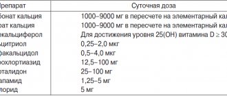
Drugs for the treatment of arthrosis
Traditional methods
It is better to learn how to treat arthrosis with these methods by consulting your doctor. The most popular methods:
- inclusion of gelatin in the diet; every day you need to eat soup (borscht) with bone broth, jellied meat, jelly - this helps restore bone tissue;
- externally: compresses based on kefir, chalk and oatmeal; mix all components in equal volumes; put the mushy mass on a napkin, apply it to the knee joint when treating it, cover the top with polyethylene, insulate it and leave it overnight; relieves pain, stimulates the turnover of cartilage tissue.
Non-drug treatments
These methods are no less important. Physical therapy (physical therapy) helps. One of the main recommendations of experts is physical activity. In this case, the correct balance between light physical activity and rest must be maintained. This is achieved by daily performing a complex of therapeutic exercises prescribed by a doctor. In order to perform exercises correctly with a gradually increasing load, they begin to be done under the supervision of a physical therapy instructor and only then proceed to be performed at home. It is better to entrust the choice of exercises to a physical therapy instructor.
Basic training rules:
- classes should be held daily;
- all movements are carried out smoothly, without sharp turns;
- If the pain intensifies, stop the exercises.
You cannot: run, jump, play football, basketball, volleyball and other outdoor sports games, lift weights, stand for a long time or be in any other static position.
Sports activities include swimming classes, yoga and Pilates exercises specially selected by the instructor.
If pain bothers you: limit physical activity for a while, provide rest to the sore limb several times a day for 20-30 minutes, but you should not rest for a long time. Rest should be replaced by light physical activity that does not cause increased pain. If the pain is severe, then it is better to move with a cane or on crutches.
Massage courses are prescribed in the absence of an inflammatory process. Massage allows you to restore blood circulation, innervation and metabolic processes, promotes the regeneration of damaged joint tissues. The tissues are warmed up, muscle spasms and associated pain are relieved.
Physiotherapy is prescribed depending on the patient’s condition and the causes of arthrosis:
- for severe pain - electrophoresis or phonophoresis with solutions of painkillers (Novocaine);
- in the absence of inflammation - warming procedures - applications with paraffin, ozokerite;
- to improve blood circulation, eliminate muscle spasms - darsonvalization - exposure to pulsed currents of high frequency and low strength;
- for the regeneration of articular tissues – laser and magnetic therapy;
- to restore muscle volume and strength - electrical myostimulation.
Hirudotherapy is the treatment of arthrosis with leeches. Leech saliva contains enzymes that help dilate blood vessels, which leads to increased metabolism. In addition, enzymes in the saliva of leeches are able to dissolve necrotic tissue and cleanse the joint cavity.
Various methods of fixing joints - the correct functioning of the limb depends on this. Patients with severe pain and joint instability are prescribed:
- wearing orthoses - special devices that support the limb in the correct position;
- the use of taping - fixing the joint in the desired position using adhesive tapes;
- wearing elastic knee pads in the treatment of arthrosis of the knee joint, fixing the knee and protecting it from injury;
- use of walking aids such as canes and crutches.
Taping procedure for arthrosis of the knee joint
Operation
In case of persistent loss of function and severe pain that is not amenable to conservative treatment, an endoprosthetic operation is performed - replacing the destroyed joint with an artificial one.
How to treat arthrosis with NANOPLAST forte therapeutic plaster
Based on the results of clinical trials, the therapeutic plaster NANOPLAST forte has shown high effectiveness in the treatment of arthrosis . This patch can be used both in the initial stages of the disease and in severe forms of arthrosis.
But it is important to understand that while in the initial stages of the disease NANOPLAST FORITE can be effective as a monotherapy, then in the advanced stage complex treatment is required. Without the use of other medications, treatment of grade 2-3 arthrosis, especially in the acute stage, will not be effective. With complex treatment, the patch enhances the effect of painkillers and anti-inflammatory drugs, which makes it possible to reduce their dosage, and if the pain syndrome decreases, cancel them altogether.
NANOPLAST forte allows you to relieve pain and inflammation, improve blood circulation in the affected area, and reduce the dose of painkillers and anti-inflammatory drugs. It has no side effects, is not addictive and combines well with other treatment methods.
For arthrosis of the joints, the therapeutic plaster NANOPLAST forte is applied to the diseased joint. The application method depends on the type of arthrosis. To relieve acute symptoms, it is recommended to use a medicinal patch for 3 to 5 days. The duration of the course of treatment for osteoarthritis of the joints is from 15 days according to clinical studies. It is usually recommended to use the patch in the morning for 12 hours, but it can also be used at night.
High efficiency, unique composition, long-term (up to 12 hours!) therapeutic effects, ease of use and affordable price make NANOPLAST forte the drug of choice in the treatment of osteoarthritis of the joints.
Read more about NANOPLAST forte
Approach to treating the disease in our clinic
We combine proven techniques of the East and innovative methods of Western medicine.
Read more about our unique method of treating arthrosis
The Paramita clinic uses an individual approach to the treatment of arthrosis. After a preliminary examination, the most appropriate course of treatment is selected for the patient, including:
- the most modern methods of drug and non-drug therapy, based on the achievements of Western medicine;
- traditional oriental methods, successfully used for centuries by doctors of Ancient China and Tibet to treat diseases of the musculoskeletal system; treatment is based on restoring the energy balance of the entire body, leading to suppression of the pathological process.
Medical doctors successfully use all the necessary methods for treating arthrosis, achieving complete relief of patients from pain and stopping the progression of the disease.
Advantages of the clinic
“Health Energy” is a multidisciplinary medical center equipped in accordance with modern standards. We offer patients:
- consultations with experienced doctors;
- examination using high-quality diagnostic equipment;
- individual approach to treatment selection;
- regular monitoring and performance monitoring.
The joints of our body can withstand enormous loads, but there are situations when they need help. Don’t let arthrosis change your life, sign up for a consultation at Health Energy.
Clinical recommendations for arthrosis
To get rid of pain and maintain normal condition of the musculoskeletal system, you need to:
- monitor your body weight – extra pounds significantly increase the risk of disease progression;
- get rid of heavy physical activity: heavy lifting, long walking, and especially long periods of standing;
- regularly perform physical therapy exercises;
- avoid injury; To do this, during training, gradually increase the load on the muscles, and also use special protective devices (knee pads, etc.);
- use special devices and techniques to restore impaired limb biomechanics (orthoses, taping);
- regularly visit your doctor and conduct courses of anti-relapse treatment.
Prognosis for arthrosis
With proper treatment of arthrosis and the patient following all the doctor’s recommendations, the progression of the disease and impairment of joint function can be stopped. It is also quite possible to completely relieve the patient of pain.
Prevention of arthrosis
To prevent arthrosis, you need to move more, avoid heavy physical activity and stick to a low-calorie diet so as not to gain weight.
Preventive measures
The basis for preventing arthrosis is reducing the risk of injury. The main measures include:
- a competent training regimen (if the sick person plays sports);
- getting rid of excess weight;
- timely diagnosis and treatment of rheumatic and endocrine diseases;
- in the case of a genetic predisposition, be attentive to your health and not overexert yourself physically, and ensure moderate physical activity.
Frequently asked questions about the disease
Arthritis and arthrosis – what is the difference?
The difference is that arthritis is an inflammatory process in the joint, and arthrosis is degenerative-dystrophic (metabolic) with gradual “drying out” and cracking of cartilage, compensatory growth of bone tissue. Chronic arthritis gradually turns into arthrosis-arthritis and arthrosis.
Is it possible to warm joints in a sauna/bath if you have arthrosis?
It is possible and it will be beneficial. But arthrosis is often complicated by inflammation (arthritis), in which warming water procedures are contraindicated. Therefore, before you start treating arthrosis with water procedures, it is better to visit a doctor.
Do they take you into the army with arthrosis?
It depends on how pronounced it is. If the function of the joint is impaired, then no.
Is it possible to develop arthrosis after coronavirus infection? If not, why not?
It is unlikely that anyone will answer this question now, there is simply no data. The development of arthrosis is a slow process.
Which doctor should I contact for arthrosis?
To a therapist. He will conduct an examination, a preliminary examination and, depending on the type of arthrosis, will refer the patient to an orthopedic traumatologist or rheumatologist.
Does arthrosis occur in children?
It happens after injuries and joint diseases.
Arthrosis is not a death sentence. It cannot be completely cured, but it can and should be treated - this will save patients from suffering. Moscow medical specialists successfully cope with the treatment of arthrosis.
Literature:
- Karateev A.E., Barskova V.G. Criteria for choosing a non-steroidal anti-inflammatory drug. Handbook of a practical doctor, 2007.- T. 5.- No. 5.- P. 13-17.
- Povoroznyuk V.V. Osteoarthritis: modern principles of treatment // News of medicine and pharmacy. - 2004. - T. 144, No. 4. - P. 10-11.
- Raisz LG Prostaglandins and ione: physiology and pathophysiology. Osteoarthritis Cartilage, 1999.- V. 79.- P. 83-94.
- Peyron JC, Altman RD Osteoarthritis: diagnosis and medical surgical management. — 2nd ed. - Philadelfia, Pa: WBSauders Company, 1992. - P. 15-37.
Themes
Arthrosis, Joints, Pain, Treatment without surgery Date of publication: 04/22/2021 Date of update: 04/11/2021
Reader rating
Rating: 4.29 / 5 (7)
Diet
Diet is one of the most important factors in the treatment of arthrosis. If you are overweight, you need to reduce it to reduce the stress on your joints. In this case, a balanced diet with a calorie deficit is prescribed. Regardless of body mass index, doctors recommend completely avoiding:
- fast carbohydrates (sugar, desserts, flour);
- alcoholic drinks;
- spices;
- legumes;
- strong tea and coffee;
- excessively fatty and spicy foods.
Canned food and offal are not excluded, but are significantly limited, as is salt. An ideal diet for osteoarthritis includes:
- lean meats;
- fish and seafood;
- eggs;
- dairy products;
- flaxseed and olive vegetable oils;
- vegetables and fruits, a large amount of greens;
- cereals in moderation, durum wheat pasta;
- products with a high content of collagen (jellied meat, jellied meat, jelly).