Joint arthroscopy > Ankle joint > Foot and ankle injuries. First aid
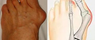
The human foot is located just below the ankle joint. The foot contains 26 small bones that are connected to the joints. The main purpose of this unit is to soften the resulting body shocks that occur during movement.
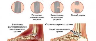
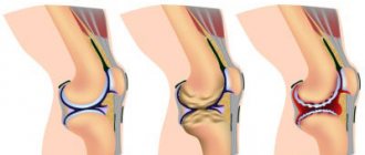
The ankle joint consists of the fibula and tibia, and below - the talus. It has a peculiar block shape, which is fixed at Christmas time.
If you hurt your foot, you always feel severe pain, since the layer of skin and muscle is quite thin, so the entire blow comes to the bone tissue. As a result, swelling quickly appears, so we are all looking for any folk ways to reduce pain.
First of all, a heating pad with ice can help; you can’t put pressure on this leg for a long time. When a foot dislocation occurs, the degree of severity can be determined by external signs. It all depends on which joint of the foot it happened in.
If the dislocation occurs in the subtalar joint inward, you may observe the following symptoms: the foot appears shifted, with the sole and heel turning inward, skin tension is observed above the ankle, and the inner one is indented. Also, a dislocated foot is accompanied by swelling, and since it makes it difficult to straighten the joint into place, there is a need to seek qualified help.
In no case can a dislocation be neutralized on your own, without the intervention of professionals and the use of anesthesia. During transportation, it is imperative to immobilize the injured leg using a splint or other available means, and also apply ice. It is forbidden to put weight on the leg, as this can lead to more serious consequences.
A dislocation in this joint outward is manifested by the following symptoms: the foot turns outward, the inner ankle protrudes and the outer one sinks in, and the skin is injured. First of all, you need to apply a bandage or splint, apply ice and go to the emergency room as quickly as possible. Dislocations in this area quickly form swelling. Therefore, you immediately need the help of a specialist.
A direct blow directly to the foot itself can lead to dislocation or severe bruising. A dislocation and a bruise are quite difficult to distinguish at first glance, so the victim needs to be sent to the hospital.
What causes a foot fracture?
Athletes and those who engage in heavy physical labor are at risk of fractures. Of the main reasons that can lead to such a foot injury, it is worth highlighting the following:
- the foot is broken as a result of being kicked on a hard surface;
- an unsuccessful jump from an impressive height can also cause a foot injury;
- if something heavy fell on your leg;
- dislocation while jogging or during normal walking.
A car accident often results in fractures. A foot injury may be due to excessive stress on the leg as a result of sudden braking. A strong blow can cause a fracture. In any case, it is important to immediately numb the area where the fracture is observed. It is also necessary to apply a cast to the injury site.
Osteochondral transplantation.
For osteochondral transplantation, cartilage tissue from the ipsilateral knee joint is most often used. Indications for transplantation are large and deep defects, often with the formation of a cyst, as well as the lack of effect from the surgical treatment methods proposed above. The possibility of using the calcaneal bone site at the Achilles-calcaneal joint as a source of tissue for osteochondral transplantation is currently being studied, with good early results.
The objective of the method is to restore the structural and biomechanical characteristics of damaged hyaline cartilage. This method is characterized by a long and difficult rehabilitation period. Morbidity of the donor site (pain in the knee joint after removal of columns of osteochondral blocks) is observed in 12% of cases. The technique itself is technically complex and time-consuming.
In clinical studies, the effectiveness of the method was estimated from 74 to 100% (average 87%).
Anatomy of the foot
The foot is the lower part of the leg. The side that steps on the floor is the sole. As for the opposite, upper side, this is the back side. Thanks to flexibility and elasticity, the arched foot structure regulates the distribution of weight. In addition, shocks are minimized while walking.
Joint anatomy - several bones in one ligament. When they are damaged, severe pain occurs.
The human feet contain a quarter of all the bones in the body. One foot has 26 bones. Babies may have extra bones at birth, which most often do not harm the baby.
In total, the foot contains 19 muscles. As a result of their interaction, leg movement becomes possible. The muscles of the sole are responsible for the movement of the toes.
With the help of tendons, muscles are attached to bones - this is their continuation. They differ:
- strength;
- elasticity.
Flexible ligaments surround the joint, support it, and serve to connect the bones. They are not elastic.
The ends of the bones are covered with cartilage tissue in the place where the joints are located. Without it, the body cannot move smoothly.
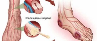
The main arteries of the foot are dorsal and posterior. Tibia should also be added to the list. Nerves control muscles. They transmit sensations to the brain center everywhere.
Thanks to the mobility of the foot, a person can easily adapt to the most unexpected surfaces on which he moves. In addition, the body moves in different directions. This part of the leg absorbs most of the stress. Therefore, it is very important to prevent a foot fracture or to begin proper treatment in time if an injury occurs.
Open surgery for osteochondral injuries of the talus.
When accessing the anterior-outer part of the ankle joint, it is necessary to remember the branches of the superficial peroneal nerve located in this area.
To access the posteromedial sections, it will be necessary to perform an osteotomy of the medial malleolus, while it is necessary to exclude damage to the tibial plateau, which bears the main axial load. Before performing an osteotomy, it is necessary to pre-drill for subsequent installation of screws fixing the osteotomized fragment.
Injury to the anterior and posterior tibial tendons, flexor pollicis longus tendon, great saphenous vein, posterior tibial artery and nerve should also be avoided. An alternative is to use two approaches at once - anteromedial and posteromedial, which allow visualization of up to 80% of the dome of the talus and avoid osteotomy of the medial malleolus.
Currently, ankle arthroscopy has become widespread. Arthroscopic approaches are much less traumatic, which avoids the formation of rough postoperative scars and greatly facilitates rehabilitation. 2.7 mm arthroscopes allow visualization of the entire dome of the talus.
What types of foot fractures are there?
There are several types:
Fractures of the tarsal bones
- Talus and navicular bones;
- Cubo- and wedge-shaped.
Metatarsal fractures
- Traumatic fracture;
- Stressful.
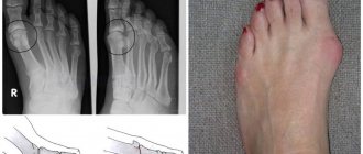
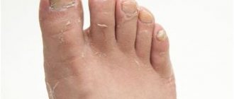
Metatarsal bones are subject to fractures more often than other bones. When a leg is injured, a characteristic crunching sound appears, and the victim clearly feels pain. A finger that is injured may even become shorter. Deviation to the side is also possible. After some time, the pain becomes weaker, but does not disappear completely.
A stress fracture occurs most often in those who are often exposed to significant physical exertion. These could be professional athletes, for example. With this fracture, a crack appears in the bone area. It is very difficult to detect. When the patient suffers from concomitant illnesses, such as osteoporosis, the situation becomes even more complicated.
If damaged, pain occurs after impressive loads. At rest it goes away. Over time, the intensity of pain reaches its peak. At this point, any action involving the foot becomes almost impossible. Even at rest, the pain now persists. There is swelling at the site of injury.
The danger of a stress fracture is that it is not immediately detectable. Therefore, the development of complications is quite often observed due to the fact that the patient does not consult a doctor in a timely manner.
Fracture - with or without displacement
Of the general symptoms accompanying a foot fracture, the main ones should be highlighted:
- pain;
- swelling of adjacent tissues.
Symptoms of a metatarsal fracture
- When you feel your foot and try to lean on it, you feel pain.
- Swelling appears on the sole of the foot. The same can happen on the back side.
- The foot is deformed.
Symptoms of a tarsal bone fracture
- Swelling appears at the site of injury. The same goes for the ankle joint.
- When you lean on your foot and turn it, a very sharp pain suddenly occurs.
How to find out that there is an offset?
- The pain in the area of the fracture is very strong.
- The entire foot swells impressively.
- Severe distortions of the foot occur.
"Stuck heel"
In an adult, the symptom of a broken foot may appear as a “stuck heel”: it is difficult for a person to tear the leg off independently from a lying position. The crunching of fragments may even be felt when palpated. Also, this condition is observed in the absence of a process of pinching of the muscles located between the fragments.
If a patient has a closed foot fracture, doctors may note pathological mobility of the foot outside the joint. It is extremely undesirable to independently check the possibility of mobility of the victim’s foot, since this can further damage the blood vessels and nerve endings, as well as displace fragments.
First aid to the victim
If you suspect that there is a fracture in your foot, you should consult a doctor by visiting a medical facility. You should call an ambulance as soon as possible if:
- the foot turned blue and cold;
- no sensitivity;
- it looks like the foot is deformed;
- there is a wound at the site of injury.
Before the ambulance arrives you should:
- remove shoes without waiting for swelling to appear;
- lift the injured foot up;
- If there is a wound on the foot, it is important to immediately apply a bandage - preferably a sterile one.
- Recommendations for the treatment of foot synovitis;
- Information on how to heal a heel after a bruise can be read here;
- An ankle sprain is a very unpleasant injury that takes a long time to resolve.
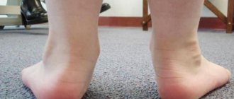
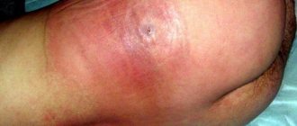
The appearance of a hematoma
A hematoma is a consequence of hemorrhage in the soft tissues of the foot. Its size and shade will be primarily influenced by the force with which the blow was struck. When the subcutaneous fatty tissue is affected, the bruise usually appears immediately 1-3 days after the injury. If the damage is very deep, the hematoma may not manifest itself at all.
Doctors also note that the hematoma will gradually change its shade: a fresh one has a bright red tint, but a little later it may acquire a purple color. On days 4-5, the hematoma has a dark blue color, and after that it becomes slightly yellowish.
How to treat a broken foot?
The choice of one or another method by which treatment will be carried out depends on how complex the nature of the injury is. There are times when it is enough to fix the damaged area with a special bandage. The same goes for shoes. In addition, in mild cases it is necessary to avoid putting stress on the injured leg. In this case, a crutch or cane will be an excellent assistant. Most often, it is necessary to ensure immobility by applying a plaster bandage. Sometimes surgery is necessary.
How long to treat and wear a cast?
Everything here is absolutely individual, depending on the type of fracture. Other factors also influence treatment and rehabilitation time.
- If we are talking about an injury to the metatarsal bones of the foot, then in this case the splint is worn for almost a month and a half. In addition, it is necessary to fix the foot for a couple of months. For this, gypsum is used.
- As for fractures of the bones of the finger phalanges, the period of wearing a plaster cast is up to six weeks.
- Fractures of the tarsal bones, if there is no displacement, require the application of a circular plaster splint for a period of 21 days to six months.
If the foot fracture is of mild severity, then treatment is quite possible, in which the application of plaster bandages is not required. The same goes for cracks. In this case, it is best to use a bandage to secure the foot. In addition, you are required to wear special safety shoes. Crutches will help minimize the load on your foot. In addition, the doctor prescribes effective medications that will need to be taken orally. These are vitamins, as well as agents that have an anti-inflammatory effect.
How long after is it allowed to step on your foot?
Localization plays a role here.
- If the talus is damaged, it will take up to three months to be able to stand on your leg and move normally.
- When the scaphoid is fractured, the limb is immobilized for two and a half or even three months. During this period, the doctor recommends wearing orthopedic shoes.
- In case of a fracture of the cuboid and sphenoid bones, after diagnosing the fracture, plaster is applied for a period of up to one and a half months. As for the rehabilitation period, it can last up to a year. During this time, you should wear an arch support.
How long does it last
To answer the question: “How long does rehabilitation last after an ankle fracture?” three criteria are always taken into account:
- the age of the injured patient and his lifestyle;
- the presence of chronic diseases that can (and will) slow down the process of tissue repair;
- the nature of the fracture, there are two of them, open and closed.
Now, in order. The older a person is, the more worn out the body is, and we emphasize that this includes his lifestyle - yes, this is relevant in our time, because now a huge number of people lead a sedentary lifestyle! Also, do not forget about one more thing - we are talking about the possible presence of bad habits that slow down the recovery process.
It’s worse when a patient, especially an elderly one, has diseases of the musculoskeletal system or diseases that can affect recovery processes - diabetes mellitus is a prime example! It has long been noted that diabetes significantly complicates treatment for other diseases and slows down the regeneration of damaged tissues.
Here is the nature of the fracture, if it is open (plus possible displacement, rupture of ligaments, blood vessels), then the recovery will become longer and more painful - about 6 months. In the best case, without complicating factors, a person will be able to get back on his feet after about 6 weeks. In children, the rehabilitation period is halved.
Rehabilitation period
How severe was the fracture, how long did it take for the fixing bandage to be removed? Depending on this, recovery may proceed differently. Rehabilitation measures include a whole range of different methods with the help of which it is possible to fully recover from such a serious injury.
Full recovery period
It depends on the type of fracture. After injury to the metatarsal bones, you will need to engage in physical therapy for a couple of months. You will need to wear a back bandage for about another month, which is made of plaster and has a thickening in the heel area. This is necessary in case of a displaced fracture.
In case of fractures of the tarsal bones, it will take a long time to recover. You will have to carry out massage and exercise therapy for a couple or three months. You must wear arch supports for at least 12 months.
After fractures of the phalanges of the fingers, a useful procedure should be carried out every day - kneading massage. In addition, you need to walk in orthopedic shoes for five months.
Physiotherapy
This method shortens recovery time. It is effective and safe. Physiotherapy has many clear benefits:
- wide spectrum of action;
- no contraindications;
- does not cause habit or allergic reaction;
- has an intense, but at the same time quite gentle effect on the patient;
- complications are excluded.
Exercise therapy after a fracture
Such exercises in the event of a foot fracture can quickly restore the functioning of the limb that was damaged. In addition, it helps strengthen the foot. This is a guarantee of positive results, subject to all requirements and recommendations regarding the implementation of physical therapy.
Foot massage after a fracture
As a result of the massage:
- atrophy of muscle tissue and blood vessels is eliminated;
- muscle tone and elasticity increases;
- massage helps restore joint mobility;
- congestion is eliminated;
- leg mobility increases.
Surgical treatment of osteochondral injuries of the talus.
Surgical treatment of OPTC depends on many factors: patient characteristics such as age, activity level, comorbidities, characteristics of the lesion itself such as its location, size, depth of the lesion, lesion morphology, duration. Basically, surgical treatment is based on one of three principles: 1) removal of the loose body with or without bone marrow stimulation through microfracture, reaming 2) preservation of cartilage through retrograde reaming, fixation of the fragment or replacement of the defect with cancellous bone 3) stimulation of the formation of new hyaline cartilage by due to osteochondral block transplantation, mosaic chondroplasty, allograft.
Prevention and recommendations for eliminating relapses
In order to prevent a fracture, it is necessary to try to maintain safety in one form or another of life activities. You should not unjustifiably risk your life, because the payment for this can be quite high.
If your foot has been injured or there is a fracture, it is important to consult a specialist in a timely manner. Only a doctor can decide how treatment should be carried out. He selects one method or another, effective drugs with the required dosage strictly individually.
Related Posts
How to identify a broken toe and provide first aid?
- Treatment of a hip fracture and why it most often affects older people
- Ankle fracture - its types, causes and treatment recommendations
Removal, curettage and bone marrow stimulation.
After debridement, several channels are produced that communicate the defect with the bone marrow. This procedure can be performed using reaming or microfracturing. The purpose of the manipulation is to destroy the barrier of sclerotic subchondral bone that prevents the migration of stem cells from the bone marrow. As a result, a clot is formed, saturated with growth factors and stem cells, which is subsequently rebuilt into hyaline-like cartilage tissue. Most often used for stages 3-4 of the disease. The size of the defect should not exceed 1.5 cm. In clinical studies, the effectiveness of the method was estimated from 46 to 100% (on average 85%).
Retrograde reaming.
This method will require the use of x-ray control and knowledge of the exact topography of the defect location. Retrograde reaming is the method of choice in the presence of a large subchondral cyst and with intact articular cartilage, as well as in cases of deep defect location, when it is difficult to reach it using the arthroscopic method. For medial defects, the entry point of the wire becomes the tarsal sinus. In clinical studies, the effectiveness of the method was estimated from 81 to 100% (average 88%).