- What is inflammation of the synovial bursa of the shoulder joint?
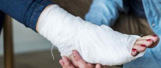
- Symptoms of shoulder bursitis
- Causes of inflammation of the mucous bursa in the shoulder
- Diagnosis and medical examination of bursitis
- Conservative treatment
- Surgical treatment of shoulder bursitis
In addition to inflammation of the mucous membrane, there are other causes of shoulder pain. Tendon inflammation, traumatic injuries or, for example, arthrosis have similar symptoms. A specialized medical examination will help establish the causes of pain and select the appropriate treatment. © yodiyim / fotolia Bursitis (Latin bursa “bag”) is a painful inflammation of the periarticular bursa of the shoulder. The bursae are located close to the joints to balance the high mechanical load between bones and other tissues. The largest bursa in the human body (Bursa subakromialis or subacromial bursa) is located in the shoulder. The cause of its inflammation may be pathologies of various structures of the complex shoulder joint. The shoulder may become inflamed due to bone spurs on the acromion of the clavicle (acromion) or structural changes in the supraspinatus tendon (eg calcium deposits and tears). Typically, these injuries are caused by traumatic injuries or excessive stress. Often the pain appears gradually, for example, when raising your arm. If a person does not attach importance to this and continues to load the shoulder, the pain intensifies and symptoms such as swelling and overheating appear. In most cases, doctors provide conservative treatment for shoulder bursitis. Experienced shoulder specialists recommend rest, anti-inflammatory medications, and biological treatment based on cell technology in this case. Quite rarely, clinics perform surgical treatment to remove the inflamed bursa.
What is inflammation of the synovial bursa of the shoulder joint?
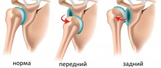
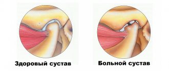
During painful bursitis, there is swelling in the area of the shoulder joint and an increase in the joint capsule, causing pressure on nearby tissues. The synovial bursa itself is an elastic, slit-like cavity filled with synovial fluid, capable of changing its shape between muscles, tendons and bone structures. In addition, the bursa allows painless movement of various tendons, bones and muscles within a highly mobile joint without friction. Bursitis in the shoulder is basically an inflammation of the periarticular bursa located between the head of the humerus and the acromion. Additionally, bursitis is the most common cause of stabbing pain in the arm. The subacromial joint capsule is the largest bursa in the human body, which is most often subject to inflammation of various types. Doctors use the term "subacromial" or "subacromial" bursitis when the area under the acromion of the collarbone (acromion) becomes inflamed.
conclusions
- Inflammation of the shoulder joint is a fairly common disease that needs to be treated promptly.
- Calcareous bursitis of the bony joint of the shoulder is a chronic form complicated by the formation of calcareous deposits.
- Treatment consists of a complex of various measures.
- In advanced cases of the disease, when traditional treatment does not work, surgery is prescribed.
We also recommend that you familiarize yourself with the symptoms of heel bursitis in this material.
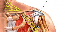
Anatomy of the shoulder joint: Position of the periarticular bursa under the acromion
Changes to the underlying supraspinatus tendon (eg, tears and calcium deposits) can distort the bursa.
Inflammation of the shoulder bursa can be caused by structural changes in the acromion, such as bone spurs. Therefore, in search of the causes of persistent bursitis, it is necessary to undergo a complete examination of all structures of the shoulder. © bilderzwerg / fotolia Inflammation of the periarticular bursa of the shoulder joint is characterized by swelling in the area of the subacromial bursa, located under the acromion. Muscles, ligaments, tendons and the bursa, that is, the soft tissues of the shoulder, are located close to each other. Thus, the largest bursa ensures the health of the supraspinatus tendon, the tendon between the shoulder blade and the head of the shoulder. If the load on the subacromial region is exceeded due to raising the arm above the head, pressure is also exerted on the supraspinatus tendon, which often causes inflammation and pain in the shoulder.
Disease prevention
You can prevent the risk of bursitis using preventive measures:
- reduce physical activity;
- use a bandage or simply bandage the shoulder area tightly;
- treat wounds in a timely manner;
- observe safety measures;
- avoid hypothermia.
Preventive measures should be carried out taking into account the general condition of the patient, therefore the doctors of our clinic recommend them to each patient individually.
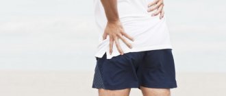
Symptoms: Shoulder pain when raising your arm up
Symptoms:
- Pain when raising your arm up
- Pain when pressing on the shoulder and increased sensitivity
- Stitching pain upon final extension of the arm
- Night pain when lying down
- Weakened muscle strength
- Swelling, redness, hyperthermia
- Pain radiating from shoulder to arm
The pain caused by bursitis of the shoulder joint begins gradually and increases depending on the nature of the movements. As a rule, at the beginning of bursitis, patients complain of discomfort when raising their arms to the top.
If the patient does not attach much importance to his complaints and continues to lead his usual lifestyle, the pain gradually becomes stronger. If the shoulder joint begins to hurt suddenly, the cause is most likely not bursitis. Specialists who treat the shoulder joint strongly recommend not to put stress on the shoulder. Otherwise, the pain will become more and more powerful and will bring you discomfort even during rest. The shoulder's increased sensitivity to pressure can lead to severe pain at night. If the patient sleeps on an unhealthy shoulder, then the shooting pain can wake him up.
Shoulder pain due to bursitis is often accompanied by increased sensitivity on the outside of the shoulder.
When the shoulder joint's range of motion is completely exhausted, patients feel an unpleasant tingling sensation in the shoulder. Such sensations are observed when a person raises his hand above his head in order, for example, to dry his hair, comb his hair, or put on a jacket. People who constantly sleep on their sides also feel the negative effects of bursitis, since while sleeping on the sore shoulder, pressure is exerted on the inflamed mucous bursa. That is why shoulder bursitis is an unpleasant disease.
Repeated monotonous movements of the shoulder only increase the pain. Due to bursitis, pain may radiate towards the outside of the shoulder towards the elbow.
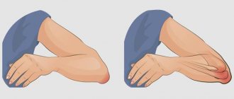
Unlike bursitis of the knee or elbow joint, inflammation of the bursa in the shoulder rarely causes external swelling or changes in the shape of the shoulder itself.
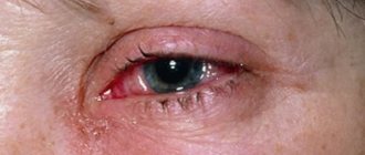
However, with extremely severe inflammation of the mucous bursa, the patient may experience redness of the shoulder joint. These not immediately noticeable signs are primarily concomitant phenomena of bacterial bursitis. Sometimes during bursitis a person's body temperature rises. Bacterial bursitis is also medically called “septic bursitis.”
Symptoms of bursitis
Acute bursitis usually begins suddenly and is accompanied by:
- sharp pain that intensifies with movement, as well as pricking or shooting pains at night;
- soreness and hypersensitivity of the skin over the joint;
- limitation of mobility in the joint;
- hyperemia and swelling of the skin over the joint;
- local or general increase in temperature;
- weakening of muscles;
- when palpating the joint, you can detect a non-rigid seal filled with liquid, which resembles a balloon filled with water to the touch;
Most often, symptoms of bursitis appear in the shoulders
,
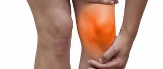
knee
,
elbow and hip joints
, as well as
joints in the area of the heel tendon
. Symptoms and treatment of bursitis depend on the intensity of the inflammation. If left untreated, the increase in body temperature can reach 40°C due to a purulent process. At this stage, there is a high probability of complications - for example, erysipelas, subcutaneous fat tissue suppuration.
Symptoms of chronic bursitis are that:
- pain becomes moderate, tolerable or absent altogether;
- the infused seal under the skin remains for a long time;
- movements are less constrained or there is no restriction on their amplitude at all.
The recurrent process is characterized by a combination of all the symptoms of bursitis: the chronic course is periodically interspersed with exacerbations of symptoms. If this condition is not treated
,
subsequently fistulas begin to form
,
which create the risk of purulent arthritis
.
Fluid accumulation in the joint cavity is a common symptom of bursitis.
Causes of shoulder bursitis
Causes
- Impact, shock, injury
- Overload, especially during arm raising
- Monotonous movements
- Mature age
- Incorrect posture
- Impingement syndrome
- Diabetes
- Calcinosis of the forearm
Several circumstances contribute to the development of shoulder bursitis.
One of the most common causes is damage to the structure of the shoulder (trauma). A fall or blow to the shoulder can cause hemorrhage into the bursa and inflammation of the mucous membrane. Despite the fact that over time the body reduces the blood content in the bursa, inflammation of the mucous membrane on the outside of the mucous bursa remains for a long time. Thus, from a common injury, chronic bursitis of the periarticular bursa of the shoulder is formed. Doctors call this form of inflammation “traumatic bursitis.”
Another cause of bursitis is repeated pressure or excessive stress on the shoulder. In this case, the mucous bursa becomes inflamed due to repeated minor injuries, leading to the same results as hitting the shoulder on a hard surface. Painters, fitters, or people whose profession involves raising their arms above their heads suffer from this pathology more often than others. Athletes who perform powerful overhead movements, such as tennis or badminton players, report similar symptoms.
With age, the likelihood of developing bursitis due to traumatic injuries or excessive stress increases.
Incorrect posture is another cause of inflammation of the shoulder bursa. When the body is strongly bent forward, the scapula is pulled towards the top, which narrows the space under the acromion. If a patient comes to the doctor with such complaints, he is diagnosed with impingement syndrome of the shoulder joint and referred for treatment. If the subacromial area is narrowed for a long time, the mechanical pressure on the subacromial periarticular bursa and the tendon apparatus responsible for shoulder mobility (rotator cuff) increases. The interaction of these factors contributes to the development of the inflammatory process inside the synovial bursa.
Bursitis that has been treated for a long time can also contribute to the appearance of inflammation of the shoulder bursa.
Metabolic disorders of the shoulder joint can also cause bursitis: Calcification of the forearm (calcification of the supraspinatus tendon) often appears simultaneously with bursitis. Calcium deposits in the supraspinatus tendon can lead to rupture of the overlying subacromial bursa and long-term inflammation of the bursa.
Impingement syndrome is characterized by friction of the rotator cuff tendons against the acromion, which is the so-called roof of the shoulder joint. In addition, during humeroscapular periarthritis, bone spurs (osteophytes) are formed on the acromial process of the clavicle, causing ruptures and inflammation of the mucous membrane of the shoulder bursa.
Diagnosis and clinical examination of bursitis
Before starting treatment, an orthopedic specialist conducts a series of examinations to rule out certain causes of the disease. Inflammation of the tendons, impingement syndrome or arthrosis of the shoulder joint have similar symptoms. In some cases, these pathologies are also accompanied by additional inflammation of the periarticular bursa of the shoulder. Thus, medicine presents several causes of shoulder pain. High-quality diagnostics at the Gelenk Klinik medical center in Freiburg in Germany, based on advanced technologies, make it possible to establish the main cause of shoulder pain.
History: diagnostic consultation with a doctor
During the anamnesis, compiling a medical history, the doctor asks the patient several questions regarding his health:
- Previous and current diseases
- General state
- Diseases caused by metabolic disorders (eg diabetes, rheumatism or gout)
- Motor vehicle accidents or other incidents involving shoulder injuries
- Overloads and shocks
- Sports loads and features of professional activity
In this way, the doctor receives a complete picture of the possible effects, deficiencies and predispositions of the shoulder joint.
Physical examination and clinical examination
During the examination, the shoulder specialist pays special attention to the following aspects:
- Pain when pressed (increased sensitivity)
- Swelling
- Hyperthermia and skin color changes
- Features of posture
Special tests to determine the strength and mobility of the arm during movement in different directions indicate possible tendon ruptures and injuries.
Imaging examination of the shoulder joint: ultrasound, x-ray, MRI
Modern medicine offers several imaging diagnostic methods, each of which shows different aspects of the disease.
Ultrasound helps the doctor test the mobility of the muscles, tendons and soft tissues of the shoulder. Also, an ultrasound examination shows swelling and accumulation of excess fluid in the bursa. Ultrasound also shows structural damage to the shoulder, for example. tendon ruptures and inflammation. © Gelenk-Klinik
Ultrasound examination (ultrasound)
Ultrasound imaging shows accumulation of excess fluid in the bursa of the shoulder joint, as well as injuries to the ligaments and tendons during movement. In addition, ultrasound shows tendon ruptures and calcium deposits.
X-ray
An x-ray does not show the condition of the soft tissues: the image shows only the position and condition of the bones of the shoulder joint. This image shows sufficient space between the head of the humerus and the acromion. It follows from this that impingement syndrome is not the cause of inflammation of the shoulder bursa. Ultrasound allows the doctor to examine muscles, tendons and soft tissues during movement. In addition, an ultrasound examination provides information about the presence of edema due to the accumulation of excess fluid in the bursa. Tendon injuries and inflammation in the shoulder also become visible after an ultrasound.
This image shows sufficient space between the head of the humerus and the acromion. It follows from this that impingement syndrome is not the cause of inflammation of the shoulder bursa. © Gelenk-Klinik
MRI (Magnetic resonance imaging)
MRI (tomographic medical images) provide information about soft tissue damage, inflammatory processes, and structural changes in the shoulder joint. For the treatment of bursitis, MRI is not the main diagnostic method. If a patient is suspected of having inflammation of the mucous membrane of the bursa, specialists at orthopedic clinics first perform an ultrasound.
Before making a diagnosis of shoulder bursitis and starting treatment, it is necessary to conduct an imaging examination to exclude tendon ruptures and injuries to bone structures. MRI is performed only in the case of a complicated form of bursitis and helps make a final decision regarding the most appropriate form of treatment for the disease. Magnetic resonance imaging helps to choose the right method of surgical treatment and prepare for surgery.
In complex cases that are not amenable to conventional treatment, a very important aspect is to exclude injuries such as tendon ruptures and bone deformations using imaging diagnostics. Magnetic resonance imaging (MRI) is one of the most important methods for diagnosing shoulder bursitis.
Laboratory analysis of blood and joint fluid
Laboratory diagnostics
- Diabetes
- Gout
- Rheumatism
- Bacterial inflammation (septic bursitis)
- Arthritis
Movement problems are not always the cause of shoulder pain. The patient may also feel discomfort in the hand due to metabolic disorders that limit mobility and cause pain. Such pathologies include, for example, rheumatoid arthritis, an inflammatory autoimmune disease. A blood test shows the presence of rheumatoid factor, which allows you to confirm rheumatoid arthritis. In addition, a clinical blood test helps to exclude bacterial infections: With an increased number of leukocytes in the blood, the likelihood of disease increases.
Diagnostics
Detection of bursitis begins with examination of the patient, study of complaints and the results of the collected anamnesis. Only a comprehensive diagnosis allows the doctor to identify the inflammatory process and its possible causes. To make an accurate diagnosis and identify the degree of articular damage, patients are prescribed the following types of studies:
- X-ray: prescribed to exclude arthrosis, malignant neoplasms and other pathologies. In the picture you can see the condition of the bones of the joint;
- Ultrasound: to clarify the nature and localization of the process, the presence of concomitant diseases of the joint, ultrasound diagnostics is used. In this case, you can examine the soft tissues, and also see the presence of edema;
- taking a puncture of synovial fluid to find out the cause of inflammation;
- CT or MRI: in some particularly complex clinical cases, the diagnosis of bursitis involves computer or magnetic resonance imaging.
Conservative treatment of shoulder bursitis
As a rule, inflammation of the periarticular bursa of the shoulder is treated conservatively, that is, without surgery.
Home remedies and home treatment
In most cases, patients can treat inflammation of the shoulder bursa on their own. However, if the pain does not stop and becomes stronger, you should immediately consult a doctor. Despite this, please note that home treatment is associated with certain risks: If you use home remedies, you may miss some important shoulder injuries or pathologies.
- Cooling compresses for acute shoulder pain.
- Anti-inflammatory medications (eg paracetamol or ibuprofen)
- Sports ointments (eg diclofenac)
- Anti-inflammatory and cooling curd compresses
- Applying aluminum acetate salt to the shoulder
In most cases, shoulder bursitis is treated with non-steroidal painkillers (NSAIDs). In addition, to restore the shoulder, orthopedic specialists recommend rest and cooling compresses to the patient. In the most difficult cases, injections of the anti-inflammatory hormone cortisone, as well as painkillers, help to immediately stop inflammation of the periarticular bursa and associated shoulder pain.
Physiotherapy and biological treatment based on cell technologies
If drug treatment does not bring the desired result and the patient continues to feel pain, the doctor refers him to physiotherapy or exercise therapy. One of the main specializations of the German medical center Gelenk Klinik in Freiburg is regulatory therapy based on cell technologies. The use of vibration therapy in medical practice helps stimulate metabolism in the inflamed synovial bursa of the shoulder joint, which helps to overcome inflammation in the shortest possible time.
Matrix therapy consists of several modules that interact to stop the development of the inflammatory process and tissue health. This image shows biomechanical stimulation (BMS) of the shoulder muscles using a vibrating device. This technique helps treat injuries, inflammation and structural disorders of the shoulder tendons. Inflammation of the mucous bursa is one of the main indications for this treatment. © gelenkreha.de
Classification of bursitis disease
As a disease, bursitis is usually divided into:
- specifics of the course
(acute, subacute, chronic or recurrent); - pathogen
(specific or nonspecific infections, inflammation without an infectious pathogen), as well as origin (primary or secondary bursitis); - type of exudate
(serous, purulent, hemorrhagic) - localization
.
Bursitis with a sudden onset and acute course can be completely cured if therapy is started early. If a visit to the doctor is constantly postponed or his recommendations are not followed, the transition to a chronic form is most likely
- in this case, the disease lasts several months, and subsequently recurs, causing irreparable consequences.
As a primary disease, bursitis occurs with direct damage to the joint capsule or local infection. Secondary - as a consequence of another chronic disease. The type of exudate can be determined only after taking a sample - based on it, treatment will be selected.
Minimally invasive surgeries of the shoulder joint for inflammation of the periarticular bursa
Only in cases where drug treatment for shoulder bursitis, as well as physical therapy, have been unsuccessful and a person continues to feel tingling in the shoulder, does there become a need for surgical treatment of subacromial bursitis. We draw your attention to the fact that treatment of bursitis of the shoulder joint is carried out using arthroscopic minimally invasive techniques. This operation, during which the surgeon completely removes the inflamed joint capsule, is performed through the so-called “keyhole”.
Shoulder arthroscopy is a minimally invasive surgery for shoulder pain, for which the surgeon uses microscopic instruments with a diameter of 0.5 - 1 cm. An arthroscopic camera helps the doctor obtain a limited but accurate image of the surgical site. This intervention consists of aspiration (suction) of the contents from the joint capsule. In addition, in parallel, the surgeon can treat diseases that cause bursitis, such as impingement syndrome or calcification of the forearm. © bilderzwerg / fotolia
Additional operations when removing a bursa (bursectomy)
Additional interventions
- Removing calcium from the supraspinatus tendon
- Acromioplasty
- Suture placement of the supraspinatus tendon
In addition to the operation, the specialist examines and, if necessary, begins treatment of all structures of the shoulder joint. Thus, if bursitis is suspected, calcium deposits from the supraspinatus tendon, as well as bone spurs under the acromion, can be removed during arthroscopy.
Shoulder arthroscopy: Postoperative treatment and prognosis
After surgical removal of the periarticular bursa of the shoulder (bursectomy), a new bursa is formed in the same place within a short time, performing the functions of the removed tissue in full.
Motor functions of the shoulder are restored a few days after the operation. The pain syndrome disappears immediately.
Almost immediately, the patient can move the shoulder as before and return to previous activities. The stitches are removed after about 10 days.
Make an appointment
Treatment
Treatment of joint bursitis is aimed at quickly relieving pain and further eliminating the disease. Therapy lasts 14 days or 1.5 months. Surgical intervention is necessary in advanced cases.
Treatment for bursitis includes:
- taking medications;
- physiotherapy;
- aspiration;
It is important to provide rest to the diseased part of the body and avoid further traumatization. If you endure pain, move or work through discomfort, then bursitis will develop into a chronic stage.
The doctor prescribes compresses and ointments to treat bursitis. Cold compresses can be applied to the sore spot for no more than 15 minutes; if kept longer, there is a risk of frostbite.
Non-steroidal medications are prescribed. These include: Ibuprofen, Dexalgin. Antibiotics if infection is present.
If liquid has accumulated, it is removed. This is done after the inflammation in the bursa has decreased, but the fluid remains. Using a syringe, the specialist pumps out 5 to 20 milliliters of liquid, then injects cortisone. Apply a pressure bandage to prevent fluid from accumulating. It is important to carry out such manipulations only in sterile conditions, minimizing the risk of infection and infection.
Injections into the sore joint help a lot. Injections for bursitis are prescribed with Diprospan. Glucocortisone, Prednisolone, Methylprednisolone, Dexamethasone, Triamcinolone are also prescribed.
Physiotherapeutic methods are used such as: ultrasound or diathermy. Physiotherapy helps relieve inflammation, relieve pain, and increase the effect of medications. At the same time, physiotherapy often has no contraindications. Perfectly complements the complex treatment of bursitis.
Other physiotherapy methods:
- balneotherapy;
- magnet therapy;
- electrophoresis;
- shock wave therapy;
A massage may be prescribed, but only during the period when the illness subsides.
Surgery is needed when there is a very large lump, and the treatment will be long and lengthy. Then the specialist can perform a puncture or drainage of fluid in the bursa.
Surgery for bursitis is performed in case of accumulation of purulent contents in the bursa. Sometimes a specialist performs tissue excision or bursectomy. This is done when the previous treatment did not have the desired effect.