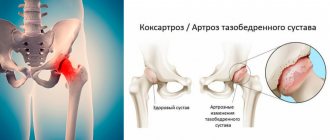
Osteoarthritis (OA) of the hip joints refers to serious degenerative-dystrophic changes that occur in the cartilaginous tissues of the articular surfaces of the hip joint. Osteoarthritis with this localization is considered one of the most common joint diseases. The pathology, which mercilessly destroys the hip joint, ranks second in occurrence after gonarthrosis of the knee joints. In terms of the risk of rapid disability in the general structure of joint diseases, OA ranks first. Another specific name for the disease, which refers specifically to damage in a given area of the musculoskeletal system, is coxarthrosis.
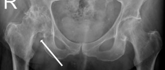
This is what an advanced diagnosis looks like on an x-ray.
Approximately 80% of elderly people (60 years and older) have radiographic evidence of hip OA to one degree or another, and a quarter of them experience intense pain and severe motor limitations. Osteoarthritis of TB joints is diagnosed almost 2.5 times more often in women. According to statistics, a sharp increase in the incidence rate is observed in the population over 45 years of age. According to authoritative medical sources, among the established causes of the development of degenerative disease of the hip joints, congenital dysplasias lead. That is, violations of the anatomical shapes of the ends of the bones that form the joint, already present from birth.
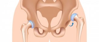
On the left is the healthy surface of the femoral head, on the right is affected by disease.
The pathology to which the entire article will be devoted is a severe variant of a medical problem with a complex orthopedic outcome. A disproportionate number of patients with this diagnosis face permanent disability accompanied by excruciating chronic pain. People very often become dependent on outside help, lose the ability to move normally, and are unable to go to work or perform usual household chores.
The most unpleasant thing is that conservative treatment does not always bring relief; it is often necessary to take extreme measures - to relieve the patient of suffering through surgery. Fortunately, in the modern age of orthopedics, surgeons have unique innovations that provide great prospects for gaining full functionality of the problem leg, which previously could only be dreamed of. We will introduce you to all the intricacies of osteoarthritis, including treatment, in order.
Knee replacement in the Czech Republic: guarantees, prices, rehabilitation, reviews and statistics.
Find out more
Minimally invasive endoprosthetics in the Czech Republic: doctors, rehabilitation, terms and prices.
Find out more
Osteoarthritis of the hip joint: symptoms and treatment regimen
Constant, aching pain, loss of ability to work, loss of the ability to move normally are the consequences of arthrosis of the hip joint. In medicine, the disease is found under such terms as coxarthrosis or degenerative disease of the hip joint. This type of pathology is usually classified as a progressive rheumatic disease, leading to damage and further destruction of cartilage tissue, which may result in disability. The disease is often diagnosed after 50 years of age, with women suffering more often. It is impossible to predict how quickly arthrosis progresses.
Bone deposits (osteophytes) almost always develop on the head of the femur, which impair the mobility of the joint and make it difficult for a person to move normally. Often, the onset of osteoarthritis of the hip joint is not recognized for years and is diagnosed already in the last stages, requiring surgical intervention.
Coxarthrosis 2nd degree
The pain intensifies and lasts longer. They do not go away after completion of physical activity and remain in an immobilized state. When moving, clicking and crunching sounds are heard in the joint. Due to the shortening of the limb, slight lameness appears and joint mobility is limited. X-ray examination shows a decrease in the gap between the bones and joints, displacement of the femoral head, and the appearance of pathological growths of bone tissue in the place of destroyed cartilage - osteophytes.
Causes and factors of arthrosis of the hip joint
The causes and provoking factors of arthrosis of the hip joint are divided into initial (primary) and subsequent (secondary). Coxarthrosis of the primary type develops after 60 years, and, as a rule, is associated with natural processes in the body - aging. In older age, the production of hyaluronic acid decreases significantly, the nutrition of the cartilage deteriorates, and the joints wear out.
Secondary arthrosis of the hip joint can develop at a young age, often patients are people under 40 years of age. Pathology of the secondary type is often a consequence of the following disorders and diseases:
Congenital defects of the hip joint, such as hip dislocation or dysplasia (inferiority of the joint).
- Growing diseases - Perthes syndrome (osteochondropathy) or Salter-Harris fracture (epiphysiolysis, destruction of the hyaline cartilage plate).
- Injuries, fractures of the acetabulum.
- Avascular necrosis.
- Rheumatoid arthritis.
- Septic arthritis.
Often the origin of the pathology is associated with mechanical damage to the pelvic area. In almost 75 percent of victims, the disease is associated with trauma.
Finally, coxarthrosis occurs due to a discrepancy between the load-bearing capacity of the hip joint and the actual tension. This imbalance can be caused by various reasons, in particular: inflammation, fractures, metabolic diseases, and long-term consequences of accidents. Increased body weight (obesity) plays an important role in the development of the disease.
Also, a provoking factor for hip arthrosis is increased physical stress on the joints. Likewise, lack of exercise is also a risk factor contributing to the occurrence of coxarthrosis.
It is currently assumed that osteoarthritis of the hip joint is a multifactorial phenomenon. In addition to risk factors for arthritis, such as age, obesity, osteoporosis, inflammatory reactions of joint joints, hip deformities, dysplasia, genetic predispositions also play a role.
Diet
With the help of nutritional correction, the patient can not only reduce body weight, but also reduce inflammatory reactions, salt deposits in tissues and metabolic disorders. It is recommended to follow a balanced menu with sufficient, but not excessive amounts of carbohydrates, proteins and fats, as well as vitamins and minerals. Particular attention should be paid to unsaturated fats (olive and flaxseed oil), omega-3 acids (found in abundance in fish), collagen (jelly, aspic). It is recommended to minimize fast carbohydrates, alcohol, strong coffee, products with artificial flavors, preservatives and flavor enhancers.
Clinical manifestation of arthrosis of the hip joint
At the stage of the inception of the clinical picture of the pathology, the first symptoms of arthrosis of the hip joint are painful sensations in the pelvic area that occur after physical stress. The pain can radiate to the groin area and knee, becoming especially pronounced when walking. After a few seconds or minutes, the intensity of the pain decreases, and after a while it increases again. Subsequently, painful sensations are observed at rest, especially at night. In the absence of conservative treatment, the patient soon develops the chroma typical of arthrosis of the hip joint and the need to use a cane or even a walker.
Symptoms of coxarthrosis
The pathological process develops slowly - clinical symptoms appear depending on the degree and severity of joint damage. Most patients suffering from coxarthrosis of the hip joint complain of the following ailments:
- Poor joint mobility
— it is difficult to move the leg to the side or, bent at the knee, to pull it towards the chest. The symptom is due to the fact that the cartilaginous layer of the articular head is thinned or partially absent. As a result, friction between the surfaces of the articular bones increases. The resulting osteophytes (bone outgrowths) increase pain when moving.
- Pain in the groin, on the front or side of the thigh, in the buttocks and knee.
The syndrome manifests itself as a dull pain or characteristic lumbago, and increases in the evening and at night.
- Periodic muscle spasms
, increasing pain and limiting joint mobility.
- Reduction of the affected limb
– typical for later stages of the disease. The leg shortens due to abrasion of cartilage and bone tissue, muscle atrophy, and a decrease in the gap between the femur and the acetabulum.
- Lameness and duck gait occur in the later stages of arthrosis
. A gait defect occurs as a consequence of limb reduction due to unbearable pain and poor mobility of the hip joint. Lameness indicates serious destruction of the joint.
In the later stages of the degenerative process, the articular surfaces, devoid of cartilage tissue, can grow together. Ankylosis (fusion) leads to complete immobility of the limb.
Coxarthrosis of the hip joint can be:
- primary (idiopathic), caused by age-related wear and tear of articular cartilage - diagnosed in older patients.
- secondary, associated with injuries and previous diseases, can occur at any age.
The pathological process goes through several stages. It may be asymmetrical or affect both joints.
Arthrosis of the hip joint 1st degree
Arthrosis of the 1st degree of the hip joint: moderate pain, no stiffness of movement. The X-ray image shows a very slight narrowing of the joint space and single pathological growths (osteophytes). The first degree of the disease responds quite well to medication; at this stage there is a high chance of stopping the progression of the process.
According to WHO estimates, 3 to 4% of people worldwide suffer from arthrosis. The disease affects about 5% of the population under the age of 30, 20% of the population over 50, 70% to 80% of those over 65 and almost everyone over 80. It is common in postmenopausal women, diabetes and obesity.
Forecasts for arthrosis of the hip joint
With effective lifelong treatment, discomfort may be completely absent or disturb the patient extremely rarely. The prospects for therapy depend on the patient's compliance with the doctor's instructions. The most accurate prognosis is made by the doctor individually.
With timely and regular treatment
In the absence of concomitant chronic diseases at stages 1 or 2 of disease progression, hip replacement can be avoided. Loss of ability to work and disability, with a competent approach, can be extended by 20 years or until old age (in rare cases).
When self-medicating
Traditional methods or independently purchasing drugs without a doctor’s prescription leads to severe exacerbation and worsening of the disease. Time is wasted.
No treatment
Without treatment, the disease progresses 2-4 times faster. The quality of life deteriorates and disability occurs as early as 45-50 years of age. Most patients have a disorder in the psychoemotional sphere.
Arthrosis of the hip joint 3rd degree
Arthrosis of the 3rd degree of the hip joint: severe pain syndrome that develops at rest, worries the patient almost around the clock, reacts poorly to NSAIDs and painkillers. Severe lameness and muscle atrophy are noted. On x-rays you can see pronounced deformation processes. The third degree of coxarthrosis is not amenable to drug therapy and practically does not respond to drugs. The only way out that will allow the patient to get rid of pain is endoprosthetics (replacement) of the hip joint.
The disease begins with nonspecific discomfort. As the pain progresses, phases of moderate discomfort alternate with phases in which the pain sharply intensifies.
Diagnosis of coxarthrosis
Timely diagnosis and proper therapy will help stop the progression of the disease, relieve pain, and normalize motor activity. Treatment of coxarthrosis of the hip joint is carried out by orthopedic doctors: orthopedists, traumatologists and rheumatologists.
For correct diagnosis, the following types of examinations are prescribed:
- visual inspection, tactile examination, specification of complaints;
- laboratory tests of blood and urine;
- X-ray;
- CT scan;
- ultrasonography;
- Magnetic resonance imaging.
Diagnosis of arthrosis of the hip joint
The diagnosis of coxarthrosis can be made by collecting the patient's medical history and a sound clinical examination by an orthopedic specialist. An x-ray shows typical signs of arthrosis - a decrease in space in the joints and the formation of osteophytes. Other cross-sectional imaging modalities, such as MRI, may provide additional information that is important in excluding nonarticular musculoskeletal pathologies. Diagnosis of arthrosis also includes:
- Blood analysis. The analysis will reveal the erythrocyte sedimentation rate, which is an indirect indicator of the inflammatory process in the body that has affected the joint tissue.
- Blood chemistry. It is carried out as a differential diagnosis of rheumatological diseases. In addition, it makes it possible to evaluate the functioning of internal organs.
- Ultrasound examination (ultrasound). It is carried out to assess the amount of synovium (synovial fluid) and the condition of the soft tissues. Ultrasound can be carried out as a dynamic study: during diagnosis, the patient makes movements, thus, joint pathology becomes obvious.
Although X-ray examination is a fundamental method for making a diagnosis, which allows you to determine the stage of the lesion and its cause, MRI is considered more informative for determining early changes in the joint.
Treatment with gymnastics
Performing a complex of therapeutic exercises, individually developed by a competent specialist for a specific patient, will help increase the range of movements in the problem area, improve the functions of the TB department, and correct muscle balance. Special physical training in an adequate mode will allow you to competently relieve the sore area of the musculoskeletal system, reduce the severity, duration and frequency of pain, increase vitality and generally improve well-being.
Exercise therapy courses should be conducted in a rehabilitation gym under the supervision of medical personnel regarding the recovery of patients with musculoskeletal disorders. In any case, until the person perfectly masters the techniques of rehabilitation exercises recommended by the doctor. Patients are recommended to have at least a certain range of aerobic training with and without machines, but with the exception of force loads on the affected joints. Loads are selected strictly taking into account the main diagnosis, concomitant diseases, weight and age of the patient, and his physical capabilities.
Activities organized in the pool - therapeutic aqua gymnastics, swimming - provide invaluable benefits. Measured walking and gentle exercise on an exercise bike are encouraged.
Therapeutic tactics: what treatment is prescribed
Effective treatment of arthrosis of the hip joint involves the use of various methods: from medication and exercise therapy to surgical intervention (endoprosthetics).
Non-surgical treatment requires quite a large financial investment, but can improve the patient’s quality of life and, in the long term, eliminate the need for endoprosthetics. If arthrosis of the hip joint was detected at the first or second stage, the first medication prescribed by the doctor will be NSAIDs and painkillers. NSAIDs are aimed at reducing the activity of the inflammatory process, and painkillers are aimed at relieving pain.
In the treatment of articular pathologies, it is important to restore nutrition to the cartilage and slow down the process of its destruction. For this purpose, the doctor will prescribe a treatment regimen that will include drugs with a vasodilator effect and chondroprotective drugs. The latter are used for therapeutic and prophylactic purposes for degenerative joint diseases.
Among chondroprotectors, you should pay attention to Artra, which contains chondroitin and glucosamine. Artra is produced in the form of tablets of 60 and 120 pieces.
Chondroitin is a glycosaminoglycan that is considered as a symptomatic slow-acting drug for osteoarthritis. Chondroitin, used with glucosamine, is indicated to relieve pain and reduce inflammation in arthrosis. This combination improves joint function and slows the progression of the disease. Chondroitin does not have carcinogenic potential and is well tolerated without serious side effects.
The next component of Arthra is glucosamine. Oral glucosamine may relieve pain in people with osteoarthritis of the knee, hip, and spine. How glucosamine works in treating disease is not entirely clear, but most research suggests that it can reduce inflammation—especially when used in conjunction with chondroitin supplements. In addition to alleviating pain, glucosamine slows down the process of destruction of cartilage tissue and prevents deterioration of the condition during arthrosis. Some studies show that patients who took glucosamine long-term were less likely to require arthroplasty and other orthopedic surgeries.
How does deforming osteoarthritis of the hip joint occur?
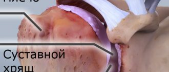
DOA (deforming osteoarthritis) is not an inflammatory, dystrophic disease of a chronic nature, with damage to the cartilage tissue of the bones that articulate in it. The disease begins with its thinning. Gradual degeneration of cartilage tissue and premature aging occurs. The elasticity of the tissues is lost, cracks and roughness appear on the articular surfaces.
Sometimes the cartilage wears away, exposing the bone. Then the articulating bones begin to rub against each other, without the presence of “cushioning”. When cartilage is lost, bone tissue grows and permanent deformation occurs with impaired functionality. When the disease affects the hip bones, we are talking about the development of coxarthrosis, which is a pathological process leading to the destruction of cartilaginous plates.
Distinctive features of DOA:
- the destruction of cartilage is degenerative-dystrophic in nature;
- bone tissue grows along the edges of the articular surfaces;
- the joint is deformed as a result of the above pathological processes.
The pathology may affect one joint, but both may be involved. Deforming osteoarthritis can be primary or secondary. In the first case, the cause of the disease is possibly genetic. In the second case, the pathological process occurs due to pathologies of the musculoskeletal system.
Regular use of Arthra helps:
- reducing the severity of pain and inflammatory response;
- stopping the process of destruction of cartilage tissue;
- increasing joint mobility;
- production of antioxidant enzymes.
To obtain the highest possible therapeutic effect, conservative treatment, including oral medications and ointments, should be combined with physical therapy. For joint pathologies, the following procedures can be used:
- Shock wave therapy. The technique is based on the use of sound waves; they improve blood supply to the diseased joint and stimulate the regenerative capabilities of cartilage tissue.
- Myostimulation. It is a pulsed current aimed at improving the muscular system of the pelvis.
- Phonophoresis. It combines two operating principles - ultrasonic waves and drug therapy. With phonophoresis, drugs penetrate deeper into the joint, thereby increasing the therapeutic effect of the use of ointments.
In the absence of a positive effect from conservative treatment, the option of surgical intervention is considered. If the patient continues to suffer from pain, and the diagnosis determines the progression of the destructive process in the cartilage, surgery remains the only effective treatment.
Complications of arthrosis
The disease is easier to treat in the initial stages of progression. In advanced forms of the disease, the following complications cannot be excluded:
- infectious and inflammatory processes – accompanied by chills, increased body temperature, pain and tissue swelling;
- subluxations – cause a sharp limitation of movements of the lower limb;
- pinching of the femoral or sciatic nerve - characterized by radiating pain along the back of the lower limb, groin and inner thigh;
- tenosynovitis - accompanied by inflammation of the inner sheath of the tendons of the muscle that attaches to the femur;
- bursitis - implies inflammation of the synovial bursae.
Prevention of arthrosis of the hip joint
The development of arthrosis of the hip joint can be prevented in various ways. Sufficient and regular exercise stimulates the supply of nutrients to cartilage tissue and helps reduce the risk of disease. Another important prevention of arthrosis is to minimize the risk of injury - blows, falls, excessive physical activity. It is also important to monitor your body weight; excess weight is a provocateur of joint pathologies. Numerous studies have found that reducing a patient’s weight by 10-15% reduces the risk of developing arthrosis lesions by 50%.