The disease provokes the appearance of necrosis in certain areas of soft tissue, resulting from lack of proper care for the patient. In advanced cases, complications develop that can lead to death. Combating bedsores requires knowledge of basic skills in caring for bedridden patients.
How to treat bedsores?
Thursday, December 9
1906
5
3
Content
- Stages of bedsores
- Why do bedsores occur?
- How to treat bedsores
- Top 10 best ointments for bedsores
- Dexpanthenol
- Naftaderm
- Betadine
- Stellanin
- Baneocin
- Levomekol
- Solcoseryl
- Sulfargin
- Dermazin
- Oflomelid
In patients who are forced to lie down for a long time, blood circulation is impaired and bedsores inevitably appear. The capillaries that saturate tissues and organs with blood and oxygen become compressed when sitting or lying for long periods of time. Because of this, blood circulation slows down and in some cases stops completely.
Typically, bedsores occur in those people who, for one reason or another, have been lying down for a long time.
What are bedsores?
Bedsores typically occur in patients who are seriously ill, bedridden for a long time, or people in a wheelchair. Bedsores are damage to the skin and underlying tissues that occur due to disruption of their nutrition, prolonged pressure on a specific area of the body. Bedsores often occur on areas of the skin located above bony protrusions: elbows, knees, hips, buttocks, sacrum. For those who use a wheelchair, the damage is localized as follows: • Buttocks, sacrum; • Shoulders, spine; • The back of the legs, arms – the places that support the chair. In bedridden patients, damage may occur on: • The back of the head, temples; • Ears; • Shoulders, shoulder blades; • Hips, lower back, sacrum; • Heels, knees, popliteal area.
Stages of bedsores
Bedsores do not appear suddenly; they develop gradually. First, a person feels pain in places that are compressed, they become red, swelling and cyanosis appear due to stagnation of venous blood. This is how ischemia develops, but this process is reversible - you need to eliminate the causes of compression and use ointment for bedsores.
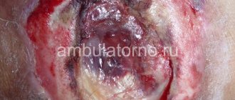
If areas of the body are compressed for a long time, the skin peels off. Then the fatty tissue dies and deep wounds appear. If the problem is advanced, you can even see exposed bone in the deepening of such a wound. The wound festers when any infection occurs.
Bedsores are easy to detect by visual inspection. If additional data is needed, the doctor may order blood or urine tests.
To cure bedsores, you need to restore normal blood flow to the damaged areas of the body as quickly as possible. If areas of necrosis have formed, local treatment will help: you need to dry the dying tissue and ensure that dry necrosis does not become wet. Wet dressings and dressings with ointment should not be applied during this period. As soon as the scab begins to come off, you can use bandages with ointment or do autodermoplasty. With deep bedsores, wet necrosis occurs. Treatment is aimed at rejecting dead tissue, and this is where ointments for bedsores help.
Stages of bedsores
Causes of bedsores
The main cause of skin pathology is compression of tissue between the bone, as well as the hard surface that the human body touches. For example, soft tissues are compressed between a bone - a wheelchair or a bone - a bed, which leads to impaired blood circulation in small vessels. As a result, oxygen and nutrients cease to flow to the cells of the compressed zone. The area of tissue becomes dead and dies. Other causes of bedsores include friction and slipping. Friction on clothes or sheets occurs if the patient changes position on his own, or is moved or turned over by family members or a medical worker. Sliding is facilitated by the high elevation of the bed in the head area. Then the patient slides down. Another reason is an attempt to stay in a semi-sitting position without support. Factors associated with incorrect patient care: • Untidy bed; • Untimely change of underwear; • Hard, uneven bed surface; • Hips, lower back, sacrum; • Neglect of hygiene procedures. Factors that are associated with the individual characteristics of the patient: • Exhaustion, obesity of the patient; • Elderly age; • Diseases of the cardiovascular system; • Disorders of the innervation of the body (for example, strokes); • Disorders of the body's metabolic processes (water-salt metabolism, drinking restrictions, diabetes mellitus); • Unbalanced diet, lack of protein foods, protein metabolism disorders; • Uncontrolled processes of defecation and urination. Also, factors that provoke bedsores include excess/underweight, smoking, an allergic reaction to skin care products, seams, folds, buttons on underwear, injuries and diseases of the brain and spinal cord, sweating due to elevated body temperature.
Why do bedsores occur?
Bedsores appear due to the following factors:
- the person’s body weight is too large, which puts pressure on the tissues;
- on the contrary, the patient’s weight is too small: the volume of fat and muscle tissue is so small that the bones press on the skin;
- the body rubs against the sheets;
- poor personal hygiene, when urine and feces come into contact with the skin for a long time;
- diseases in which blood circulation in tissues is impaired (for example, anemia);
- too sensitive skin;
- little protein in the body (muscle mass is reduced).
Typically, bedsores appear due to a combination of several of these factors. To avoid problems, you need to keep problematic areas of the body of a bedridden person dry and clean, treat the skin with ointments for bedsores, frequently change body position, and monitor general well-being and temperature. A nutritious diet with enough protein is also important.
Read also: 10 most effective acne remedies 10 creams and gels to combat acne and acne.
The main objectives of the treatment of pressure ulcers
Specialists at the Yusupov Hospital rehabilitation clinic select medications against bedsores on an individual basis, after conducting a thorough examination and determining the stage of the disease. If conservative therapy is ineffective, surgical intervention may be prescribed.
Drug treatment of bedsores has the following objectives:
- stimulate blood circulation in affected tissues;
- eliminate necrotic masses from the surface of the affected skin area;
- activate regenerative processes in wounds.
In parallel with treatment, the rehabilitation clinic of the Yusupov Hospital carries out mandatory measures to prevent bedsores.
How to treat bedsores
At the first signs of pathology, a doctor’s consultation is necessary. If the skin is not damaged, gentle products are suitable. If the integrity of the skin is compromised, these areas must be treated with an antiseptic every day. A hydrogel bandage or ointment for bedsores will help.
In advanced cases, the wound will need to be cleaned, which can only be done by a specialist. To prevent infection, you will need a bandage with a special agent. If fluid is leaking from the wound, an absorbent bandage will be needed.
IMPORTANT!
Bedsores are a dangerous condition, so do not use traditional methods to treat them, they often only worsen the problem.
Do not use soap with an antibacterial effect - it destroys not only harmful bacteria, but also useful ones - the skin loses local immunity and becomes unprotected.
Alcohol-based products (camphor alcohol, lotion) can only be used by those with oily skin. Do not rub the skin when washing, so use soft sponges so as not to injure the epidermis. Dry the skin with blotting movements. Air baths are also important to allow the skin to “breathe.”
If bedsores are treated incorrectly, the situation can quickly get worse. If there are abscesses and fistulas, the risk of sepsis increases.
Today, pharmacies sell many remedies for bedsores - even at the most advanced stage, ointments for bedsores can heal wounds on the body.
Where do bedsores usually occur?
Prevention of bedsores
To eliminate the first signs of a bedsore it is necessary: • Change the patient's position once every 2 hours. If there are no contraindications, it is recommended to use special pillows that will change the position of the limbs and body relative to the surface of the bed and create gaps between the bed and the skin; • Monitor the head of the patient's bed. It should be level or lower with it; • Control the moisture of the patient’s skin using hygiene products - washing cream, solution, foam, spray, warm baths (do not use hot water). These procedures should be done at least 2 times a day. If there is uncontrolled defecation, then it is necessary to remove contamination as quickly as possible; • Promptly remove excess moisture from the epidermis and skin folds (remains of liquid food, water, urine, sweat, wound exudate). For this purpose, special absorbent pads, napkins, and towels are used; • Remake your bed regularly. Bed linen should be changed at least once a day; • Use anti-decubitus mattresses of cellular or balloon type. They are equipped with silent compressors, which is necessary to maintain and change the rigidity of the base. The mattresses also have adjustable and programmable inflation in different areas. • Do not perform intense massage. Only light stroking of the area of skin with signs of a bedsore is allowed. There should be no friction during the massage; • Use special pillows for patients in wheelchairs, which can be filled with gel, air, or foam. It is also important to monitor changes in body position in the chair. Bedsores are pathologies whose occurrence is best avoided. But if this was not possible, then when affected skin lesions occur, development occurs very quickly and is characterized by long-term treatment of the wound. The outcomes of bedsores are quite dangerous. The resulting defects, as well as the consequences of bedsores, are treated exclusively in a hospital setting.
Dexpanthenol
This ointment based on dexpanthenol is indicated both for the treatment of the initial manifestations of bedsores, when there is no infection of the wounds, and for the completion of therapy, when it is necessary to relieve swelling, inflammation and redness. "Dexpanthenol" accelerates healing and protects against dryness. You usually need to apply the ointment for bedsores once a day: the drug perfectly heals wounds in the initial stages. This drug has a low price, but the quality is not inferior to expensive analogues. The only negative is that you can’t buy Dexpanthenol in every pharmacy.
Dexpanthenol
JSC "Tatkhimfarmpreparaty", Russia
Dexpanthenol is a drug for external use that has a regenerating and metabolic effect, as well as some anti-inflammatory effect.
Dexpanthenol is a B vitamin, a derivative of pantothenic acid. In tissues, dexpanthenol is converted into pantothenic acid, which is part of coenzyme A. As part of coenzyme A, pantothenic acid takes part in the processes of acetylation, lipid and carbohydrate metabolism, the formation of acetylcholine, porphyrins and corticosteroids. from 66
5.0 1 review
939
- Like
- Write a review
Classical medicine
Therapeutic and surgical treatment takes a lot of time and effort from the patient and the people serving him. For a quick and, most importantly, effective cure, it is necessary to constantly monitor the patient’s condition, evaluate the appearance of the skin, carefully remove keratinized, diseased tissue, carry out air baths and measures to maintain blood circulation at a normal level, observe preventive measures, use special mattresses for bedridden patients from bedsores, sheets, pillows and so on. The use of medications prescribed by a doctor is also mandatory.
Diaper rash cream
These are not therapeutic, but prophylactic agents. If bedsores have already formed, then there is no point in using them. They are used after hygiene procedures for bedridden patients. They are applied to areas and zones that are often and for a long time exposed to high humidity or are subject to compression. They help increase tissue turgor, dry the skin, and reduce areas of maceration.
Ointment
This dosage form is used to fight infections, as a stimulant to accelerate regeneration processes, and so on. They are used at stages III-IV of the disease to treat deep wounds and ulcers. The most commonly used medications of this type include the following.
- Vishnevsky ointment
- Levomikol
- Solcoseryl
- Oak bark
- Field chamomile
- Sea buckthorn oil
- Onion
- Potato
- Baking soda
It was developed in the middle of the last century by a group of Soviet surgeons. Can be used to eliminate foci of inflammatory processes caused by various microbes. To treat bedsores, it is applied to the affected areas twice a day and fixed with a medical gauze bandage.
A more modern product with an antimicrobial, dehydrating effect based on chloramphenicol. During the day, a single application is sufficient, after which the affected area is covered with a cloth or gauze.
A medicinal product that improves regeneration processes and blood microcirculation, which is due to the presence of calf blood extracts, vitamins, microelements, and nutrients. Used 2 times a day.
If there is positive dynamics, the prescribed ointment is used until complete recovery is achieved. Otherwise, it should be replaced with another, more effective drug or used in combination with another ointment.
Medicines
In addition to acting directly on the affected areas themselves, the doctor must also prescribe systemic drugs: antibiotics, antithrombosis, anti-inflammatory, to improve microcirculation, to relieve pain. The duration of the course and the dose of the drug taken are determined by the doctor depending on the patient’s condition. The course of treatment with antibiotics is usually from one to one and a half weeks, anti-inflammatory - up to 2 months.
Medicines are taken before the healing process begins, while the ointment is applied until the final epithelization of the affected surface.
Naftaderm
This is one of the most natural ointments for bedsores; it contains purified naftalan oil (a completely natural product). You can buy Naftaderm in the form of a liquid ointment or liniment. This drug is prescribed for many problems, including bedsores. The product has a complex effect, therefore it perfectly relieves itching and inflammation, heals and disinfects the wound. The ointment has a specific smell and can stain the bed, but almost all reviews about it are positive.
Naftaderm
Retinoidyi FNPP, Russia
- psoriasis; - eczema;
-neurodermatitis; -seborrhea; - pityriasis rosea; -boils; -sycosis; - pyoderma; -pruritus; -wounds; -bedsores; - poorly healing ulcers; -erysipelas. from 406
5.0 7 reviews
770
- Like
- Write a review
How to recognize bedsores?
Patients who predominantly lie on their backs complain of the formation of bedsores in the area of the shoulder blades and sacrum. Also, areas damaged by necrosis are found on the back of the head and heels. When lying on your side, the following areas may be affected:
- hips;
- ears;
- whiskey;
- shoulders;
- knees;
- ankle
Symptoms of necrosis:
- formation of red or blue edema;
- increased sensitivity in the affected area and pain;
- blisters, which, when opened, provide an overview of superficial wounds;
- formation of a crater with purulent discharge.
Also, yellowish areas of necrosis form at the edges of the wound, which gradually acquire a dark tint.
Betadine
Betadine is an effective iodine-based antiseptic. The product can be purchased in the form of an ointment and in the form of a solution. Thanks to its strong bactericidal effect, the drug heals bedsores in the shortest possible time, destroying bacteria and fungi in the wounds. "Betadine" is prescribed both for obvious infection of wounds and for prevention. The ointment helps cleanse the wound of necrotic tissue and pus; it quickly heals and heals. During treatment of bedsores, the patient does not feel a burning sensation. It is worth considering that Betadine can cause allergic reactions.
Betadine
Egis, Hungary
Betadine is an antiseptic and disinfectant.
It has a bactericidal effect on gram-positive and gram-negative bacteria (with the exception of Mycobacterium tuberculosis). Active against bacteria, fungi, viruses, protozoa. Suppositories are made on a water-soluble basis and do not have an irritating effect. from 132
5.0 1 review
1384
- Like
- Write a review
How to carry out prevention
Early prevention of bedsores helps to significantly slow down the development of the necrotic process. It is advisable for the patient’s relatives to take care of a special two- or three-section bed, an anti-decubitus mattress and pillows or rubber cushions placed in potentially dangerous areas.
To avoid bedsores, the patient should not remain in the same body position for more than 2-2.5 hours.
It is necessary to carry out genital hygiene daily and change diapers in a timely manner, since bedsores and diaper rash primarily form in the sacral area due to urine leakage.
Daily skin care to prevent bedsores includes:
- thorough inspection;
- air baths;
- wiping with a soft damp sponge;
- cleansing with physiological medical solution;
- treatment with means for the prevention of bedsores (“wet” areas should be dried with powder, dry areas should be moistened with cream);
- light massage with camphor oil on vulnerable areas of the body.
The patient's bed linen and underwear should be cotton and kept in exemplary cleanliness. It is important to maintain a comfortable temperature in the room, since hypothermia or overheating interferes with blood circulation.
It is necessary to control the patient's diet. Doctors recommend focusing on foods rich in protein, vitamins, calcium and iron, and minimizing the amount of simple carbohydrates as much as possible.
Stellanin
An innovative remedy for the treatment of bedsores, which literally from the first use restores blood vessels and improves microcirculation in tissues. "Stellanin" reduces inflammation, disinfects wounds, clearing them of pus. The healing process of bedsores is accelerated. The duration of treatment depends on the extent of skin damage. Even in the most advanced stages, positive dynamics are observed. And although the course of treatment is expensive, many patients highly praise Stellanin ointment.
Stellanin
JSC "Tatkhimfarmpreparaty", Russia
trophic ulcers of the lower extremities;
- bedsores; — burns of the 1st and 2nd degrees; - abrasions, cuts, scratches, cracks, scratches, incl. after insect bites; - additional treatment of aseptic postoperative wounds (after excision, coagulation, episiotomy, for the treatment of skin cracks, wounds and sutures); — acceleration of skin graft engraftment. from 362
314
- Like
- Write a review
Symptoms
There are several stages of necrosis:
- 1. Redness appears, which gradually disappears after changing position and massage.
- 2. There is persistent redness. Various skin disorders are possible - superficial growths, blisters, erosions, etc.
- 3. Obvious lesions of the skin. The ulcers have indentations, are filled with yellow liquid, and emit an unpleasant odor.
- 4. Deep dead areas. Necrosis reaches the muscle mass.
Baneocin
This ointment contains two antibiotics that effectively fight infection of bedsores. "Baneocin" reduces inflammation, relieves swelling, cleans the wound and helps it heal faster. The drug does not enter the bloodstream, acting only locally; addiction to the active substances does not develop. You can buy Baneocin in ointment and powder form. The drug is applied to the wound under a bandage, Baneocin should be used 2-3 times a day, the course of treatment is about a week. During this time, significant improvement occurs. Among the disadvantages of the drug are its high price and possible allergic reactions.
Baneocin
Sandoz, Germany
Infectious and inflammatory skin diseases caused by microorganisms sensitive to the drug: - bacterial skin infections of limited prevalence, incl.
weeping contagious impetigo, infected trophic ulcers of the lower extremities, infected eczema, bacterial diaper dermatitis, secondary bacterial infection in diseases caused by Herpes simplex, Varicella zoster (including infection of vesicles with chickenpox); —prevention of umbilical infection in newborns; - prevention of infection after surgical (dermatological) procedures: for additional treatment in the postoperative period (after tissue excision, cauterization, episiotomy, treatment of cracks, perineal rupture, weeping wounds and sutures). from 183
810
- Like
- Write a review
Prevention of bedsores
Complications from bedsores
Incorrect diagnosis, incorrectly determined stage of development of the lesion, ineffective treatment, and insufficient care on the part of service personnel can lead to the development of quite serious complications against the background of bedsores, including the death of the patient. The most serious consequences include the development of the following diseases.
- Erysipelas
- Phlegmon
- Sepsis
- Gangrene
Or erysipelas is typical for older patients with stage II bedsores. This is an infectious disease caused by streptococcus, which penetrates the wound through damaged skin. Treatment involves the use of sulfonamides, penicillin antibiotics, and nitrofurans.
It is an acute inflammatory process of the cellular space as a result of penetration of E. coli and staphylococcus through the wound surface. Among the main symptoms of infection: high temperature, redness, pain, swelling, purulent contents. Broad-spectrum antibiotics are prescribed. The source of the lesion must be opened.
It is also an inflammatory process that can occur at stages III-IV of bedsores. This disease affects the entire body, therefore, if treatment is not started in a timely manner, it often leads to death. Anti-inflammatory treatment is carried out using glucocorticoids. Detoxification of the body is mandatory.
At stage IV of development, bedsores can lead to the development of gangrene. The complication is accompanied by darkening of the epidermis, rapid tissue necrosis, and a putrid odor. Upon palpation, a specific crunch is diagnosed. Intramuscular injections are prescribed to protect the tissue adjacent to the wound from infection. Treatment is surgical and involves amputation if the limb is affected.
To prevent the development of these complications, it is enough to provide the patient with proper systematic care, regular hygiene procedures, and strictly follow the instructions of the attending physician.
Levomekol
One of the most popular ointments for bedsores. Levomekol is inexpensive and has a complex effect. The ointment relieves inflammation, removes pus, and cleans the wound. The antibacterial effect of the drug helps get rid of the infection that often develops in wounds with bedsores. Reviews of Levomekol are mostly positive, especially since the product has a very reasonable price. "Levomekol" helps at the prevention stage and with any stages of bedsores (wounds need to be treated two to three times a day). Among the disadvantages are an inconvenient tube and a too liquid texture when heated.
Levomekol
OJSC "Nizhpharm", Russia
— purulent wounds (including those infected with mixed microflora) in the first (purulent-necrotic) phase of the wound process.
from 38
1077
- Like
- Write a review
Types of bedsores
It may seem strange to most people who have not encountered this problem, but in a sick person, whose body is weakened by the fight against a serious illness, bedsores can appear after just two hours of lying or sitting in one position. The process from minor redness to the appearance of the first signs of necrosis, and then serious wounds, can occur very quickly.
In accordance with the symptoms accompanying formations at various stages of their development and transformation, 4 stages of the disease are distinguished.
- Areas of greatest pressure, which are the most likely areas of damage, are characterized by slight redness, a slight local increase in temperature, and soreness to the touch. At this stage, there is still no structural damage to the skin. If you start treatment at this stage, it will not be difficult to get rid of the first symptoms.
- If treatment is not started, the formation of bedsores will continue. The formation of blisters, loss of integrity and detachment of the skin will begin, the temperature will rise, and tissue swelling will increase. The lesion may reach the layers of subcutaneous tissue.
- This stage is characterized by more serious lesions, up to the first signs of necrosis (death) of areas of the epidermis; muscle and fatty tissue can be observed through areas with dead cells; the wounds are filled with pus.
- The destruction process involves not only skin tissue, but also muscle, bone, fat, and tendons. The wounds are completely filled with purulent contents, the area of necrosis expands, necrotic processes affect all layers. In this situation, therapeutic treatment will no longer bring relief to the patient; surgical intervention is mandatory.
Solcoseryl
Solcoseryl contains deproteinized calf blood extract. Nucleotides, amino acids and glycoproteins contained in the drug have a good effect on the skin. In particular, cells receive glucose and oxygen, damaged tissues are restored, and vascular walls are strengthened. You can buy Solcoseryl in the form of ointment and gel. The gel does not contain fats, so it is easier to wash off. As soon as granulations begin to appear on the wound and it stops getting wet, it is better to switch from the gel to Solcoseryl ointment, which forms a protective film on the wound. Solcoseryl is prescribed for minor cuts, scratches and wounds that do not heal well (trophic ulcers, bedsores). The gel is suitable for weeping wounds, the ointment for dry ones. Before applying Solcoseryl, the wound is cleaned with chlorhexidine or hydrogen peroxide. A sterile bandage should be applied on top. Since Solcoseryl does not have an antimicrobial effect, it should not be applied to an uncleaned wound.
Solcoseryl
Solko Basel, Switzerland
Solcoseryl is an activator of metabolism in tissues.
It is a deproteinized hemodialysate of the blood of healthy dairy calves. Solcoseryl increases reparative and regenerative processes, promotes activation of aerobic metabolic processes and oxidative phosphorylation, increases oxygen consumption in vitro and stimulates glucose transport into cells under hypoxic conditions and into metabolically depleted cells, increases collagen synthesis (in vitro), stimulates proliferation and migration cells (in vitro). from 172
2432
- Like
- Write a review
Bedsores: prevention and treatment
The term bedsore (decubitus), derived from the Latin word decumbere (to lie), is not entirely correct in the light of modern ideas, since it suggests that bedsores form only when the patient is lying down. Bedsores are a serious complication in patients with impaired tissue nutrition, not only under the influence of external compression, but also as a result of various systemic diseases.
To date, it has been established that the most important factors contributing to the formation of pressure sores, and subsequently pressure ulcers, are: continuous pressure, shear forces, friction and humidity.
Limited motor activity of patients, insufficient nutrition and care, and urinary and fecal incontinence also play a major role in the development of ulcers. In addition, comorbidities such as diabetes mellitus, Parkinson's disease, paraplegia and malnutrition are significant risk factors. Among the social risk factors, it should be noted: being male, the age of patients over 70 years old and the lack of staff in hospitals. Prolonged exposure to continuous pressure leads to local tissue ischemia. As a result of special studies, it has been demonstrated that a continuous pressure of 70 mm Hg. Art. within 2 hours causes irreversible changes in tissues. At the same time, when the pressure is removed every 5 minutes, minimal changes occur in the tissues without any consequences. Muscle fibers are more sensitive to the ischemic factor than the skin. Changes in response to pressure develop primarily in the muscle layer over the bony prominence. They subsequently spread towards the skin. Displacement forces play a decisive role in the formation of ulcers. When the head of the bed is raised, when the patient's torso slides down, pressure moves to the sacrum and deep fascia. Displacement forces lead to tension and bending of the vessels, causing thrombosis and skin damage. The combined effect of shear forces and continuous pressure can lead to the development of pressure ulcers even at low external pressure. Friction also plays an important role because it causes the protective outer stratum corneum of the skin to peel off. Classification of pressure ulcers and assessment of the risk of their formation
Currently, there are many classifications of pressure ulcers, both by individual authors and accepted in large medical forums. In domestic literature and medical practice, the classification proposed by V.P. has been widely used for a long time. Balich and O.G. Kogan. It includes 5 stages:
– superficial bedsore, – deep bedsore, – deep bedsore with side pockets, – deep bedsore with osteomyelitis of the underlying bones, – scar bedsore.
Although this classification corresponds to the stages of the clinical course, it does not meet all of the above requirements and cannot be a guide to determining tactics in the treatment of pressure ulcers.
There are exogenous and endogenous bedsores. Bedsores can be superficial, caused by local irritation of the skin, or deep, when changes occur in the underlying tissues. Deep bedsores often go undetected until they affect the upper layers of the skin.
In the development of exogenous pressure ulcers, the main role is played by the factor of intense long-term compression of soft tissues. There are external and internal exogenous bedsores. External pressure ulcers most often occur in places where there is no muscle between the skin under pressure and the underlying bone (for example, in the area of the back of the head, shoulder blades, femoral condyles, olecranon, sacrum, etc.). As a rule, such bedsores are observed in operated or trauma patients who are in a forced immobilized position for a long time. The immediate causes of exogenous bedsores are incorrectly applied plaster casts or splints, inaccurately fitted prostheses, corsets and medical orthopedic devices, as well as folds of clothing and sheets, tight bandages, etc. Internal exogenous bedsores are formed under hard drains, catheters, etc., which remain in place for a long time. wound, cavity or organ. Endogenous bedsores develop with severe neurotrophic disorders and circulatory disorders. Conventionally, mixed and neurotrophic endogenous pressure ulcers are distinguished. Mixed bedsores occur in weakened, emaciated patients who are unable to independently change the position of the body or limb. Prolonged immobility leads to impaired microcirculation, skin ischemia in the area of bone protrusions and the formation of bedsores.
Endogenous neurotrophic pressure ulcers occur in patients with damage to the spinal cord or major nerves, stroke, or brain tumor. Due to disruption of innervation, severe neurotrophic disorders develop in tissues, including the skin. For the formation of neurotrophic pressure ulcers, the mass of one’s own skin over bony protrusions (for example, over the upper anterior iliac spines, over the costal arches, etc.) is sufficient.
In the dynamics of processes with external pressure ulcers, three stages are distinguished. The stage of circulatory disorders (stage I) is characterized by local blanching of the skin, which is replaced by cyanosis, the tissues become swollen and cold to the touch. Then bubbles appear, which, merging, lead to detachment of the epidermis.
In the stage of necrotic changes and suppuration (stage II), necrosis of the skin and underlying tissues (fiber, fascia, etc.) occurs. With exogenous pressure ulcers, dry necrosis often develops, and with endogenous pressure ulcers, wet gangrene usually develops with progressive necrosis of surrounding tissues.
In the healing stage (stage III), with favorable development of the process, the wound is cleared of necrotic tissue, covered with granulations, followed by scarring or epithelization.
Clinical manifestations of bedsores develop against the background of an underlying, often very severe disease and depend on the type of pathogenic microflora and the nature of necrosis. Early signs of superficial bedsores include shiny, reddened skin on areas of the body that are under pressure. In stage I, mild local pain and a feeling of numbness are noted. If the spinal cord is damaged, areas of necrosis may appear within 20-24 hours; in other cases, the transition to stage II of the process occurs more slowly. With the development of bedsores of the dry necrosis type, the patient’s condition is not noticeably aggravated, since intoxication is not pronounced. The mummified area is limited to the demarcation line, since dry necrosis does not tend to spread. A different clinical picture is observed with the development of bedsores of the wet necrosis type. A foul-smelling liquid is released from under the necrotic tissues, and as a result of the rapid proliferation of pyogenic and putrefactive flora, the purulent-necrotic process quickly spreads. Developed decubital gangrene causes purulent-resorptive fever and severe intoxication. There is a rise in body temperature to 39-40°, depression of consciousness, delirium, chills, shallow breathing, tachycardia, decreased blood pressure, enlarged liver, etc. Severe intoxication is accompanied by pyuria, proteinuria, progressive dysproteinemia and anemia. Leukocytosis with neutrophilia and an increase in ESR are detected in the blood. Bedsores often lead to complications. The most serious of these is sepsis. In many cases, a putrefactive and anaerobic non-clostridial soft tissue infection develops (necrotizing cellulitis, necrotizing fasciitis, necrotizing myositis). Cellulitis, abscess, purulent tendovaginitis, arthritis, cortical osteomyelitis, etc. are possible.
Treatment of bedsores
Treatment of bedsores should be comprehensive. It consists of three main components: 1) stopping constant pressure on the area of pressure sores; 2) local treatment; 3) treatment of the underlying disease.
If the color of the skin changes, any pressure on this area is stopped by changing the position of the body, placing an inflatable rubber circle, the skin is treated with camphor alcohol, and washed with cold water.
Patients with stage I pressure ulcers do not require surgical treatment, but the presence of such ulcers should mobilize medical personnel to prevent the progression of the process. In this case, it is necessary to re-evaluate the patient’s health status, paying special attention to identifying or eliminating various external and internal risk factors for the development of pressure ulcers. The main goal of treatment at this stage is to protect the wound from infection and further exposure to damaging factors. In addition to special preventive measures, mandatory treatment of any concomitant diseases and syndromes that contribute to the formation of bedsores of various locations is necessary (diabetes mellitus, occlusive arterial diseases, adequate pain relief and correction of water and electrolyte balance).
The leading factor in the successful conservative treatment of pressure ulcers is the elimination of prolonged continuous pressure. Turning the patient over in bed every 2 hours can completely prevent the formation of bedsores, but this poses great difficulties due to the workload of medical personnel. In this regard, products aimed at reducing the force of pressure, as well as ensuring its discontinuity (plastic splints, special beds, as well as mattresses, pillows and pads that are filled with foam, water, gel, air or a combination of these) have become widespread. materials). The discontinuity of the pressure factor is successfully provided by systems with adjustable pressure and vibration, which reduce local pressure on the skin. Local treatment of a developing pressure ulcer includes thorough toileting of the area of changed skin. Hexachlorophene, chlorhexidine, povidone-iodine are drugs that disrupt the permeability of cell membranes and suppress the ability of cells to resist bacterial invasion. Sometimes, by killing leukocytes in the wound, they create favorable conditions for the development of microflora. Therefore, in the presence of a clean pressure ulcer or inflamed skin surface, the toilet is made with saline solution or preparations that do not have ion-exchange properties. After using the toilet, with the integrity of the skin intact, its surface is thoroughly dried and treated with agents that improve local blood circulation.
In order to protect the inflamed skin from the bacterial factor, adhesive polyurethane film dressings (transparent films) are applied, which provide access of oxygen from the atmosphere to the ulcer and evaporation of moisture from the ulcer surface. At the same time, the fairly small pores of the dressing prevent bacterial flora from entering the ulcer, and the transparency of the dressing allows visual monitoring of the condition of the skin.
Stage II is transitional and is characterized by small superficial skin lesions. From the point of view of surgical intervention in the second stage, it is enough to limit yourself to cleaning the wound in a dressing room. This removes the epidermis in places where blisters form, as well as general contamination. Areas of skin devoid of epidermis should not be treated with ion-exchange antiseptics; special dressings are used to cover altered areas of skin. To heal superficial skin lesions, the following can also be used: – transparent adhesive film dressings; wafer hydrocolloid or hydrogel dressings; semi-permeable foam dressings. Foam semi-permeable dressings should be preferred as they meet all the requirements for the treatment of pressure ulcers. The ulcer should be carefully monitored until the epithelial layer is restored. If any signs of inflammation appear, the patient should be immediately prescribed antibiotic therapy in combination with more frequent dressing changes.
Stage III is characterized by necrotic skin lesions throughout the entire depth with the involvement of subcutaneous fatty tissue up to the fascia. The goal of treatment is to remove necrosis surgically, cleanse the pressure ulcer from purulent exudate and necrosis residues, absorb the discharge and protect the healing wound from drying out.
Timely necrectomy and opening of purulent leaks and cavities make it possible to quickly clear a bedsore and reduce intoxication. The wet necrosis that forms during bedsores has no demarcation and quickly spreads to adjacent, poorly supplied tissues. Under these conditions, it is wrong to expect spontaneous rejection of necrotic tissue, so it is advisable to excise the tissue before the appearance of capillary bleeding. Even with the external picture of dry necrosis, the mixed form predominates, when wet necrosis and purulent fusion are revealed under the scab. For mixed forms, the optimal method is sequential necrectomy.
The basis of further treatment is the sanitation of the resulting pressure ulcer in the inflammation stage using local antiseptics and other drugs. In addition to antibacterial drugs for local treatment of bedsores (bactericidal and fungicidal agents), the following are used: a) necrolytic drugs (collagenase, deoxyribonuclease, trypsin, chymotrypsin, terrilitin); b) dehydrating – hyperosmolar drugs; c) products that improve microcirculation and endothelial function (Actovegin, forms for intravenous injection and forms for topical use - Actovegin 20% gel, 5% cream and 5% ointment); d) anti-inflammatory drugs (dexamethasone, hydrocortisone, prednisolone); e) stimulants of reparative processes (Actovegin, methyluracil). The combined use of these drugs with antibacterial therapy makes it possible to stabilize the patient's condition, relieve the septic condition and quickly clear the ulcer.
Ointments on a water-soluble basis (Actovegin 5% ointment) are especially effective, since they provide a pronounced dehydration effect and have a positive effect on the healing process. If there is copious discharge from the ulcer, foam dressings are used, as in stage II. For ulcers with minimal discharge, hydrogel dressings (Actovegin 20% gel) are used, which allow patients to be bandaged less frequently, changing the dressings once every 3–5 days.
Stage IV is characterized not only by extensive damage to the skin and subcutaneous tissue, but also by necrosis of deeper tissues: muscles, bones, tendons and joint capsules. The goal of treatment at this stage after excision of necrosis is the absorption of discharge and proper hydration of the healing ulcer. Complete excision of all necrotic tissue during surgical treatment of pressure ulcers is impossible and in some cases impractical (it is not always possible to determine the boundaries of tissue necrosis). It is especially important to preserve viable tissue as much as possible in the area of the neurovascular bundles and articular capsules. In addition to the medications used in stage III, various methods of physical influence are used during surgical treatment and stimulation of healing processes. To minimize microbial contamination, ultrasonic treatment of the ulcer, exposure to UHF in a thermal dose, phonophoresis with antiseptics and electrophoresis of antibiotics are performed. In order to stimulate reparative processes, tissues are exposed to low-intensity laser radiation, post-sanvalization of the pressure sore circumference, direct current stimulation of the wound surface, mud applications and electroacupuncture are performed. If the size of a deep pressure ulcer does not decrease by 30% with conservative treatment within 2 weeks, the issue of re-evaluating the patient's condition and changing the initially adopted treatment method should be considered.
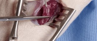
Surgical treatment of pressure ulcers
Surgical treatment of pressure ulcers is determined by the stage and size of the pressure ulcer. Incorrectly performed surgery can only increase the area of the ulcer. Therefore, preliminary assessment of the effectiveness of various surgical interventions in the treatment of pressure ulcers is extremely important. This assessment avoids complications in the majority of patients. Various methods of non-invasive and invasive assessment of the state of blood circulation in the skin under different external pressures are used. One of the simplest and most effective methods is skin pressure plethysmography, which determines the amount of skin blood flow at different pressures. In this case, the skin blood flow sensor can be installed on any area of the skin.
Assessing tissue viability during surgery is a satisfactory method, but lacks the ability to be quantitatively characterized. A more effective method is examination with a Wood lamp 10 minutes after administration of the fluorescein ampoule. Principles of surgical treatment of pressure ulcers:
1) there must be no acute infectious process in general and in the area of the bedsore in particular; 2) inflammation in the area of the bedsore should be minimal; 3) during surgery, the patient should be positioned in such a way that the tension when closing the defect is maximum; 4) all infected areas of the pressure ulcer, necrotic, poorly vascularized and scar tissue should be excised; 5) resection of soft tissues should be minimally sufficient, leaving anatomically important formations; 6) bone resection is used to remove infected areas and reduce skin tension over protruding areas; 7) in the presence of osteomyelitis, after removal of altered bone tissue, it is necessary to use a drainage system with constant washing of the wound with antiseptic solutions; after excision of a pressure ulcer in the presence of a large tissue defect, it should be filled with well-vascularized tissue. For this, it is best to use a muscle flap; 9) when cutting out flaps, it is necessary to remember the maximum preservation of the great vessels and vascular collaterals; 10) skin tension in the area of excision of the bedsore should be minimal; 11) the area of the sutures should not be located above protruding bone formations; 12) in the postoperative period, fluid accumulation under the flap should not be allowed; 13) flap necrosis can be prevented by correct positioning of the patient and using special beds; 14) antibiotics should be used during surgery and in the early postoperative period; 15) stitches must be kept in place until the wound is completely healed - at least 14–15 days.
The question of the sterility of bedsores is controversial. According to A.V. Baskova (2001), all bedsores without exception are infected. Proteus and Staphylococcus aureus are most often cultured from the surface of pressure ulcers. It is more appropriate in this regard to judge not about the infection of ulcers, but about the signs of an acute inflammatory process.
Postoperative complications, their prevention and treatment
Early complications include:
1) accumulation of fluid under the skin flap, 2) failure of the sutures, 3) formation of marginal necrosis of the flap, 4) wound suppuration and bleeding.
By late - the formation of a fistula with the formation of a cavity and relapse of bedsores. The latter are the result of an operation that was insufficiently effective or failed for various reasons. If early complications appear immediately and, as a rule, are eliminated as a result of additional therapeutic measures within 1–2 months, then “late” ones are a continuation of early complications that cannot be treated. Fluid accumulation most often occurs under the displaced flap when there is insufficient outflow of rinsing water or exudate as a result of inadequate drainage of the space under the displaced flap. Flushing the drainage and periodic punctures after removing the drainage lead to the elimination of this accumulation. Bleeding during the closure of pressure ulcers occurs quite rarely. Hemostasis is preferably carried out by electrocoagulation. When ligating blood vessels, only absorbable suture material is used, since the use of non-absorbable material leads to the formation of ligature fistulas. A high risk of further wound suppuration is tamponade of the subflap space with a blood clot. To prevent suppuration, the following are necessary: careful handling of tissues during surgery, careful necrectomy, the use of electrocoagulation for hemostasis and the widespread use of reserve antiseptics both during and after surgery. Suture failure occurs as a result of excessive tension on the edges of the wound. Prevention is based on the use of special sutures that reduce the risk of tissue eruption, sufficient mobilization of the wound edges, resection of bone protrusions in the area of the pressure ulcer and the use of drugs that reduce muscle spasm. Necrosis of the skin flap develops when a pressure ulcer is repaired with a displaced flap as a result of disruption of its blood supply. More often, marginal necrosis occurs to a small extent. Prevention of marginal necrosis of the transferred flap is the use in the postoperative period of drugs that improve microcirculation processes.
The most effective drug that improves tissue trophism and promotes the engraftment of skin flaps and the healing of bedsores is the intravenous administration of Actovegin solution for infusion 250–500 ml (1,000 mg–2,000 mg) or intramuscular injection of 5 ml of Actovegin (200 mg ) within 30 days before and after surgery.
Sulfargin
“Sulfargin” improves the condition of stage 2 and 3 bedsores literally the next day of use. This ointment for the prevention and treatment of wounds contains silver. The active ingredients of the drug destroy bacteria in the wound, relieve pain, swelling, cleanse and heal. Before applying Sulfargin, it is necessary to clean the wound of pus. The product can be used both under a bandage and without. If under a bandage, then treat once a day. And although this ointment for bedsores is expensive, all patients speak very highly of it.
Sulfargin
Tallinn HFZ, Estonia
In adults and children over 1 year of age for the prevention and treatment of purulent wounds and burns with mild exudation, as well as trophic ulcers, bedsores: - when treating fresh burn surfaces to prevent the development of infection;
- for the treatment of wound and burn surfaces in the first stage of the wound process with mild exudation; - for the treatment of wounds and burns of I-IIIA degrees in the second and third stages of the wound process; - for the treatment of trophic ulcers, bedsores, long-term non-healing wounds (including wounds of the stump). from 238
402
- Like
- Write a review
Reviews
Anna
I would like to express my deep gratitude to Alexander for his professionalism, responsiveness and concern for patients and their relatives, which, unfortunately, is not typical of modern doctors. Alexander promptly came to my mother’s home for a consultation on the treatment of a bedsore, which we tried to cope with for 3 months, involving different doctors. Before this, the healing process was very slow with varying success. Alexander arrived with all the necessary sterile instruments and means for treatment, told and showed how to properly care for a bedsore. He even left the tools and processing equipment so that we had the opportunity, without interrupting the treatment, to order and buy everything we needed. After that, if necessary, he adjusted our treatment. Together with Alexander we dealt with this pain! I will definitely recommend this wonderful doctor! Thank you, Alexander!
Yablonin Boris
Dear Alexander Shadzhievich! I would like to express my sincere gratitude to you for your help. It's hard to overestimate her. From the moment of your first dressing, there was hope that something could be done with such a severe wound. A very important point was the dressing technique, building a wound care system, and the thoroughness and systematic implementation of all subsequent actions. Deep professional knowledge of the processes occurring in the wound, which are noticeable only to a doctor with your experience and knowledge, and most importantly the ability to communicate with a sick person. It helped me a lot that you systematically and consistently explained to me every day what processes occur in the wound, how they manifest themselves and how they can occur. Rarely do any doctors devote such filming specifically to transferring their knowledge to those who must continue caring for the wound at the stage of further rehabilitation. Without seeing the dressing technique with your own eyes, without trying to do it yourself, using your technique with new dressing materials, it would be impossible to continue caring for the wound yourself. It is not surprising that with such treatment, even I was able to see and adopt your thoroughness, attention to the smallest changes in the wound, consistency and commitment to all the small details of your technique. Separately, I simply must thank you for the psychological help that you provided to both me and my sick mother, simultaneously with the treatment of the wound itself. This help allowed me to believe in the possibility of recovery and not give up halfway through this very difficult and lengthy process. Your ability to communicate with the patient and his environment calmly, kindly, without false optimism, and very patiently was a huge moral support for me, instilling faith in the possibility of my mother’s recovery. With a feeling of sincere gratitude and appreciation for the assistance provided.
Shamsutdinova Olesya
I, Olesya Shamsutdinova, am eternally grateful to the surgeon Alexander Shadzhievich for a qualified operation (removal of necrosis) on my mother. For his sensitivity, professionalism and desire to endure, understand, hope... because together it is easier to endure sorrow. Low bow to you.
Irina
In the summer of 2021, we were faced with a very unpleasant and scary problem. Due to a long stay in the hospital and the inability to move due to a broken leg, a bedsore developed on the back (in the lumbar region). In the hospital, unfortunately, it was neglected and at the time of discharge it was assessed at grade 4 severity. Friends advised me to see a doctor, surgeon Alexander Shadzhievich Garmaev. And I want to say that we didn’t regret it for a second. Alexander arrived on the same day, performed all the necessary procedures on the wound, and observed him for several days in a row. The doctor showed how to properly treat the wound and apply bandages. The problem was solved quite quickly thanks to well-chosen treatment and modern drugs. Alexander, thank you for your professionalism, for always being in touch and coming when necessary. The psychological support of the patient and family was invaluable.
Boris
Tatiana
Andrey
Dermazin
This ointment effectively destroys almost all bacteria that cause wound infection. At the same time, Dermazin has excellent penetrating ability; it treats bedsores well even with tissue necrosis. It contains silver sulfadiazine, which acts effectively and delicately at the same time. For maximum effect, the ointment is applied under the bandage once or twice a day (just before doing this, you need to thoroughly clean the wound). Sometimes an allergic reaction to Dermazin is possible, but this is rare. Reviews about the drug are mostly good, the cost is quite affordable.
Dermazin
Lek d.d., Slovenia
— treatment and prevention of burn infections (including before autodermoplasty);
- treatment and prevention of infection of trophic ulcers and wounds. from 180
440
- Like
- Write a review
Reasons for education
Poor circulation, continuous compression of soft tissues and blood vessels leads to the occurrence of stagnation in them and their gradual death (necrosis). This complication occurs especially often (7 out of 10 cases) in older patients. This is caused by the fact that in older people the functioning of the circulatory system deteriorates and the process of tissue self-healing slows down.
Timely implementation of preventive measures and competent treatment can prevent the formation of bedsores. If the medical personnel caring for the patient or family members did not pay attention to the first signs of an approaching problem, then spots on the skin can quickly turn into deep festering wounds, and fistulas can form in their place. The development of severe complications can even lead to the death of the patient.
Bedsores are very difficult to treat, the wounds are cleared slowly, they may re-suppurate, and the formation of new necrotic areas. The fact that the patient is still in a supine position also complicates healing.
But tissue necrosis can occur not only in patients who have been on bed rest for a long time. Any prolonged external compression of tissues, even if the body is in normal condition, can lead to the formation of bedsores. Such a complication can be caused, for example, by an incorrectly applied plaster cast, the patient’s constant presence in a wheelchair, impaired tissue nutrition as a result of severe damage to the spinal cord, and so on.
Most often, bedsores form in areas where there are bony protrusions. There is usually no fat layer there, so there is two-way pressure on the skin: from the outside and the inside. Depending on the position of the body, these may be the surfaces of areas such as
- hips, knees, ankles if the patient is lying on his side;
- cheekbones, iliacus and chest bones, pubis, when the patient is forced to lie on his stomach;
- ischial tuberosity, shoulder blades, elbows, sacrum, back of the head, heels when lying on the back;
- shoulder blades, buttocks, ischial tuberosity, foot bones in people who spend a significant part of the time in a wheelchair.
Among the reasons contributing to the formation of bedsores, several main factors can be identified.
- Overdrying of the skin
- Excessive moisture
- Compression of blood vessels and nerve fibers
- No microvibration
- Decreased muscle mass
- Every 1.5 or 2 hours the patient needs to be turned over and changed position so that external influence (pressure) is applied to other parts of the body. If areas with persistent redness of the skin or maceration develop, you should refrain from placing the patient on this area.
- Try to minimize the force of pressure on the surface of the patient’s body from the outside. Currently, online stores and pharmacies offer a wide selection of devices that provide pressure continuity and reduce its value. These include beds with a built-in special mechanism, anti-bedsore mattresses and pillows filled with water, helium, and air.
- The patient’s diet is also of great importance. First of all, it is necessary to provide the body with “building material” for the formation of new cells. Therefore, products offered to bedridden patients should contain sufficient quantities of not only vitamins and microelements, but mainly protein. An insufficient amount of it can lead to disruption of the immune and regenerative functions of the body.
- Strict adherence to all necessary hygiene procedures, especially after urination, defecation, use of medical diapers, absorbent diapers for bedridden patients, and so on. Several times a day, not only in the morning and evening, wipe the entire body with special products or even just wet wipes to prevent sweat from appearing on the skin.
- Antibacterial soap cannot be used to care for bedridden patients, as it “kills” not only harmful but also beneficial microorganisms for the skin. Alcohol-containing mixtures can cause drying of the skin, so they can only be used for oily skin.
- During manipulations, you should not rub the skin too much; it is best to use soft cotton washcloths, and after performing hygiene procedures, do not wipe the body, but gently blot it or let it dry on its own.
- Massage is also a must. To carry it out, you can use a soft terry mitten, which you can buy or make yourself. The red spots that appear cannot be massaged; you only need to treat the intact skin around it.
As a result, the superficial stratum corneum peels off faster, protecting the skin from external influences.
This problem can occur as a result of excessive sweating, urinary and fecal incontinence. An aggressive environment causes skin irritation, it swells, and friction increases. In addition, high humidity promotes the development of infection.
Blood vessels cannot fully perform their functions: provide tissue cells with nutrients and oxygen. Hypoxia (oxygen deficiency), lack of nutrition, deterioration of metabolism in tissues, as a result of the inability of nerve fibers to perform their functions, lead to a decrease in cell viability and their death.
Compression of the lymphatic vessels negatively affects the functioning of the excretory system. Used and dead cells, along with toxins, processed substances, and waste products, accumulate in the intercellular space and tissues. This, in turn, leads to a decrease in immunity. Microbes and viruses freely penetrate the body, inflammatory processes develop, purulent complications, even blood poisoning.
Even when the body is at rest, muscle cells continue to produce energy due to microvibrations of muscle cells. It promotes more efficient flow of biochemical, metabolic, and physiological processes in the body. The level of microvibrations is maintained at a sufficient height due to physical activity and sports. That is why a sedentary lifestyle leads to a deficiency of this energy, and, as a result, to inhibition of the basic functions of the body.
A decrease or complete absence of muscle load, a sedentary (or completely immobile) lifestyle are the causes of atrophy - a decrease in muscle mass, which in turn leads to a reduction in microvibrations.
Taking into account all of the above factors, the formation of bedsores in bedridden patients and disabled people leading a sedentary lifestyle is a very important problem. The formation of such defects occurs very quickly, and, despite the fact that this complication has been known for a long time, there is not yet a sufficiently effective method of treating and preventing the disease that can quickly restore all functions of the body. The search for new modern therapeutic agents continues, the task still remains relevant.
Oflomelid
This ointment for bedsores contains anti-inflammatory, disinfectant and analgesic substances - all this together helps to quickly cope with the problem. Oflomelid usually needs to be applied once a day to reduce the risk of infection of the wound and clean it. Significant relief occurs already in the first days of using the drug, and bedsores can heal completely after two weeks of constant use. Reviews about Oflomelid ointment are only positive, despite its cost (although the tube of the product is large). The only disadvantage of Oflomelid is that the ointment is not available in all pharmacies.
Oflomelid
Novopharm-Biosintez LLC, Ukraine
Infected purulent wounds of various localization and etiology in the first (purulent-necrotic) phase of the wound process, incl.
accompanied by severe pain: - infected burns of II-IV degree; - bedsores; - trophic ulcers; — postoperative and post-traumatic wounds and fistulas; - wounds after opening abscesses, phlegmons, after surgical treatment of abscessed boils, carbuncles, hidradenitis, suppurating atheromas, lipomas. from 93
5.0 2 reviews
218
- Like
- Write a review
How does necrosis develop?
The process of bedsore formation is a gradual deterioration in the condition of those parts of the body that are constantly pressed against the bed. In medicine, the severity of pressure ulcers is assessed in four stages:
- Permanent, persistent redness of the epidermis (outer layer of skin), without damage to the structure. May be accompanied by local hyperthermia, soreness, and mild itching.
- Violation of the integrity of the skin, including the layer of the dermis located between the epidermis and subcutaneous tissue. At the same time, swelling and a subcutaneous bubble (blister) with liquid contents form.
Deepening of tissue damage, with the formation of a non-healing wound in the subcutaneous tissue and a high risk of suppuration. At the same time, the connective tissue membrane and muscle fibers still remain intact.- Global destruction of skin and muscle structures, involving articular and bone tissue in the necrotic process. As the purulent infection progresses, there is a danger of blood poisoning and fatal outcome.
Successful treatment of bedsores in bedridden patients at home largely depends on the severity of the pathological process. In most cases, relatives manage to cope with the problem on their own only at stages 1 and 2 of the disease.