Bedsores are skin ulcers that occur under certain conditions due to poor circulation in places where the skin is poorly supplied with blood due to compression of blood vessels, and there are bony protrusions. There are three main factors of their development: compression, shear and displacement, friction. Typically, areas of ischemia and tissue necrosis appear in patients who are forced to remain in one position for a long time. The main locations of bedsores are:
- when lying on the back - big toes, heels, sacrum, spine, elbows, back of the head;
- when lying on the side - ankle, hips, shoulders, ears;
- when in a wheelchair - shoulder blades, tailbone, buttocks, back of the knee joint and heels.
The development of bedsores can be avoided if you properly care for the patient’s skin using the services of professional nurses at a private boarding house. The Silver Age nursing home was created so that bedridden patients suffering from serious illnesses feel needed and are sure that help is always nearby - 24 hours a day, so that they are not afraid of their illnesses and receive proper care. We try to make the lives of older people more fulfilling and give them back the joy of every day. We are very pleased to see when bedridden patients begin to walk again, and relatives have confidence that their loved one is in good hands.
Through our activities, we try to refute the negative stereotype that has developed in our country about nursing homes and create a positive image when old age and serious illnesses cease to frighten the patient and his relatives.
For each severely bedridden patient, an individual program for the prevention and treatment of bedsores is developed, and a schedule for changing position in bed is developed.
How to treat bedsores
The causes of the appearance and development of bedsores are usually a weakening of the protective properties of the skin. Advanced age and dementia are aggravating and irreversible internal risk factors.
Failure to promptly treat bedsores is a direct threat to life for the following categories of elderly people:
- paralyzed patients;
- people in a coma;
- patients with a hip fracture;
- bedridden patients after a stroke;
- patients with reduced sensitivity in the later stages of dementia;
- patients with incontinence;
- people who are poorly nourished.
Dry skin is more susceptible to the action of irritating factors, which can disrupt the state of balance in it, leading to irritation and the appearance of dangerous phenomena.
Complications
Complications of bedsores (especially pressure ulcers) can be life-threatening and include:
- Phlegmon
Acute purulent inflammation of the skin and soft tissues. People with nerve damage (neuropathy) or a spinal injury often feel no pain in the area affected by inflammation and pressure sores. Cellulitis can cause severe intoxication and even general blood poisoning (sepsis).
- Heel ulcer
A chronic heel wound that does not heal for a long time significantly worsens the patient’s quality of life, as it leads to impaired support and walking function. A trophic heel ulcer can occur as a result of a bedsore, not only in spinal patients, but also in postoperative and post-resuscitation patients due to their prolonged immobilization.
- Damage to bones and joints
Infection from an ulcer can affect joints and bones. Infection in the joint cavity can cause general blood poisoning. Bone infections (osteomyelitis) can lead to destruction of the heel bone.
- Cancer
Wounds that do not heal over time can eventually develop into squamous cell skin cancer.
- Sepsis
General blood poisoning develops with weakened immunity, diabetes mellitus and other severe concomitant diseases and often causes death.
Prevention of bedsores
Bedsores, like any other disease, are easier to prevent than to treat. Our nursing home employs professional caregivers who use the most modern methods of preventing and treating bedsores:
- changing the patient's position every 2 hours during the day and 3.5 hours at night according to the scheme;
- daily hygienic treatment and cosmetic skin care;
- treatment of areas at highest risk of pressure ulcers;
- special massage;
- balanced diet rich in protein and drinking regimen.
When carrying out hygiene procedures for bedridden patients, special products are usually used - gels, foams, wipes, which do not require rinsing with water and allow you to wash the patient in bed.
To prevent the formation and treatment of bedsores, our sanatorium uses cellular-type anti-bedsore mattresses, special splints, pillows and pads. But their use does not reduce the vigilance of caregivers. They prevent the formation of folds on bed linen and underwear, which avoids friction on vulnerable areas of the body. If the patient moves, he should be encouraged to change the points of greatest pressure. In addition, we provide training to older adults in self-help techniques for mobility.
Condition/skin assessment
Each patient with motor activity disorders has known risk factors, which begin to exert their influence under a combination of individual unfavorable circumstances. If the patient has the risk factors listed in the previous chapter, then proceed as follows:
- Inspect and assess the skin condition of a bedridden patient (especially high-risk areas) as often as possible. Do this during grooming procedures - when changing your underwear, doing your morning toilet, or changing diapers.
- Keep your skin dry and clean.
- If the patient complains of pain, a burning sensation or pressure in any area, pay attention to it immediately. This may be the first symptom of a bedsore.
- If redness occurs on the skin, which does not disappear after the pressure is released, then this can be considered the first sign of a bedsore.
- Do not massage the reddened area under any circumstances! It has been found that massaging the injured area can lower the temperature of the skin and lead to tissue degeneration, which in turn can give rise to bedsores.
How to treat bedsores in bedridden patients?
If bedsores have already appeared, then with the help of experienced specialists, treatment of bedsores will be much faster and more comfortable for the patient than at home. Treatment of bedsores in bedridden patients is selected depending on the degree of development of the disease:
- The first stage of bedsores: visible redness of the skin that occurs quickly, sometimes after several hours of sitting still.
- Second stage: persistent redness, superficial abrasion, and the appearance of a bubble filled with liquid.
- Third stage: deep damage to the muscle layer, to the border with the subcutaneous tissues, the appearance of liquid discharge from the wound.
- Stage four: necrosis, damage reaches the fascia of the bone, cavity formation.
- Fifth stage: a state of general infection, sepsis, necrosis spreads towards the fascia and muscles.
Treatment of bedsores at stages 1 and 2 is treated conservatively using special means, at stages 3, 4 and 5 - only surgically.
Degrees and symptoms of bedsores
The first subjective sensations that a patient may experience: • Tingling on the skin where a bedsore is likely to develop. This is due to stagnation of blood and lymph that feed the nerve endings; • Numbness (loss of sensation). It occurs on the affected area of the body after about 3 hours. Visible signs of a bedsore: • Stagnation of peripheral blood and lymph. Initially it appears in the form of venous erythema, blue-red in color, and has no clear boundaries. Localization sites are areas of bone and muscle protrusions in contact with the bed. The intensity of skin coloring gradually increases; • Desquamation of the epidermis with or without preliminary formation of vesicles with pus. These are the main signs of a developing bedsore. It is urgent to take measures to prevent further deterioration of the pathology. The development of bedsores occurs in 4 stages, which are accompanied by different symptoms. Stage I – initial. Its signs are as follows: • The skin is not damaged; • There is redness (light skin). The skin does not change color when pressed; • Dark skin color often does not change. The skin does not turn white when pressed. Sometimes appears irritated, cyanotic or purple; • The affected area of the skin may be sensitive, painful, softer, cooler/warmer compared to other areas. Stage II – The bedsore turns into an open wound. Signs: • The epidermis, part of the dermis, is either damaged or missing; • Bedsore – a swollen, red-pink sore (similar to an ulcer); • May have the appearance of an intact or burst bubble of fluid. Stage III – deep wound. Appearance: • Necrosis, as a rule, reaches not only the skin, but also adipose tissue; • The ulcer becomes like a crater; • The bottom of the wound may have dead, yellowish tissue; • Damage may spread between layers of still healthy dermis. Stage IV – extensive tissue necrosis. Appearance: • Tendons, muscles, bones may protrude into the wound; • The bottom of the wound is dark, hard dead tissue; • The lesion extends far beyond the lesion.
How to recognize bedsores?
Patients who predominantly lie on their backs complain of the formation of bedsores in the area of the shoulder blades and sacrum. Also, areas damaged by necrosis are found on the back of the head and heels. When lying on your side, the following areas may be affected:
- hips;
- ears;
- whiskey;
- shoulders;
- knees;
- ankle
Symptoms of necrosis:
- formation of red or blue edema;
- increased sensitivity in the affected area and pain;
- blisters, which, when opened, provide an overview of superficial wounds;
- formation of a crater with purulent discharge.
Also, yellowish areas of necrosis form at the edges of the wound, which gradually acquire a dark tint.
Risk category and place of formation
Bedsores occur in people who are permanently bedridden or use a wheelchair. Pathology occurs due to constant pressure on individual areas of soft tissue, which leads to disruption of local blood circulation and their gradual destruction.
The points of formation of bedsores are associated with the patient’s usual body position:
- When lying on your back, the area of the sacrum, heels, and elbows is affected. The occipital and scapular region is affected.
- Positioning on the side leads to injuries in the area of the femur, greater trochanter, and mastoid process. The area of the temporal bone is involved in the process.
- A sitting position causes compression of the tissues in the feet and shoulder blades. The ischial tuberosities are affected.
The risk of necrosis formation and the stage of pressure ulcers are determined using the Waterlow scale. The calculations take into account the general condition of the patient.
The appearance of redness, itching and pain with an unpleasant odor in places where soft tissues come into contact with hard surfaces indicates an infection. The patient's relatives should consult a doctor. Professional help is necessary if bleeding or foreign fluid appears from a bedsore.
Reasons for appearance
Key reasons for the formation of bedsores:
- absence of a change in position, which causes the development of necrotic processes in the epidermal layers;
- the use of rough underwear, the folds of which injure the skin;
- poor quality patient care;
- sudden change in weight;
- the presence of concomitant chronic diseases, due to which the functioning of blood vessels is disrupted or hormonal disruptions occur in the body.
Knowing the reasons for the formation of bedsores can reduce the risk of wound formation and prevent the spread of the disease.
Condition assessment
The first step is to get an overview of the extent of the damage. To do this, you need to free the damaged area from pressure and clothing. Examine the patient's skin in a well-lit area. Test the damaged area with your hand. If the area turns red and the redness does not disappear after the pressure is released, then most likely we are dealing with an incipient bedsore. You can conduct a test near the damaged area: press your finger on healthy skin - after removing your fingers, a lighter area appears on the skin, which over time will again become the same as the rest of the skin. If the blood supply is disrupted in the area being examined, then such a reaction does not occur. If the skin in the damaged area is still healthy, then there is no need to cover it with anything. If the skin is too dry or if there is a danger that the skin will come into contact with urine, then it is enough to just moisturize the skin. If only one area of injury has been identified, move the patient to a position where the reddened area remains free of pressure. If the skin begins to crack, you should immediately contact a specialist, such as a family nurse or home health nurse, or, if necessary, a family doctor. The choice of bedsore care products is very wide and to make the right choice you need the help of a specialist. When choosing a care product, you need to take into account the stages of development of a pressure ulcer (size, depth, presence of dead tissue, signs of infection, amount of discharge), its location, frequency of changing ulcer care products, and patient preferences. If the caregiver does not have special training in caring for pressure ulcers, it is not recommended to carry out the care yourself. Ask for advice and help from a family nurse and home health nurse! If a bedsore has already appeared, then it can be cured as soon as possible in 2 weeks. If a bedsore does not go away, you should definitely contact a specialist to evaluate the correctness of treatment methods.
Stages of bedsores
Depending on the degree of necrosis, the method of treating the patient is selected. Sometimes it is enough to treat the wound with a disinfectant solution, which can be done at home, but sometimes surgical intervention is required.
Prevention and treatment of stage 1 bedsores
More often, this degree of the disease is treated quickly and without any problems. The disadvantage of pathology of this degree is mild symptoms, which makes it impossible to start treatment in a timely manner and prevent the development of the disease. The main symptoms of stage 1 bedsores:
- change in the shade of the affected area;
- skin irritation;
- pain at the site of injury.
To treat necrosis, it is recommended to examine the patient to identify the listed changes. Only in this case will it be possible to prevent the development of ulcerations.
Prevention and treatment of stage 1 bedsores are the same. Therapy involves:
- treating the affected area with camphor alcohol or other solutions;
- removing secretions and drying the skin surface;
- treating the affected area with ointments that accelerate blood flow;
- cold and hot shower.
Doctors recommend combining treatment with the use of polyurethane film. The patch will not be able to cope with the problem. It is advisable to use it only to prevent microdamage.
Prevention and treatment of bedsores Stage 2, non-infected wound
At this stage, the skin damage resembles an ulcer or small wound. Feature: swollen edges. If a person is conscious, then he can talk about the following symptoms of the second stage of bedsores:
- pain at the site of skin lesions;
- burning;
- itching.
Patients with stage 2 bedsores try to avoid contact of the wound with bedding, and also complain about uncomfortable clothing.
Treatment of bedsores in the form of small wounds should be carried out using medications recommended by a doctor. To prevent the development of the disease, it is recommended to clean the wound from effusion and necrotic cells, and also treat the affected area with an antiseptic.
Additionally, doctors recommend applying antiseptic bandages and regularly inspecting the area for inflammation.
If a wound becomes infected during treatment, it is necessary to add treatment with an antibacterial drug.
Prevention and treatment of bedsores Stage 3, non-infected wound
The damaged area looks like this:
- necrosis affects all layers of the epidermis;
- on the skin the wound resembles a crater with red, swollen edges;
- Clumps of dead yellow tissue form on the bottom and walls of the damaged area.
At this stage, the nerve processes die off, which reduces the level of pain. Treatment is carried out in the same way as in the case of the second stage of necrosis. The only caveat is that more thorough treatment of the wound and cleansing it of dead cells is required.
Prevention and treatment of bedsores Stage 4, non-infected wound
Such bedsores are typical for patients with weak immunity. Symptoms of the disease:
- great depth of the wound;
- darkened crater floor;
- pale or reddened skin around the wound.
In the most severe cases, when it comes to extensive necrosis, large areas of dead tissue form. It is noteworthy that grade 4 necrosis does not bring discomfort or pain to the patient. This is explained by the destruction of nerve endings.
In this case, treatment is carried out under the supervision of a surgeon. It is also necessary to organize care for bedridden patients with bedsores. Before using antiseptics, it is recommended to promptly excise necrotic tissue to prevent the development of infection and speed up the regeneration process. During the treatment process they use:
- necrolytics;
- tissue repair stimulators;
- stimulants that accelerate blood flow;
- anti-inflammatory drugs.
Dead skin areas are removed through surgery.
Prevention and treatment of bedsores Stage 4, infected wound
Accompanied by an unpleasant odor and the formation of purulent contents. In this case, serious medical attention is required. In this case, urgent surgery is necessary to remove the affected cells.
Definition
Bedsore
(ulcer, pressure injury, pressure ulcer, decubitus ulcer, bedsore) – local damage to the skin and (or) underlying tissues. Bedsores are formed as a result of pressure or a combination of pressure with other damaging factors (friction, shear, humidity and others). They are located, as a rule, over bony prominences (sacrum, heels, ankles, fingers, elbows, sit bones, spinous processes of the spine, coccyx, hip joints and the back of the head).
Weakened bedridden and sedentary patients with impaired blood circulation, sensitivity (strokes, spinal cord injuries) and concomitant diseases most often suffer from this problem. A significant role in the occurrence and development of local damage to the skin and underlying tissues is played by improper care, urinary and fecal incontinence. Bedsores are a very unpleasant, dangerous and expensive complication that worsens the quality of life, the patient’s condition and prognosis, slows down the treatment process, and requires additional care, medical supervision and expenses. This complication must be kept under control.
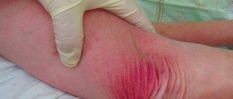
Bedsore photo
Risk factors for pressure ulcer formation
Risk factors include the patient's high or very low weight, immobility, and poor care. But there are other additional points that are important to consider. There is also a risk of bedsores in people who have a low pain threshold or whose body reacts less well to pain due to painkillers. In such a situation, the prevention of bedsores consists of constantly inspecting the body, without waiting for the patient to complain of discomfort.
Another risk of bedsores is postoperative and other pain, which even forces a person with good mobility to constantly be in one position. Most often in such cases, the problem is detected at the first stage of bedsores, so it can still be corrected with less effort.
An additional problem may be taking medications that disrupt the water-salt balance in the body. If the instructions contain such data, the formation of bedsores can be avoided through careful prevention and constant monitoring of the situation.
