Anatomical features
According to its structure, the ankle joint is a trochlear joint: between a kind of “fork” of two ankles, a block of the articular surface of the talus is inserted. Stability is provided by numerous ligaments: they are arranged in several layers, powerful and durable.
Fig.1. Ankle structure
Freedom of movement is somewhat limited: the joint allows the foot to flex and extend with good amplitude only in one plane.
The ability to move the foot to the right and left is small, but the supporting ability allows it to cope with loads reaching ten times the body weight. True, sometimes even such a safety margin is not enough.
Diagnostics
The doctor will discuss with the patient the medical history (how long the pain has been, what happened before the injury) and symptoms. He will also ask how the injury occurred and examine the affected area. If your trauma surgeon thinks you may have broken your ankle, he or she will order a series of tests to more fully understand the injury.
X-rays : X-rays can show whether the ankle bone has been broken and how many pieces of broken bone are there. They can also determine if there is displacement (a gap between broken bones). The doctor may also take X-rays of other parts of the leg or foot to make sure nothing was damaged as a result of the injury.
Stress test : A stress test is performed to determine whether surgical procedures are necessary to heal the injury. The doctor will apply some pressure to the ankle and take a special x-ray to determine the severity of the injury.
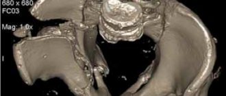
CT scan (computed tomography): If the fracture extends beyond the ankle, a CT scan may be needed to further examine the injury. A CT scan allows you to obtain a series of images in different planes, from which the doctor can determine the severity of the injury. Source: Comprehensive diagnosis of ankle joint injuries. Kim L.I., Dyachkova G.V. Genius of Orthopedics, 2013. p.20-24.
MRI (Magnetic Resonance Imaging) Scan: If your doctor suspects ligament damage has occurred, he or she may order an MRI scan to get a better look at the affected area. MRI can look deeper into bones, soft tissues, and ligaments to create higher-resolution images than most other tests.
Mechanism of injury
Fractures of the ankle joints occur under the influence of multidirectional forces:
- Axial force perpendicular to the sole (causing wedging of the talus between the ankles).
- Excessive bending (flexion) and extension (extension).
- Turning inward (supination) and turning outward (pronation).
- Rotation of the foot inward (inversion) and outward (eversion).
If a sufficiently large amount of energy is applied in any of these directions, the joint structure may fail and break.
Stages of fusion
At the moment of fracture, local hemorrhage occurs, and for the first five or seven days there is an inflammatory process with the formation of a soft compaction of fibrous tissue (resorption). Then the creation of collagen connecting threads (reversion) from special cells - osteoclasts and osteoblasts - begins. After this, as a result of cell mineralization, a callus is formed between the fragments within a month. Over the next three to four weeks, the formed structure ossifies, due to its saturation with calcium.
Complete restoration of the damaged bone and its surroundings, which ensures the full functioning of the ankle joint, is possible after 4-6 months of rehabilitation.
Varieties and types
The classification of ankle fractures is quite complex. Separately classified:
- Fractures of the fibula (outer malleolus).
- Tibial (inner malleolus).
- Damage to the tibiofibular syndesmosis.
- Talus.
Each of these categories has sub-items. In case of injuries, they are combined and rarely occur in isolation. Knowing all the nuances of each type of fracture and understanding the mechanism of injury allows you to eliminate the damage as completely and effectively as possible.
Usually, ankles break off: external (lateral, - the process of the fibula) or internal (medial - part of the tibia). Both can break at once.
Rice. 2. a. displaced fracture of both ankles; b. fracture of the ankles and posterior edge of the tibia.
The peculiarity of such fractures is that sometimes the ankles seem to be torn off from the bones of the leg: when the leg is twisted, strong ligaments may not break, but pull the ankle along with them, tearing it off completely, or causing a breakage and displacement of a small fragment of the bone.
Severe high-energy injuries may not be limited to the ankles and are accompanied by fracture of the articular surface of the tibia.
The talus fractures noticeably less frequently. But if it is damaged, intra-articular fragments may occur. They do not always produce pronounced symptoms and often end in the development of false joints.
We have to deal with fracture-dislocations: when one or both ankles of the leg are broken off, they lose the ability to fix the block, the talus dislocates and shifts, complicating treatment.
Fig3. A. fracture of the outer malleolus, rupture of the syndesmosis and deltoid ligament, subluxation of the foot outward. b. fracture of the talus.
Such injuries are dangerous due to disruption of the blood supply to bone tissue, which causes aseptic necrosis (talus bone) and greatly complicates the work of doctors.
A frequent complication is rupture of the external and deep ligaments and displacement of bone fragments.
Injury
A common bruise is the result of a foot hitting a hard object. In this case, as a rule, serious destruction of body tissues is not observed. Bruises of the lower leg and ankles are very painful, since the bones in this area are devoid of protective fatty cover. Recovery from a minor injury is often quick. Extensive damage can cause massive swelling, hematomas, or even injuries to blood vessels and nerves. However, more often than not, a “regular” bruise will involve only moderate pain and minor swelling.
Symptoms
Certain aspects of external manifestations depend on the type and extent of injury. Attention should be paid to the following symptoms:
- Pain in the injured joint.
- Swelling in one or both ankles.
- Atypical foot orientation.
- Joint deformity.
The tricky part is the risk of low-symptomatic variants. You may encounter a break-off of a small fragment of bone, which does not lead to gross external manifestations, which means there is a delay in seeking medical attention after a fracture and, accordingly, treatment.
Dietary recommendations
Recovery after a fracture is a long process, which can be accelerated by organizing proper nutrition. The diet includes calcium-containing foods:
- poppy;
- sesame;
- various nuts;
- dairy and fermented milk products;
- legumes (beans, soybeans);
- different types of fish;
- green apples and vegetables (white cabbage, Peking cabbage, etc.);
- jellied meat (eating this dish will speed up the restoration of cartilage tissue).
Ground eggshells, which are highly digestible, perfectly compensate for calcium deficiency. It can be added to dishes or consumed in its natural form.
The balance of vitamins and microelements will be maintained in optimal condition by a mixture prepared from a glass of raisins, walnuts, honey, dried apricots and 2 lemons. Thoroughly washed raisins and dried apricots are briefly soaked in water, after which they are rolled through a meat grinder along with the nuts. Next, the mixture is mixed with honey and lemon juice. The tasty medicine should be taken 3 teaspoons three times a day.
Diagnostic measures
Questioning and inspection cannot provide the necessary information. The key role is played by x-ray methods. As a rule, a radiograph of the injured limb is required in two projections.
Moreover, for better visualization of individual parts of the joint, different techniques are used (displacement, rotation, tilt of the foot). When necessary, a photo of the healthy leg is taken for comparison. There may be a need for CT or MRI.
Fractures of the talus require a more careful choice of diagnostic technique, as they are more complex in terms of treatment.
Fig4. A. CT scan shows a fracture of the talus and tibia, b. MRI revealed a fracture of the talus
Therapeutic measures
Uncomplicated ankle fractures without significant displacement allow conservative treatment. Manual closed reduction is performed, the details of which depend on the type of injury.
The basic principles are:
- Carrying out the most accurate reposition before applying a plaster immobilization bandage.
- Rehabilitation after removal of the cast, restoration of movement and joint function.
After this, a plaster-based fabric bandage is applied: immobilization is necessary, its duration depends on the degree of the defect and ranges from 4 weeks.
We will help you restore bone integrity, joint function and return to your usual active lifestyle. We have an individual approach to each patient.
Possible complications
If you visit a doctor late, self-medicate, or violate the rules and timing of wearing a fixation device, the bones and their fragments can grow together in an unnatural position, which will interfere with the normal functioning of the joint and provoke dislocations and the development of flat feet.
An improperly formed callus can impinge on nerve fibers and impede or block the innervation of the adductor muscles of the foot and the sensitivity of the skin. Untimely treatment of a postoperative wound can cause the development of an inflammatory process or infectious disease of muscle tissue, bones and blood vessels.
Surgical measures
If it is obvious that closed reduction will not be effective, plaster is not used: they resort to open osteosynthesis surgery and installation of metal structures directly on the bone tissue.
Violations of the integrity of the talus are preferable to be treated surgically due to the risk of aseptic necrosis. Reduction and reposition of her fracture-dislocations must be carried out by repeating the mechanism of injury in the opposite direction.
Fig.5 a. osteosynthesis of the talus and calcaneus with screws, b. fracture of both ankles; osteosynthesis with plate and screws.
First aid for a fracture of the outer ankle
If the ankle is broken, the victim must be taken to the emergency room. It's better to call an ambulance. When this is not possible, you will have to organize a stretcher from available materials and deliver the victim to the nearest medical facility.
Before the doctors arrive, the patient needs to be provided with pre-medical care:
- Free the leg from traumatic factors, of course, if this is possible without additional damage to the limb.
- Elevate the affected leg. To do this, you can make a roller out of clothes.
- If bleeding occurs, apply ice or something cold, if possible - a hemostatic tourniquet, which should be loosened for 20 seconds, every 20 minutes.
- If the integrity of the skin is damaged, under no circumstances should you try to set bone fragments.
- If the pain is severe, give the victim an analgesic.
- When there is no specialized splint for immobilization, and the patient needs to be taken to a medical facility independently, you need to apply a splint from improvised means.
Gently bend the injured limb at the knee joint and position the foot so that the heel is at a right angle to the shin. Apply an improvised splint and secure it with a bandage, belt, belt, etc.
Consequences
If you do not consult a doctor in time, even a simple ankle fracture can complicate your future life.
A common consequence of delayed treatment is ankle pseudarthrosis, fusion in the wrong position, post-traumatic arthrosis.
Insufficiently thought-out treatment leads to impaired ankle stability and deformation. In old cases, eliminating such consequences is not easy.
Gross defects are much more dangerous:
- Persistent impairment of the supporting function of the foot.
- Due to blood supply disorders, aseptic necrosis of bones occurs.
- Infectious complications in the affected area.
The loss of physiological gait affects the entire human skeleton, primarily the spine. In some cases, ankle replacement may be required. .
Causes
Traumatic ankle fractures occur from a strong blow or other excessive external force on the ankle during sports, falls, or traffic accidents. Twisting your foot on a slippery, uneven surface or wearing ill-fitting shoes often leads to this type of injury. Unsuccessful falls can be caused by underdeveloped muscles and poor coordination of movements, especially if you are overweight. Due to disruptions in the normal process of bone tissue restoration, adolescents, pregnant women and older people are at risk.
Congenital or acquired degenerative changes, as well as various diseases: arthritis, osteopathy, osteoporosis, tuberculosis, oncology increase the likelihood of injury. An unbalanced diet, lack of calcium and other microelements reduce the strength of bones and the elasticity of ligaments.