Publications in the media
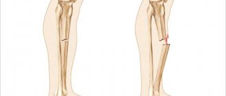
Pelvic fractures account for 4–7% of all fractures. Classification • Marginal fracture: fractures of the iliac spines, ischial tuberosities, coccyx, transverse fracture of the sacrum below the sacroiliac joint, ilium • Fracture of the pelvic ring without breaking its continuity •• Unilateral or bilateral fracture of the same branch of the pubic bone •• Unilateral or bilateral fracture of the ischium •• Fracture of one branch of the pubis on one side and the ischium on the other • Injuries with disruption of the continuity of the pelvic ring •• Vertical fracture of the sacrum or fracture of the lateral mass of the sacrum •• Rupture of the sacroiliac joint •• Vertical fracture ilium •• Fracture of both branches of the pubic bone on one or both sides •• Fracture of the pubis and ischium on one or both sides (butterfly type fracture) •• Rupture of the symphysis • Damage with simultaneous disruption of the continuity of the anterior and posterior half rings (Malgenia type ). and posterior on the other •• Dislocation of the innominate bone - rupture of the sacroiliac joint and symphysis •• Combination of rupture of the symphysis with a fracture of the posterior semi-ring or combination of rupture of the sacroiliac joint with a fracture of the anterior semi-ring of the pelvis • Fracture of the acetabulum •• Fracture of the edge of the acetabulum; may be accompanied by posterosuperior hip dislocation •• Fracture of the floor of the acetabulum; may be accompanied by a central dislocation of the hip - a displacement of its head inward towards the pelvic cavity.
Fracture of the marginal pelvic bones • Causes: direct trauma, short-term compression of the pelvis, sudden muscle contraction. • Detachment of the anterosuperior spine •• Clinical picture: pain, swelling, displacement of the fragment downwards and outwards, which creates the impression of a shortened limb; Lozinski's symptom of reverse movement - the appearance of sharp pain when flexing the hip while taking a step forward (moving the leg back causes less pain); the patient walks backwards •• Treatment: pain relief, the leg is placed on a Beler splint in a position of slight abduction for 3 weeks. Sometimes osteosynthesis is used. • Duvernay's fracture is a fracture of the ilium and the upper part of the acetabulum •• Clinical picture: pain in the area of the iliac wing, aggravated by tension of the abdominal muscles and percussion, limitation of movements in the hip joint. When the wing is displaced upward, a shortening of the distance from the xiphoid process to the anterosuperior spine is detected •• Treatment: pain relief - intrapelvic blockade according to Shkolnikov-Selivanov, Beler splint, exercise therapy, physiotherapy; if there is significant displacement, the patient is placed in a hammock with compression of the lateral surfaces of the pelvis for 4 weeks. • Transverse fracture of the sacrum and coccyx •• Clinical picture : pain, aggravated in a sitting position, pressing on the distal part of the sacrum during rectal examination, difficulty in defecation, swelling in the area of the sacrum (coccyx), pathological mobility of the distal fragment of the coccyx (sacrum). If the sacral nerves are damaged, urinary incontinence and anesthesia of the buttock area develop •• Treatment: pain relief, the patient is placed in bed on a backboard (a wide cushion is placed under the lower back so that the sacrum does not touch the bed) or in a hammock (the hammock is placed under the back from the angle of the shoulder blades to fracture area so that the distal fragment of the sacrum does not touch the bed) for 3–5 weeks, belladonna suppositories, warm enemas, physiotherapy; if pain persists, presacral blockades and physical therapy are repeated; if conservative treatment is ineffective, the fragment is removed.
Fracture of the pelvic ring • Fracture of the pelvic ring without breaking its continuity •• Causes : direct trauma, compression of the pelvis in the anteroposterior direction (fracture of the ischium) or load on the greater trochanter (fracture of the pubis) •• Clinical picture : pain in the pubic area (with a fracture pubic bones) or in the perineum (with a fracture of the ischial bones), aggravated by palpation, leg movements, compression of the pelvis from the sides; sometimes - a symptom of a stuck heel •• Treatment: anesthesia, the patient is placed on a hard bed with a shield; for a unilateral fracture - a Beler splint in the position of abduction of the leg; for bilateral fractures - the “frog” position. Bed rest - 4-5 weeks. • Fracture of the pelvic bones with disruption of the continuity of the pelvic ring •• Frequency : up to 50% of all pelvic injuries •• Complications : shock, damage to the pelvic organs (rectum, bladder, urethra) •• Causes: indirect injury - anteroposterior or lateral compression of the pelvis , fall from a height, birth injury (rupture of the symphysis) •• Damage to the anterior semi-ring of the pelvis ••• Clinical picture: pain in the pelvis and perineum, aggravated by leg movement, anteroposterior and lateral compression, an attempt to separate the iliac bones; Volkovich's symptom - with a fracture of the upper branch of the pubic and ischial bones, the patient is in a frog position; with a fracture near the symphysis and its rupture, the thighs are brought together and slightly bent, an attempt to separate them causes sharp pain; stuck heel symptom; when the symphysis is torn, the space between the bones is sometimes palpable ••• X-ray examination. It must be remembered that the width of the symphysis pubis at 18 years of age is 6 mm, later it decreases to 2 mm ••• Treatment: fractures without displacement - Volkovich position for 5-6 weeks, from the end of 1 week - exercise therapy, physiotherapy; fracture of the pubic and ischial bones on both sides (butterfly-type fracture) with displacement - Volkovich position; when the X-shaped fragment is displaced upward, additional pillows are placed under the back to bring together the attachment points of the rectus abdominis muscles; if ineffective, skeletal traction with a load of 4–5 kg; symphysis rupture - treatment on a hammock with cross traction; osteosynthesis is used when conservative treatment is unsuccessful; in the postoperative period - treatment on a hammock with cross traction for 6 weeks •• Damage to the posterior semi-ring ••• Clinical picture: the patient lies on the healthy side, active movements of the leg on the injured side are limited, painful, pain increases with palpation; if the sacroiliac joint is ruptured, the posteriorly displaced edge of the ilium is palpated ••• Treatment: the patient is placed on a backboard in a hammock without cross traction for 8–9 weeks; for displaced fractures - skeletal traction; if it is impossible to reposition a rupture of the sacroiliac joint with displacement, arthrodesis of the sacroiliac joint is used •• Malgenya fracture - simultaneous disruption of the continuity of the anterior and posterior half rings ••• Causes: compression of the pelvis, fall from a height ••• Clinical picture: pain, dysfunction of the lower limbs, bruises in the area of the scrotum, perineum and inguinal ligament, asymmetry of the pelvis, upward displacement of one of its halves by 2–3 cm - a decrease in the distance from the xiphoid process to the anterosuperior spine; with lateral compression or an attempt to separate the halves of the pelvis, pathological mobility is revealed ••• Treatment. Antishock therapy - intrapelvic anesthesia, compensation of blood loss; for a non-displaced fracture - treatment on a hammock, skeletal traction with a load of 4-5 kg on each leg for 8 weeks; when half of the pelvis is displaced upward and inward on the side of displacement, the load of skeletal traction is increased to 10–14 kg, traction is carried out in the abduction position for 8–10 weeks; the hammock is used only after reposition; in case of a bilateral vertical fracture with upward and inward displacement - skeletal traction with a load of 10–14 kg for both legs in the abduction position for 8–10 weeks; in case of symphysis rupture - treatment on a hammock with cross traction after reposition for 10–12 weeks.
Fracture of the acetabulum • Causes : lateral compression of the pelvis in the area of the greater trochanters, load on the greater trochanter • Clinical picture: pain in the hip joint and dysfunction (pain intensifies with axial load and tapping on the thigh); with combined dislocation, the apex of the greater trochanter is above the Roser–Nelaton line, the limb is adducted, slightly bent and internally rotated; with central dislocation of the hip, the greater trochanter falls • Treatment •• Anesthesia: intrapelvic blockade according to Shkolnikov-Selivanov •• Fracture without displacement - skeletal traction with a load of up to 5-7 kg, if closed reduction is unsuccessful - osteosynthesis of the broken edge of the acetabulum •• For fractures of the acetabular floor depressions accompanied by central dislocation of the hip, skeletal traction along the axis of the hip and beyond the greater trochanter or subtrochanteric region, transosseous compression-distraction osteosynthesis; if closed reduction is ineffective, open reduction of the central dislocation of the hip and osteosynthesis of fragments of the acetabulum.
ICD-10 • S32 Fracture of the lumbosacral spine and pelvic bones
Mechanogenesis of rotationally unstable fractures and injuries of the pelvic ring
R.Ya. KHABIBYANOV
Republican Clinical Hospital of the Ministry of Health of the Republic of Tatarstan, 420064, Kazan, Orenburgsky tract, 138
Khabibyanov Ravil Yarhamovich - Candidate of Medical Sciences, Head of the Research Department, tel. (843) 237-34-46, e-mail
The article presents the mechanogenesis of rotationally unstable fractures and injuries of the pelvic ring with anatomical determination of the nature of displacements during disintegration in the sacroiliac joints.
Key words: unstable fractures and injuries, pelvic ring, disintegration.
R.Ya. KHABIBYANOV
Republican Clinical Hospital of the Ministry of Healthcare of the Republic of Tatarstan, 138 Orenburgskiy Trakt, Kazan, Russian Federation 420064
Mechanogenesis of rotation-unstable fractures and lesions of pelvic bones
Khabibyanov R.Ya. — Cand. Med. Sc., Head of Scientific-Research Department, tel., e-mail: [email protected]
The article presents the mechanogenesis of rotation-unstable fractions and lesions of pelvic bones with the anatomic determination of dislocations in case of disintegration in the sacroiliac symphyses.
Key words: unstable fractions and lesions, pelvic bones, disintegration.
Most often, unstable fractures and injuries of the pelvic ring occur as a result of road traffic accidents, industrial accidents, and falls from a height [1-4]. Under direct influence of an external agent, the nature of fractures and displacements and their combination can be varied (Fig. 1).
Picture 1.
Polyfocal fracture of the pelvic bones. Altitude injury
Treatment of unstable pelvic fractures (types B and C - classification according to the accepted international system AO/ASIF) with rotational and rotational-vertical instability, especially with significant displacements or in not fresh cases, is difficult. If you do not take into account the patient’s initial condition (stable or unstable), the difficulties lie in performing preliminary and final reposition with subsequent reliable stabilization of the pelvic ring [5].
The traditionally accepted term “rotational instability” of the pelvis has an extremely limited relationship to rotation (Rotation [lat. rotatio circular rotational movement] - in functional anatomy - circular movement, for example, in the shoulder, intervertebral, etc. joints). Low-amplitude passive rotational movements of a sliding nature in the SIJ normally occur around the frontal axis below the second sacral segment S-2 [6] and are related to a change in the physiological positions of the sacrum from counternutation to nutation. In case of fractures and injuries of the pelvic ring with rotational instability, the hemipelvis is displaced relative to the sacrum in the lateral or contralateral direction in the horizontal plane. According to our observations, injuries to the pelvic ring with rotational instability and lateral displacement (Fig. 2) account for 84% of injuries to the pelvic ring with rotational instability. Moreover, rupture of the symphysis with discrepancy and damage to the SIJ in 96% of cases is the result of an acceleration injury (accident) with an indirect mechanism of action. In this case, the victims, as a rule, are passengers of vehicles. Their position at the time of the collision is sitting, the hips are moderately apart; in a frontal collision of a car, by inertia, both knee joints symmetrically or asymmetrically rest against an obstacle in the form of a car panel or the backs of the seats in front. Other cases include a fall from a small height, including the height of one’s own height, soil collapses in trenches, a motorcycle injury when colliding with an obstacle, a symphysis discrepancy of more than 1.5-2 cm during childbirth, etc.
Taking into account the articular surfaces of the SIJ that converge downward, inward and posteriorly (Fig. 3, 4), with rotational instability with lateral displacement, a more or less uneven sliding of the articular surface of the ilium occurs posteriorly relative to the auricular surface of the sacrum. The lumboiliac ligaments provide a lesser degree of sliding of the upper parts of the articular surface of the ilium relative to the auricular surface of the sacrum posteriorly. Slipping of the lower parts of the articular surface of the iliac
Figure 2.
Rotational unstable injury to the pelvic ring with lateral displacement
bone growth occurs to a greater extent in accordance with the posteriorly converging articular surfaces of the sacroiliac joint (SIJ). Moreover, slipping in the lower parts of the SIJ is caused by the influence of the psoas muscle on the anterior pelvis. Moreover, the amount of posterior displacement depends on the degree of damage to the anterior and interosseous ligaments of the SIJ. The greater the degree of posterior displacement of the articular surface of the ilium in the SIJ, the greater the discrepancy in the anterior semi-ring of the pelvis. In case of bilateral damage to the SIJ with lateral displacement of the hemipelvis, these displacements have nothing to do with the position of the sacrum - nutation (movement of the sacrum, similar to nodding the head), in which tensile forces affect the symphysis. Disintegration of the posterior sections of the pelvis leads to the fact that, under the influence of the lumbar muscles, there is an increase in the angle of inclination of the pelvis anteriorly, which in itself normally should lead to a relative verticalization of the sacrum - counternutation, which ensures stable balance of the pelvic ring.
Figure 3.
R CT scan of the pelvis of patient B. after osteosynthesis of a transacetabular fracture with transition to the body and wing of the ilium, frontal section at level S -2
Figure 4.
R CT scan of the pelvis of patient B. after osteosynthesis of a transacetabular fracture with transition to the body and wing of the ilium, horizontal section at level S -2
In case of rotationally unstable fractures and injuries of the pelvic ring with lateral displacement, displacements occur in the frontal, sagittal and horizontal planes.
Rotational-unstable injuries with contralateral displacement have a different pattern of destruction. The SIJ ligaments generally remain intact. Fractures of the pubic and ischial bones occur on one or both sides, as well as a marginal fracture of the Ala Sacralis, related to the SIJ. Moreover, the nature of marginal fractures of Ala Sacralis with contralateral displacement of the hemipelvis depends on the position of the sacrum at the time of exposure to the external agent. A marginal fracture at the level of S1-S2 vertebrae (Fig. 5a, c) occurs when the sacrum is in counternutation or close to it, since it is in this zone that there is close contact and emphasis of the anterior edge of the sacropelvic surface of the ilium with Ala sacralis (Fig. 6).
A marginal fracture at the level of S2-S3 vertebrae (Fig. 7a, c) occurs when the sacrum is in nutation or close to it.
When the sacrum is nutated, the upper parts of its auricular surface, corresponding to the articular surfaces of the SIJ converging backward, slide anteriorly. In this case, the interosseous sacroiliac ligament twists, which leads to migration of the axis of rotation into the SIJ, and unevenly “pulls” back the auricular surface of the ilium in accordance with the articular surfaces converging backwards. The lower parts of the auricular surface of the sacrum slide posteriorly to a lesser extent. A decrease in the anterior tilt angle of the pelvis leads to contact of the lower parts of the auricular surface of the sacrum with the anterior lower parts of the sacropelvic surface of the ilium (Fig. 8).
Figure 5.
Marginal fracture of Ala sacralis at the level of S 1- S 2 vertebrae
Figure 6.
Pelvic ring. The position of the sacrum is counternutation. The boundary line of the pelvis is continuous
When exposed to an external agent in this position of the sacrum, a marginal fracture occurs in the area of the S2-S3 vertebrae.
Since 1997, NICT "VTO" and subsequently in the trauma center of the Republican Clinical Hospital of the Ministry of Health of the Republic of Tatarstan have provided assistance to 87 patients with rotationally unstable injuries of the pelvic ring with disintegration in the SIJ in the form of marginal fractures of the Ala sacralis or fractures of the lateral masses of the sacrum, and the relationship of the fractures at the level S1-S2 vertebrae and S2-S3 vertebrae were 3 to 1.
Figure 7.
Marginal fracture of Ala sacralis at the level of S 2- S 3 vertebrae
Figure 8.
Pelvic ring. The position of the sacrum is nutation. The pelvic boundary line is interrupted at the level of the SIJ
LITERATURE
1. Odynsky B. (ed.). Fractures of the pelvic ring. - M.: Folium, 2003. - 206 p.
2. Cherkes-Zade D.I. The use of external fixation devices to optimize the conditions of reparative regeneration for pelvic fractures / D.I. Cherkes-Zade, A.F. Lazarev // Bulletin of traumatology. and orthopedist, named after. N.N. Priorova. - 1996. - No. 1. - P. 52-56.
3. Govender S. Open pelvic fractures / S. Govender, A. Sham, B. Singh // Injury. - 1990. - Vol. 21, No. 6. - P. 373-376.
4. Witschger P. Beckenzwinge zur Schockbekampfung der hinteren Beckenringverletzung / P. Witschger, P. Heini, R. Ganz // Orthopade. - 1992. - Vol. 21. - P. 339-399.
5. Khabibyanov R.Ya. Closed osteosynthesis of displaced pelvic fractures. Method. recommendations. - Kazan, 2013. - 14 p.
6. Lesgaft P.F. Selected works on anatomy. - M.: Medicine, 1968. - 370 p.
REFERENCES
1. Odynskiy B. (ed.). Perelomy tazovogo kol'tsa. Moscow: Folium, 2003. 206 p.
2. Cherkes-Zade DI, Lazarev AF The use of an external fixator to optimize conditions for reparative regeneration of bone fractures of the pelvis. Vestnik traumatol. i orthoped, im. NN Priorova, 1996, no. 1, pp. 52-56 (in Russ.).
3. Govender S., Sham A., Singh B. Open pelvic fractures. Injury, 1990, vol. 21, no. 6, pp. 373-376.
4. Witschger P., Heini P., Ganz R. Beckenzwinge zur Schockbekampfung der hinteren Beckenringverletzung. Orthopade, 1992, vol. 21, pp. 339-399.
5. Khabib'yanov R.Ya. Zakrytyy osteosintez smeshchennykh perelomov kostey taza. Method. Recommendations. Kazan, 2013. 14 p.
6. Lesgaft PF Izbrannye trudy po anatomii. Moscow: Meditsina, 1968. 370 p.