“The number of leprosy cases has increased in Russia”, “More than 6 cases of leprosy in 2021” - such news headlines have excited people. After all, it seemed to many that such a disease had long since sunk into the past. And they often know little about it, and often more of something from the “mythological” category.
About what this disease is, how it is defined and whether it is possible to protect against it, AiF.ru was told by Ph.D., dermatovenerologist, associate professor of the Department of Hospital Pediatrics named after. Academician V. A. Tabolin, Faculty of Pediatrics, Federal State Autonomous Educational Institution of Higher Education, Russian National Research Medical University named after. N.I. Pirogov Alexandra Botkina.
Article on the topic Leprosy: a history of the disease and medieval misconceptions
Case history of leprosy
Leprosy has been known since ancient times. Information about the disease was recorded already in the 15th - 10th centuries BC. Leprosy is mentioned in the Bible and the Ebers papyri. Hippocrates described the disease. In ancient times, leprosy was rampant in the east. The widespread spread of the disease was noted in the Middle Ages. At the beginning of the 13th century, epidemics began in Europe. Presumably the disease was brought to this territory by the Crusaders. The deformation of appearance and deformity of leprosy patients caused disgust and fear. Such patients were called “lepers.” They were turned into real outcasts. In order to prevent the spread of infection, many places of settlement for “lepers” were created - leper colonies, where patients were kept until the end of their lives. The first leprosariums were located within the monasteries and contributed to the non-spread of infection, acting as quarantine institutions. In the middle of the 13th century, there were about 19 thousand leper colonies in Europe. Despite the fact that leprosy has been known since ancient times, the causative agent of the disease was discovered by Gerhard Hansen only in 1873. In 1948, the French journalist Raoul Follero founded the Order of Charity, and in 1966 the Federation of European Antileprozoal Associations. January 30th is Leprosy Rights Day.
Henry XIV, Louis XI, the Byzantine Emperor Constantine, and the impressionist artist Paul Gauguin suffered from leprosy.
Rice. 2. Patients with leprosy (“lepers”) were expelled from settlements in ancient times.
Prevalence of the disease
Leprosy is common mainly in countries with tropical climates. The maximum number of patients is recorded in countries such as Brazil, India, Burma, Indonesia, the Philippines, Nepal, Tanzania and the Republic of Congo.
Leprosy in Russia
In the territory of the former USSR, leprosy was recorded in the Lower Volga region, the Far East, Karakalpakia, the North Caucasus, Kazakhstan and the Baltic states. In modern Russia, the disease is rare - isolated cases every year. In total, there are about 700 patients who live in 4 leper colonies located in Astrakhan, Tver, Moscow region and Krasnodar Territory.
Rice. 3. Consequences of leprosy. Loss of fingers.
Historical facts
- The spread of leprosy is associated with the Crusades, when knights participating in them became infected with leprosy in conquered countries and brought the disease to Europe.
- Leprosy was stopped by the plague. During the epidemic of this disease in Europe, weakened and exhausted people, including those with leprosy, fell ill first.
- In France, the king issued a decree according to which all lepers were subjected to a “religious tribunal”, according to which they were taken to the church, where they were laid in a coffin and had a funeral service, and then taken to the cemetery and lowered into the grave. After the coffin was lowered into the grave, the words were said: “You are dead to us, not alive,” and several shovels of earth were thrown onto the coffin. Then the “dead man” was removed and sent to the leper colony. He had no right to return, and for his family and friends he was considered officially dead.
- Lepers in the Middle Ages were deprived of social rights. They were not supposed to visit churches, taverns, fairs and other public places, as well as wash in open water, drink running water, eat with healthy people, touch their things and talk to them.
- Leprosy in the Catholic Church was a legal ground for divorce, although the Catholic faith prohibits the latter.
- Other names for leprosy in the Middle Ages were: black sickness, Phoenician disease, lazy death, slow death, mournful disease. In Russia, leprosy began to be called leprosy, from the Old Russian word “kazyt”, which means to distort, disfigure.
The causative agent of leprosy. Microbiology
Despite the fact that leprosy has been known since ancient times, the causative agent of the disease was first discovered by the Norwegian scientist Gerhard Hansen only in 1873.
Taxonomy of the pathogen
The cause of leprosy turned out to be a bacterium (bacillus) belonging to the Mycobacteriacea family, called Mycobacterium Leprae hominis (Hansen's bacillus, Hansen's bacillus).
In 2008, a second type of bacterium that causes leprosy, Mycobacterium lepromatosis, was discovered. These bacteria have been found to cause diffuse lepromatous leprosy, prevalent mainly in the Caribbean. The disease is characterized by extensive ulcerations on the skin, damage to nerves and internal organs.
The structure of the causative agent of leprosy
Mycobacterium leprosy is similar to tuberculosis bacilli in a number of ways.
- The bacteria are cylindrical in shape. Straight or slightly curved. Their length ranges from 1 to 8 microns, width - from 0.3 to 0.5 microns. The ends are rounded or slightly pointed.
- They are located singly or in clusters, resembling cigarette packs or balls (leptospirosis balls). Motionless.
- The causative agents of leprosy do not form spores.
- Bacteria are obligate intracellular parasites. They are ingested by macrophages, but are not digested (incomplete phagocytosis).
- Pathogens have the property of pleocytosis (diversity of forms). In leprosy you can find granular, club-shaped, budding, branched, filamentous, coccoid and dumbbell-shaped forms of bacteria.
- The cytoplasm of mycobacteria is surrounded by several membranes:
- the first (innermost) shell is represented by a three-layer cytoplasmic membrane, which includes lipoprotein complexes and enzyme systems;
- Next is the cell wall, which ensures the stability of the shape and size of the pathogen and provides mechanical, chemical and osmotic protection. The wall contains lipids, mycolic and leprosic acids, and wax (leprosin). Its thickness is 3 - 10 nm;
- A microcapsule is closely associated with the cell wall - the outermost wall, which is a polysaccharide shell, consists of 3 - 4 layers, its thickness is 5 - 10 nm. The microcapsule protects the cell from the effects of negative environmental factors.
Rice. 4. Mycobacteria leprosy are cylindrical, straight or slightly curved.
Pathogenicity factors
The virulence factor of Mycobacterium Leprae is the phosphatide fraction, which is part of the cell wall lipids.
Antigens of the causative agent of leprosy
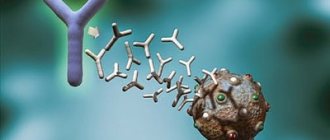
The microcapsule of the leprosy bacterium has antigenic properties. Two antigens have been identified: heat-stable and heat-labile. The heat-stable antigen (polysaccharide) is common to all mycobacteria; the heat-labile antigen (protein) is highly specific for leprosy mycobacteria.
Pathogen genome
The genome of the causative agent of leprosy was first deciphered in 2001. It consists of 3,268,203 nucleotide base pairs. Genetics from the University of Tübingen (Germany) have proven that the genome of Mycobacterium leprae has not changed over the past 500 years.
Reproduction
Mycobacterium leprosy reproduces by transverse fission. The process of reproduction occurs only in the cytoplasm of tissue cells.
Sustainability
In the external environment, leprosy pathogens exhibit increased resistance, but at the same time their virulent properties are quickly lost.
Microscopy
According to Ziehl-Neelsen, mycobacteria leprae are stained pink (Gram-positive, acid-fast). In recent years, the technique of staining smears using the Marcinowski method has become widespread.
Cultivation
It is not possible to grow Mycobacterium Leprae on artificial media. For cultivation, special media are used that contain human serum. In vitro, lineages of isolated mycobacteria are maintained in armadillos and on the paws of infected mice.
Rice. 5. The causative agents of leprosy (Mycobacterium Leprae) are located singly or in clusters, reminiscent of cigarette packs or balloons.
List of sources
- Yushchenko A.A., Irtuganova O.A., Duiko V.V. and others. Modern methods of diagnosis, treatment and prevention of leprosy (a manual for doctors). Astrakhan, 1997. 33 p.
- Grigorieva Yu.G., Belopasov V.V. Pathogenetic mechanisms and the role of serum markers in the development of leprosy neuropathies // Current issues in clinical and experimental leprology: Proceedings of the International Scientific and Practical Conference. Astrakhan, 2011. pp. 53–67.
- Belopasov V.V. Leprosy. Chronic neuroinfections. 2nd ed. M.: GEOTAR-Media, 2021. pp. 392–415.
- Leprosy. Fact Sheet No. 101. World Health Organization (October 2016).
- Duiko V.V. Quality of life and features of management of patients with leprosy. Russian Journal of Skin and Venereal Diseases 2013; 2. P.54-57.
Epidemiology
Leprosy is a low-contagious disease. When infected, the disease develops in 10 - 20% of cases. There is a high natural resistance to leprosy infection. A strong immune system guarantees that a person will not get leprosy.
Risk group
Those at high risk for leprosy include those living in endemic areas. There is a high risk of getting sick among armadillo hunters, those who make crafts from the animals' shells, and when eating animal meat. Living in unsanitary conditions, malnutrition, alcoholism, hard physical labor, and diseases that weaken the immune system contribute to the disease. Leprosy is common in economically underdeveloped countries among the poorest segments of the population.
Source of infection
The reservoir and source of leprosy pathogens is a sick person and the armadillo animal. A person becomes infected from an animal through scratches received while hunting or by eating meat. Armadillos live in Central and South America and belong to the class of mammals. Every year, up to 150 people become ill with leprosy in the United States. All of them either hunted armadillos, or ate their meat, or made fakes from the armor of the animals.
How is leprosy transmitted?
Airborne droplets and contact are the main routes of transmission. The disease is not transmitted in utero.
- A sick person who is not undergoing treatment spreads Mycobacterium leprosy by coughing and sneezing. When talking, bacteria spread within a radius of up to 1.5 meters.
- Somewhat less frequently, pathogens enter the human body by contact through microcracks, scratches, wounds or scratches. The patient's belongings are also a factor in the transmission of infection. Mycobacterium leprosy in patients during periods of exacerbation is found in urine, tears, semen, urethral discharge, blood and breast milk.
Rice. 6. The photo shows an armadillo. The animal has a shell consisting of horny scutes, movably connected to each other.
Dr. Gerhard Hansen (1841−1912)
0
Microbiologists have established that no global changes have occurred with leprosy and the mycobacterium has completely retained its genome over the centuries. But humanity has changed. The population of Europe has become much more resistant to Hansen's disease, and in addition, people who are not at all susceptible to the infection have appeared. The thousands of years of dangerous proximity had an effect, which helped to adapt, as well as the lifelong isolation of patients who did not leave offspring. Before Hansen discovered the causative agent of leprosy, many scientists believed that the disease was inherited. The discoverer of M. leprae gave a scientific basis for the isolation of leprosy patients, convincing everyone that the disease is not given from above and is not transmitted from parents, but passes from sick to healthy through contact.
Pathogenesis
Leprosy pathogens enter the human body through the mucous membrane of the upper respiratory tract and damaged skin. The full picture of the disease develops after an incubation period of years or decades. The penetration of Mycobacterium leprosy into the human body causes disease only in 10 - 20% of cases.
Lepromatous granulomas
Mycobacteria affect the skin, mucous membranes, superficially located peripheral nerves and internal organs, where granulomas form. In the lepromatous type of leprosy, granulomas consist of foamy macrophages stuffed with mycobacteria. Clusters of granulomas form lepromas. In the tuberculoid type of leprosy, granulomas consist of epithelioid cells and contain a small number of pathogens.
Rice. 7. Patients with leprosy. Lepromatous granulomas are visible on the face and neck.
Lepromatous neuritis
Leprosy affects the peripheral nervous system. There is a pronounced tropism of pathogens for lemmocytes - Schwann cells, located along the axons of nerve cells (long process), which perform supporting and trophic functions. Inflammation of peripheral nerves and associated neurological disorders develop more quickly in the tuberculoid type. In lepromatous leprosy, peripheral nerves are affected late. Lepromatous neuritis has an ascending nature. Gradually, the nerve fibers are destroyed and replaced by connective tissue. Damage to peripheral nerves leads to the development of motor and trophic disorders.
Rice. 8. Damage to peripheral nerves. Claw-shaped brush.
Damage to internal organs
More pronounced inflammation of internal organs is observed in the lepromatous type of leprosy. In the mucous membrane of the upper respiratory tract, lymph nodes, liver, spleen, testicles, adrenal glands, kidneys, heart, lungs, granulomas appear, consisting of macrophages filled with a huge number of leprosy pathogens.
Types of leprosy
Depending on the degree of immunological reactivity (cellular immunity), the patient develops one or another type of leprosy.
- The tuberculoid type of leprosy (the most favorable) is formed in individuals with a high degree of immunological reactivity. The disease is less dangerous for others. The course is not severe, with primary damage to the skin and peripheral nerves. The rashes on the skin look like small papules and are accompanied by anesthesia. The infiltrates contain a small number of pathogens.
- The lepromatous type of leprosy occurs in individuals with a low degree of immunological reactivity. Antibodies produced during illness do not protect the body from infection. The disease affects the skin and mucous membranes, nerve trunks, lymph nodes and internal organs. Leproma infiltrates contain a huge number of mycobacteria. Over time, lepromas disintegrate. The resulting ulcers heal slowly. The lepromatous type of leprosy may have blurred and atypical forms.
- The undifferentiated type of leprosy develops when the type of immunological activity has not yet been formed in patients. With a favorable course, the tuberculoid type of leprosy develops in the future; with a low degree of immunological reactivity, the lepromatous type of leprosy develops.
Rice. 9. Tuberculoid (photo on the left) and lepromatous (photo on the right) type of leprosy.
Answers to popular questions
- What is the prognosis for leprosy?
If a person begins to receive treatment at an early stage of the disease, the prognosis is favorable. When leprosy is in an advanced form, there is a high probability that the patient will become disabled.
- Are there leper colonies in Russia?
Yes, in Russia there are 4 leper colonies, which are located in Astrakhan, Krasnodar Territory, Stavropol Territory and Sergiev Posad. Sick people in leper colonies have their own houses and run their households. Doctors live next to the leper colony.
- After recovery, are already formed deformities reversible?
No, if a person has lost his hands or fingers, they will not grow back. Treatment is aimed at eliminating leprosy bacteria in the body. To cope with paralysis, paresis and contractures, the patient is recommended physiotherapy, exercise therapy, and sometimes surgery is performed.
- What are the complications of leprosy?
The main complications include: trophic ulcers, damage to the organs of vision with the development of complete blindness, loss of voice, deformation of the nose, loss of fingers, paralysis. If there is no therapy, the person dies from cachexia, asphyxia or amyloidosis.
- Is it possible to get vaccinated against leprosy? What are the measures to prevent the disease?
There is no vaccine for leprosy. There is evidence that BCG placement significantly reduces the likelihood of infection with Mycobacterium leprosy. To prevent infection, you need to support your immune system and also avoid contact with sick people. If a person with leprosy lives in a family, then he should have his own personal belongings, from a comb to dishes. All family members should be regularly examined for detection of Mycobacterium leprosy in their body, and also carefully observe the rules of personal hygiene.
Lepromatous type. Signs and symptoms
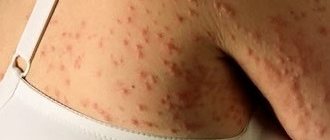
The lepromatous type of leprosy is characterized by the development of various elements of skin lesions - fuzzy spots, plaques, infiltrates and nodes. When the disease occurs, the mucous membranes and internal organs are involved in the pathological process quite early, and the nervous system quite late. This type of leprosy is difficult to treat. A huge number of pathogens are localized in the elements of skin lesions. The lepromine reaction is negative due to the developed anergy.
Leprosy skin lesions
The disease most often affects the skin of the face, ears, wrists, elbows, knees and buttocks. Lepromas appear extremely rarely on the scalp, inner parts of the eyelids, armpits, elbows and popliteal fossae.
Spots and plaques
At the beginning of the disease, barely noticeable, blurred, reddish spots appear on the skin, transforming into plaques. Plaques do not have clear boundaries. Over time, as a result of the development of vascular paresis and hemosiderosis, spots and plaques acquire a brown or copper (rusty) tint. Due to increased sebum secretion, their surface becomes smooth, shiny, glossy (greasy). Enlarged excretory ducts of sweat glands and vellus hair follicles in the area of infiltrates give the skin an “orange peel” appearance. Over time, sweating decreases and stops altogether. Sensitivity in the affected areas is not impaired. After 3 - 5 years, spots and plaques transform into lepromas.
Lepromas
Initially, lepromas resemble small single or multiple dense nodules ranging in size from 1 - 2 mm to 2 - 3 cm, painless, with a greasy surface, sharply demarcated from the surrounding tissues, and contain many leprosy pathogens. Over time, the tubercles transform into powerful infiltrates - nodes. Lepromas are dermal and hypodermal.
Hypodermal lepromas are located under the dermis (the skin itself) and at first the diseases are detected only by palpation. When they reach the dermis they become visible to the eye.
Dermal lepromas initially look like oval-shaped papules, gradually turning into bumps that have a reddish-rusty color, with a shiny surface, and rise above the surface of the skin.
Sometimes lepromas resolve. A pigmented spot remains in their place. Sometimes lepromas ulcerate. The formation of ulcers occurs from the center. They have steep, steep, raised edges. The bottom of the ulcers is covered with a yellowish-gray coating. Ulcers can merge to form extensive ulcerative surfaces. The bloody discharge contains a huge number of mycobacteria (up to 1 billion per 1 cm3). Gradually the ulcers scar. In their place, a sunken (sometimes keloid) scar remains. When ulceration of deep-lying lepromas occurs, joints and small bones are involved in the pathological process, which are destroyed and fall off (mutilatio), leading to deformities and disfigurement.
Signs and symptoms of leprosy affecting the face
Diffuse infiltration of facial skin leads to deepening of natural wrinkles and folds. The brow ridges begin to protrude sharply forward. The earlobes are growing. The nose, lips, cheeks and chin thicken. They take on a lobed appearance and a fierce expression, reminiscent of a “lion's face” (fades leonina).
Rice. 10. Photos of people suffering from leprosy. Multiple lepromas are visible on the skin of the face.
Signs and symptoms of leprosy with nasal lesions
The nasal mucosa is always affected in leprosy and long before the skin is affected. Rhinitis and nosebleeds are the first signs of leprosy. The nasal mucosa turns red and swells, then small erosions appear and massive crusts form, which leads to difficulty breathing. A picture of leprosy rhinitis develops.
Leprosy ulceration leads to destruction of the nasal septum. First, the tip of the nose rises upward, then the nose sinks (“saddle nose”).
Mycobacterium leprosy is detected in mucosal staples. Especially many pathogens are localized in the mucous membrane of the cartilaginous part of the septum.
Rice. 11. People suffering from lepromatous leprosy were called lepers. The photo shows patients whose faces are disfigured by leprosy.
Signs and symptoms of leprosy in the mouth
In severe leprosy, damage to the mucous membrane of the oral cavity and larynx is noted. The soft and hard palate, the back of the tongue and the red border of the lips are affected.
Damage to the vocal cords and epiglottis leads to a narrowing of the glottis and aphonia (loss of sonority of the voice).
Rice. 12. With leprosy, the earlobes grow.
Signs and symptoms of leprosy in the eyes
With lepromatous leprosy, the organs of vision are often affected: the episclera becomes inflamed (episcleritis), the cornea of the eye (focal keratitis), the iris (iridocyclitis), the outer layer of the eye (conjunctivitis), clouding of the lens is noted, and the edges of the eyelids become inflamed.
The appearance of lepromas on the surface of the iris leads to disruption of accommodation and deformation of the pupil. Lepronose keratitis and iridocyclitis without treatment lead to complete loss of vision. When the edges of the eyelids become inflamed, eyelashes fall out. Leprosy keratitis is characterized by the absence of discharge and mild symptoms of inflammation.
Rice. 13. Damage to the organs of vision due to leprosy.
Damage to hair and nails due to leprosy
After 3 - 5 years from the onset of the disease, hair loss is observed in areas of infiltration. Hair falls out from the eyebrows, eyelashes, beard and mustache areas. On the eyebrows, hair begins to fall out from the outer edge. In areas of infiltration, vellus hair loss is noted.
Nails with leprosy lose their shine, become dull, thickened, brittle, and acquire a grayish tint.
Rice. 14. As a result of trophic disorders in leprosy, mutilation (rejection) of the hands and feet develops.
Signs and symptoms of leprosy affecting internal organs
In lepromatous leprosy, damage to internal organs is noted.
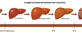
- The liver and spleen increase in size and become denser. Many millet-like lepromas appear in the tissues of these organs. Chronic hepatitis develops.
- All groups of lymph nodes (except for the thoracic and mesenteric ones) are enlarged. They acquire a dense consistency, are mobile and painless.
- Leprosy affects the endocrine glands. A person ages quickly. Women experience menstrual irregularities and early menopause. In men, the testicles (orchitis) and appendages (epididymitis) are affected. Testicular function decreases. The sclerotic process leads to azospermia. Gynecomastia and infantilism develop. Potency decreases to the point of impotence.
Rice. 15. With atrophy of the circular muscles, the eyes do not close completely.
Signs and symptoms of leprosy affecting the nervous system
In leprosy, the peripheral nervous system is affected late. This is due to the high resistance of the axial cylinders to lepromatous infection. Basically, patients develop symmetrical polyneuritis. Nerve trunks affected by infection thicken, become dense and smooth, and are easily palpated.
In areas where leprosy infiltrates develop, sensitivity persists for a long time, but is lost over time until complete anesthesia is achieved, which leads to frequent burns and injuries that patients do not notice. Its causes are compression of the nerves by cellular infiltrates.
Deep sensitivity, periosteal and tendon reflexes are preserved.
When the central nervous system is damaged, neuroses and psychoses develop.
When the peripheral nervous system is damaged, patients with leprosy develop motor and trophic disorders, and sensitivity is impaired.
Trophic disorders:
- Skin pigmentation is disrupted.
- Trophic ulcers appear on the feet.
- Mutilation (rejection) of the hands and feet is noted. At first, the hands and feet become soft, like the legs of frogs or seals, and then spontaneously tear off. The hands and feet are shortened.
- The function of the sweat and sebaceous glands is impaired. Over time, hypofunction is replaced by a complete absence of sweating and sebum secretion. The skin becomes rough and further cracks.
Movement disorders:
- As a result of the predominance of flexor tone, contractures develop. The fingers and toes bend (claw-shaped hand, cauda equina).
- As a result of muscle atrophy, the interosseous spaces collapse. The hand becomes flattened and resembles a monkey's paw.
- With atrophy of the orbicularis muscles, the eyes cannot close completely (lagophthalmos).
- Damage to the facial nerve leads to atrophy of the facial muscles. The face becomes mask-like and takes on a sad expression.
- With the disease, paresis of the masticatory muscles develops.
Rice. 16. Consequences of leprosy. When the nervous system is damaged, contractures, muscle atrophy and trophic disorders develop.
Forecast
The prognosis of leprosy is determined by such factors as the clinical form of the disease, the duration of its course, and the time of initiation of treatment. In most cases, with timely early diagnosis of the disease (within a year from the onset of characteristic symptoms), it is possible to minimize the risk of developing disabling consequences. With late diagnosis of the disease, paresis, sensory disturbances, and disfiguring deformities that are difficult to treat may develop and persist. In the absence of specific therapy, there remains a high risk of death of patients from asphyxia , intercurrent diseases , leprosy cachexia , and amyloidosis .
Signs and symptoms of leprosy. Tuberculoid type
Tuberculoid leprosy is relatively benign and slow. The disease affects the skin and peripheral nerves. Internal organs are slightly affected. Cases of spontaneous regression are often observed. In most cases, the prognosis of the disease is favorable. Tuberculoid leprosy has a low degree of contagiousness.
Skin lesions
In the tuberculoid form of the disease, depigmented or reddish spots with central blanching and sharply defined boundaries appear on the skin. The spots increase in size over time, and their edges rise. Along the edges of the spots there are dense and flat papules, delimiting the damaged area from healthy skin. When spots merge on the skin, patterns of a bizarre ring-shaped configuration are formed. The central part of the spots undergoes depigmentation and atrophy, and fades over time. Within the affected area, there is no skin sensitivity, sweating and sebum secretion stops. The resulting injuries, damage and burns lead to the development of secondary flora.
Rice. 17. Tuberculoid leprosy.
Damage to the peripheral nerves develops in the same way as in the lepromatous form of the disease, but already in the first stages of the disease and more easily. The affected nerve trunks (usually the radial, ulnar, facial and parotid nerves) become painful over time, thicken and begin to be palpated. Contractures and muscle atrophy develop, causing the limbs and face to take on a characteristic appearance (mask-like face, claw-shaped hand, horse foot). The skin appendages are affected - the work of the sebaceous and sweat glands is disrupted, hair and eyelashes fall out, nails thicken and crumble. The lepromine reaction is sharply positive.
Rice. 18. Tuberculoid type of leprosy.
How they tried to make life easier for patients
Medieval medicine could not rid a person of leprosy, but whenever possible they tried to soften the manifestations of the disease and protect the patient from other diseases. The charter of the Antwerp leper colony of the 15th century stated: if the guests, in addition to leprosy, are struck by any curable disease, the workers are obliged to call a doctor to see them. Judging by the documents, the patients were indeed provided with assistance, although sometimes qualified doctors tried to shift the work to practicing surgeons.
To relieve symptoms, medical treatises sometimes suggested very exotic remedies. For example, the theologian and expert on natural sciences Albertus Magnus in the 13th century believed that hair could grow back on damaged areas of the skin. To do this, he recommended a potion made from the skin of a hedgehog, burned along with the needles. And Moses Maimonides, a thinker, rabbi and doctor of the 12th century, believed that the manifestations of the disease could be moderated with a remedy made from dried porcupine meat. However, these drugs were hardly popular.
The main practical means was considered to be a correct lifestyle. Patients were advised to avoid cold and heat, as well as spicy, sour and salty foods. Doctors also advised sick people - both men and women - to abstain from sex (contrary to the then widespread popular belief that it, on the contrary, helps and even heals completely). Doctors with the most radical views recommended castration - whether anyone actually decided to do this is unknown.
All these restrictions grow from the most important “theory of four bodily fluids (humors)” for medieval medicine. According to it, health depends on whether the body manages to maintain the balance of four substances: blood, phlegm, yellow and black bile. They were attributed certain qualities - “temperature” (hot or cold) and “condition” (dry or wet). For example, blood was considered “wet” and “hot.” To maintain or restore the balance of humors, medieval doctors prescribed a specially selected regimen that compensated for the missing “qualities.”
Diseases were seen as a strong shift in the balance in one direction or another. Even in Antiquity, the idea appeared that leprosy was caused by the accumulation of black or yellow bile in the veins. Both biles were considered "dry", so a regimen capable of retaining moisture in the patient's body was required. Doctors believed that sex warms the body, heats the blood and “dries out” existing moisture - therefore, it is harmful to the already “dry” bodies of patients, just like extreme temperatures or spicy food. Lost water had to be replenished with “wet” food, which primarily included meat, fish and eggs.
Many leper colonies did provide patients with a meat diet. Chickens, pigs and even cows were kept at the “houses”; fishing was allowed in local rivers. Residents of the surrounding area did not forget about the sick. Helping the suffering was considered a charitable deed that increases the chances of saving the soul, so wealthy laymen supplied hospitals with food - donations became smaller only in cases where hunger threatened the donors themselves. Archaeologist Christopher Sylvester studied the remains of people buried in the cemetery at the leper colony in Chichester, England, and found out that his patients actually ate meat much more often than poor townspeople. This was indicated by the condition of the teeth of the deceased.
It is known that the urban poor ate a lot of cheap bread, into which small fragments of stone millstones fell with poorly sifted flour. Therefore, their teeth were noticeably worn out, and the residents of the leper colony had much less such marks.
In addition to diet, measures were also taken to “cleanse” the body. In accordance with medieval ideas, the bodies of patients were from time to time riddled of “bad” blood through bloodletting. But water procedures helped much better: washing and exfoliating the affected skin. They could even temporarily restore the body's sensitivity lost due to illness.
Undifferentiated type of leprosy
The undifferentiated (borderline) type of leprosy develops when the patient has not yet developed a type of immunological activity. With a favorable course, the tuberculoid type of leprosy subsequently develops; with a low degree of immunological reactivity, the lepromatous type of leprosy develops. The lepromine reaction, depending on the direction of the pathological process, can be negative or positive. The undifferentiated (borderline) type of leprosy combines the clinical manifestations of 2 main types of the disease, but with a milder course.
Rice. 19. The undifferentiated (borderline) type of leprosy develops in cases where the type of immunological activity has not yet formed.
Treatment
Medicine has made great strides forward and therefore leprosy today is curable, especially if the disease is “captured” at an early stage, when the person has not yet become disabled. Treatment of such patients is carried out in leper colonies - special anti-leprosy institutions or on an outpatient basis. Leper colonies have been known since the Middle Ages, when society tried to limit the contact of lepers with healthy people.
- Currently, patients with multiple skin rashes and positive results of bacterioscopic examination are admitted to anti-leprosy medical institutions for the purpose of conducting the primary stage of treatment.
- Also, patients who are under dispensary registration are sent to the leper colony if they develop a relapse of the disease.
- Patients with a small number of rashes and negative results of bacterioscopic examination are treated on an outpatient basis.
Treatment of leprosy should be carried out comprehensively and include the simultaneous administration of two to three anti-leprosy drugs with the parallel administration of stimulants and restoratives (methyluracil, vitamins, autohemotransfusion, pyrogenal, gamma globulin and other drugs).
- The main anti-leprosy drugs include sulfone drugs (diaphenylsulfone, solusolphone and diucifone).
- Antibiotics are used together with them: rifampicin, lampren, ofloxacin, ethionamide, clofazimine.
- The duration of one course of treatment with anti-leprosy drugs is 6 months. If the patient tolerates the therapy well, there are no breaks between courses. Complex treatment in one course includes the prescription of one sulfone drug and 1 - 2 antibiotics. To prevent the development of drug resistance, drugs are alternated every 2 courses of therapy.
Treatment of patients with leprosy is long-term and lasts from 12 months to 2 – 3 years.