Publication date: September 18, 2020
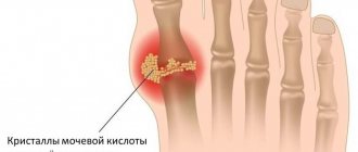
A broken big toe is a fairly common injury that a person can get while playing sports, at work, while walking, and even at home. Pain sensations differ from the severity of the injury and its size. For example, if we are talking about a fracture of only one nail phalanx, then it is quite easy to miss it or even confuse it with a regular bruise. And it is precisely because of the latter that a slight fracture can lead to quite serious consequences and improper healing. We strongly recommend that after a strong blow or injury, you can contact a medical center to rule out a fracture or begin its immediate treatment.
Signs of a broken toe
Depending on the location and severity of the fracture, symptoms can vary greatly. When a phalanx is cracked, pain is practically not felt, and the victim may not even be aware of the problem; very often the bone heals without plaster or fixation. When the first phalanx of a finger is fractured, severe, aching pain is felt.
When a toe is broken, the general symptoms are as follows:
- a hematoma forms at the fracture site;
- possible hemorrhage;
- the skin becomes dark blue, the skin around the damaged bone swells;
- severe and sharp pain with any attempt to touch or move a finger;
- unnatural mobility of the injured finger;
- inability to lean on the affected leg;
- immobility or partially limited movement of the finger;
- increased temperature and redness at the site;
- with a comminuted fracture, shortening of the finger is possible;
- with an open fracture, a wound with bone fragments;
- pronounced twitching or pulsating of the finger.
Upon physical examination, crunching of bone fragments is observed if little time has passed since the injury. The crunch is the result of the friction of broken bones against each other. Finger fractures are combined with damage to the ligamentous apparatus, sprains and dislocations of the phalangeal joints.
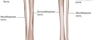
Briefly about the anatomy of the foot
The foot is part of the human musculoskeletal system, the part of the lower limb most distant from the body, responsible for stable position, balance, and movement of the body in space.
The foot has a flexible, elastic and movable arched design, which allows you to distribute the load on the limb, adapt to unevenness, and achieve a smooth gait thanks to the powerful ligamentous apparatus. In interaction with other parts of the leg, it is responsible for moving the body.
Twenty-six bones structurally form the three sections of the foot: the tarsus, metatarsus, and phalanges (toes).
The first section includes seven bones arranged in two rows:
- posterior – calcaneus and talus,
- anterior - navicular, three wedge-shaped and cuboid.
All of them are connected by joints, which allows a person to bend, straighten and make circular movements with the foot.
The second section is the metatarsus, consisting of five short tubular bones, located between the tarsus and the fingers.
The third section is the phalanges - tubular bones (14 pieces), connected by muscles and joints to form the five toes.
At the same time, the thumb consists of two phalanges, and the rest - of three. All of them are significantly shorter compared to the fingers, and the middle phalanx of the little finger is often inseparable from the nail. The phalanges are connected to each other by joints.
How can you distinguish a possible bruise from a fracture?
A bruise or fracture can be determined by several parameters:
- Features of the pain syndrome.
- Finger movement.
- Skin color at the site of swelling (bruise).
- Presence of hemorrhage.
- Phalanx shape.
The clinical picture of a bruised finger is as follows:
- The victim experiences sharp pain, which begins to subside over time. The nature of the pain is “aching.” Using a cold compress can speed up the process of relieving pain symptoms.
- When bruised, the finger is not deformed. Immediately after a bruise, all movements are accompanied by sharp pain (pulsation is possible); as the pain subsides, the motor activity of the finger is gradually restored.
- Depending on the nature of the bruise, the color of the skin at the site of the injury may be dark red, pink, or pale pink. Swelling may appear immediately, after a day, or not appear at all. The blood at the site of the injury spreads diffusely (scattered), a bruise may appear.
What is a splint and what is it for?
The splint is a plaster-based bandage, which is secured on top with a regular bandage. Using this device, the damaged area of the leg is fixed in an anatomically correct position. It reliably protects and covers the injured segment of the leg. It is used not only for healing fractures, but also for correcting congenital pathologies of bones and joints of the legs. Correction of congenital leg pathologies is carried out in early childhood, when bone structures are just forming and can be corrected.
The splint is a removable structure, which, unlike a simple plaster cast, is very convenient because it makes it easy to carry out sanitary and hygienic procedures. People call a splint a splint.
An elastic bandage is placed on top of the plaster splint, covering the completely damaged area. For pathologies or damage to the joint, only elastic bandages are used. The bone structures are fixed with other splints.
These structures are made from different materials. There are devices called clamps that are made of plastic. It better immobilizes the limb and keeps it in the required position.
How to diagnose a broken finger
- Upon palpation, the pain sharply intensifies and does not go away for a long time (an hour or two).
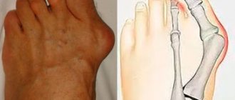
- When a fracture occurs, a sharp pain is felt, which can radiate to the nearest parts of the foot. Deformation of the phalanx, unnatural position of the finger. Swelling and strong (acute) pulsation at the fracture site.
- If there is a fracture, the victim cannot move the injured finger. Any attempt to stand on the affected leg causes the patient to experience severe pain. To relieve pain symptoms, the affected finger is fixed in one position.
- Hemorrhages form under the nail, hematoma and swelling appear, and the skin becomes bluish.
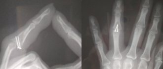
Diagnostics
Making a diagnosis in most cases is not difficult. The gold standard for diagnosis is x-ray examination. Pictures are taken in two projections, which allows you to see even small cracks.
The diagnostic process consists of several stages:
- The main point in collecting anamnesis is the fact of injury. A fall on the lower limb of a heavy object, a jump from a height, or a blow during sports may suggest damage to the thumb;
- Among the complaints, pain at the site of injury plays an important role, movements in the interphalangeal joints are limited;
- On examination, swelling and redness of the skin are observed. The finger is in an unnatural position. Upon palpation, the doctor determines the presence of fragments and crepitus.
- X-ray examination allows you to confirm the diagnosis;
- Computed tomography is used in rare cases when the clinical picture does not correspond to the radiological one.
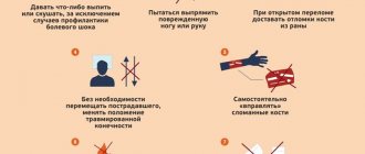
First aid for a broken finger
If you have symptoms indicating a possible fracture, there is no need to panic in the first place. The first thing you should do is call an emergency ambulance. Before the doctor arrives, all efforts of the victim should be aimed at stopping the bleeding (in the case of an open fracture), fixing the limb and anesthetizing the injury site. To prevent negative consequences, the following rules must be followed:
- even in the absence of pronounced symptoms, one should not refuse to consult a doctor;
- fixation of the limb is one of the main stages of effective treatment;
- the broken finger should not touch foreign objects;
- non-displaced fractures may not be fixed until the ambulance arrives;
- pain can be relieved with nimesil, analgin and ibuprofen;
- A cold compress is another effective way to relieve pain.
It is important to note that when applying an ice pack, keep the compress for no more than 10 minutes to prevent possible frostbite. A repeat procedure is possible after a 3-4 minute break. Even if you suspect a fracture, experts recommend immediately contacting a specialized medical institution for qualified help.
Rehabilitation period
Rehabilitation measures include massage, physiotherapeutic procedures, exercise therapy. Wearing a cast for a long time is not pleasant, but it is necessary for proper bone healing. If the bones do not heal properly, this will lead to improper distribution of body weight on the foot in the future and more problems when walking.
It is recommended to walk as little as possible during rehabilitation, wear comfortable, roomy shoes without high heels, and keep the affected leg elevated during rest, for example, place it on a pillow. To reduce severe swelling, you can apply cold compresses in the first 2-3 days, but do not overuse them.
You should eat rationally, eat enough vegetables and fruits, especially pay attention to foods rich in calcium: cheese, cottage cheese, etc. At the same time, limit or completely abandon strong coffee, carbonated and alcoholic drinks, as they remove calcium from body. It is also recommended to take multivitamins, C, D and B12.
Author of the article:
Kaplan Alexander Sergeevich |
Orthopedist Education: diploma in General Medicine received in 2009 at the Medical Academy named after. I. M. Sechenov. In 2012, she completed postgraduate studies in the specialty “Traumatology and Orthopedics” at the City Clinical Hospital named after. Botkin at the Department of Traumatology, Orthopedics and Disaster Surgery. Our authors
Treatment methods for a broken finger
| Name | Description |
| Closed reduction | This method is used for closed fractures and in the absence of displacement. Antiseptic agents are applied to the damaged finger, after which the doctor uses mechanical force (pulling) to return the finger to its normal position. A significant disadvantage of this method is the need to repeat this procedure. |
| Skeletal traction | This method is used for displaced fractures. This procedure requires a metal needle, which is passed through the finger with a small load, which allows the bones to be in a normal position. At the end of the procedure, the doctor performs immobilization. |
| Public Methods | The surgeon performs osteosynthesis. Secures bone fragments with special metal elements. The broken parts of the bone are united and its correct shape is restored. Open reduction is performed for all open and comminuted closed fractures. The operation also eliminates complications that arose during the treatment. |
| Surgical intervention | The operation is indicated for patients who have an open fracture of the big toe or in case of crushed phalanx. During surgery, the doctor restores the physiological position of the finger. To fix fragments, knitting needles, plates, and screws are used. |
Classification
Types of toe bone fractures:
By localization:
- I, II, III, IV, V fingers;
- main, middle, nail phalanges;
- damage to the diaphysis, marginal fracture, epiphysiolysis.
By the presence or absence of a wound:
- open (with violation of the integrity of the skin);
- closed (without such violation).
Based on the presence of offset:
- with offset;
- without displacement.
Along the fracture line:
- transverse;
- oblique;
- longitudinal;
- helical;
- splintered.
Making a splint with your own hands
This fixing device can be made by hand. To do this, you need to have gauze cloth soaked in plaster and dry plaster itself. Gypsum is mixed with water in equal proportions, then after a few minutes it is checked how hard it has hardened. To do this, squeeze the material in your hands. The plaster must remain intact, not break or crumble - in this case, everything is done correctly. If the material is unstable, breaks and crumbles in your hands, and also has an unpleasant odor, then mistakes were made in its preparation.
After the solution has been made, take a bandage three meters long and cut off its edge. Such bandages are better modeled because they are more elastic. The prepared mixture is applied to a certain area of the bandage, several layers are made (4-7) and the ends are folded towards the middle. Then, using water, vigorously smooth them out with your hands. The resulting bandage is applied to the damaged limb, and small incisions are made on the protruding areas (on the heel).
Use of additional materials
When spraining ligaments and dislocating a joint, use not gauze with a bandage, but an elastic bandage with plaster. The use of an elastic bandage has a number of advantages, since it is more flexible and reusable. These characteristics contribute to the rapid healing and restoration of injuries, since the device is easily removable and allows you to treat swelling with the help of special creams and ointments. Doctors advise using a removable device with an elastic bandage only a week after the injury. If there is such a possibility, then it is better to use synthetic polymer gypsum, which does not break or crumble when hardened.
Causes
There are many situations in which a person can suffer a foot injury. Such injuries occur in domestic conditions or as part of the production process.
Often the cause of fractures is unsuccessful falls, careless sports, and violation of safety rules at the enterprise.
Provoking factors for finger injuries include the following:
- An intense blow or a fall on the leg from a heavy object;
- Sports injury that caused forced hyperextension of the foot;
- Sloppy movements;
- Hitting your foot on a hard object.
Drug therapy
Three types of medications are used in the treatment of fractures:
- anti-inflammatory;
- painkillers;
- chondroprotective.
They are taken orally and topically on the painful area.
The action of the drugs provides:
- stimulation of the processes of restoration of damaged tissues;
- improvement of salt metabolism in bones (calcium-phosphorus);
- replenishment of calcium deficiency;
- elimination of inflammation.
The patient is prescribed: Chondroitin, Teraflex, Calcemin, Vitamin D3, Calcium Gluconate, Nise, Nurofen, Dexalgin. For local impact on the inflamed area: Traumeel-gel, Nise-gel, Voltaren.
The gel is applied three times a day.
The advantage of the splint
It lies in the fact that when wearing it you can monitor the condition of the skin. You can sometimes remove it, perform various hygienic manipulations, give the skin the opportunity to “breathe,” take a shower, do scrubs and peels on the damaged area. If increasing swelling is observed, it expands the edge of the bandage, which prevents the development of tissue ischemia. It can also be modeled and modified, for example, converted into a circulatory bandage using additional bandage volume.
SYNERGEL
— the healing power of 4 natural poisons will quickly relieve joint pain!
Get Synergel on promotion only
Old price: 4790₽
New price: 147₽
Contactless delivery and safe receipt of your order!
The classic bandage is applied in a thick, dull layer. Of course, it creates high-quality immobilization and is used when there is a need for long-term immobilization. A traditional circular plaster cast involves bandaging the leg from the foot to the center, with each previous round overlapped by the next by 2/3. The shape of the bandage is then changed using plaster. With such fixation, the growing swelling is unable to expand the boundaries of the bandage, which provokes serious compression of the tissues up to their necrosis. In this context, the splint has a greater advantage over the traditional circular bandage. A classic plaster cast requires medical supervision in a hospital for 1-2 days.
Ligamentous apparatus
The entire joint system provides the necessary functionality of the foot.
- Tarometatarsal - Small, flat joints with limited mobility. They form the base of the foot, with the help of ligaments in the tarsus.
- Interphalangeal - Provides immobility of the phalangeal bones.
- Subtalar - Inactive, located in the hindfoot, providing the arch of the talus and calcaneus.
- Metatarsophalangeal - A ball-and-socket joint that allows the fingers to flex and extend.
- Talocaleonavicular - Connects three bones to the axis of rotation. The foot can be rotated outward and inward.
- Ankle - A large joint connecting three bones. Forms a block between the tibia and the talus. The joint is attached to cartilage and forms ligaments on the side.
- All rotational and flexion movements of the leg occur at the expense of the ankle. The entire load while walking or running falls on the ankle joint.
The foot performs 3 functions:
- Supportive
- the ability to prevent pressure from the supporting surface. If the function is impaired, the person experiences severe pain when running or jumping. When walking, the foot performs a pushing function - accelerating movement. - Cushioning
- smoothes out shocks when walking and running. It protects joints from damage. If the arch of the foot is low, then the function is reduced, diseases of the bones, joints, and sometimes internal organs develop. - Balancing
– provides full coverage of the support surface and maintains the position of the human body when moving.