An examination by a doctor and the medical history collected during an appointment are often not enough to make an accurate diagnosis and prescribe treatment. Especially when it comes to the abdominal cavity. The location of internal vital organs does not make it possible to make an accurate diagnosis, or confirm it without instrumental diagnostics. To obtain the most informative results from the study, it is necessary to properly prepare for the examination procedure. We tell you how to do this.
Research effectiveness
Many years of practice have shown a high detection rate of pathologies during ultrasound examination. The procedure is performed on adults and children, including the smallest and infants. During the study, the doctor conducts a detailed examination of the spleen, liver, and pancreas. He examines the vessels of the abdominal cavity along their course, the portal vessels of the liver, identifies enlarged lymph nodes throughout the intestine, assesses the condition and size of the gallbladder and ducts. High detection of diseases is associated with high density of ABP.
Assessment of the intestine is performed with ultrasound only indirectly: by the uneven thickness of the intestinal walls, by the accumulation of gases in its loops and other signs.
To assess the condition of the stomach in detail, fibrogastroscopy is prescribed; therefore, during an ultrasound examination of the gastrointestinal tract, the condition of the stomach is not assessed and this organ is not described in detail.
Indications for ultrasound examination:
- for prevention - carried out once a year for early diagnosis of tumors;
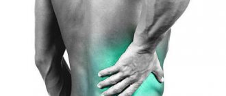
- pain in the abdomen and lower back;
- nausea and vomiting, painful defecation, loss of appetite, changes in the character of feces, jaundice, palpable neoplasms of the abdominal cavity, etc.;
- blunt abdominal trauma - in order to exclude internal bleeding, rupture of parenchymal organs;
- results of laboratory tests - clinical tests and biochemical indicators indicating pathological processes in the liver, spleen, and other organs of the PD;
- dynamic monitoring of previously identified neoplasms of various nature;
- monitoring the condition of the organs of patients receiving long-term hepatotoxic treatment;
- dynamic monitoring of patients during the rehabilitation period after surgery.
Ultrasound of the OBP can reveal various pathologies. For example, when examining the liver, cirrhosis, fatty hepatosis, that is, fatty degeneration, can be diagnosed. Stones, an inflammatory process (cholecystitis), and also biliary obstruction or blockage of the bile ducts can be detected in the gallbladder. An ultrasound shows the condition of the spleen: it can be reduced or increased in size, and upon examination, neoplasms are revealed.
The pancreas may be enlarged or reduced, which is also visible on ultrasound. Its diffuse increase occurs during inflammation as a result of the appearance of edema. Focal enlargement is typical for cysts and tumors. This test helps diagnose abdominal aortic aneurysm.
Who performs the ultrasound
At the Tibet clinic, ultrasound of the abdominal cavity and retroperitoneal space is performed by a highly qualified ultrasound diagnostics doctor, Maria Aleksandrovna Vasilyeva. Her exceptional professionalism and extensive experience are a guarantee of the high quality of the examination, the reliability of the data obtained and the correctness of the diagnosis.
How to use class=”aligncenter” width=”992″ height=”247″[/img]
or call +7 Moscow
Free consultation Survey, examination, pulse diagnostics from 30 minutes
Diagnostics Ultrasound, MRI, Laboratory tests (as prescribed)
Treatment Individual plan
What to do before the study
Before performing an ultrasound examination of the abdominal cavity, it is necessary to consult a gastroenterologist. He collects anamnesis, finds out the presence of chronic diseases, genetic predisposition, and allergies to drugs. If necessary, prescribes additional studies and tests.
Ultrasound is a non-invasive diagnostic method and does not require additional tests. You only need to undergo chest fluorography (FLG), donate urine and blood for a general analysis. However, in order to make an accurate diagnosis, other studies are prescribed along with ultrasound, the number and list of which depends on the preliminary diagnosis.
Not only a gastroenterologist, but also other specialists can prescribe an examination. For example, an ultrasound examination helps a neurologist monitor the condition of the liver during long-term use of drugs that affect this organ; an allergist prescribes a prescription to identify pathologies of the liver and gallbladder that can cause an autoimmune disease; a hematologist is interested in the condition of the hematopoietic organs: the spleen and liver. They also prescribe a whole range of additional tests and studies.
Contraindications
As with almost any medical procedure, there are a number of contraindications for performing an abdominal ultrasound. These include:
- hyperthermia;
- infectious diseases in the acute phase;
- open wounds in the peritoneum;
- skin diseases with exudation localized in the area under study;
- severe general condition of the patient.
Pregnancy, contrary to popular belief, is not a contraindication to the procedure. The study is completely safe for the health of the woman and fetus and cannot cause deterioration in well-being.
Constant improvement of diagnostic equipment has made it possible to carry out the procedure within pediatric practice over the past two decades. If pathology of the abdominal organs is suspected, the examination can be carried out even on newborn children.
Ultrasound of the abdominal organs in a medical setting provides the opportunity to most accurately identify the cause of the existing pathology, make the correct diagnosis and, as a result, receive prescriptions for effective therapy.
Basic rules of preparation
Ultrasound examinations of the ABP should be done on an empty stomach, and are prescribed both in the morning (on an empty stomach) and in the afternoon. It is necessary to adhere to some rules and recommendations before an ultrasound examination:
- Follow a diet 2-3 days before the procedure.
- You must not smoke for two hours before the examination.
- It is not recommended to conduct ultrasound examination after FGDS, FCS.
- If studies were carried out with the introduction of a radiopaque substance into the body - a barium preparation, for example, during irrigoscopy, fluoroscopy of the esophagus, stomach, then an ultrasound scan should be performed two days later.
This is a reminder for those who are preparing for an ultrasound scan of the OBP.
Adviсe
Before an ultrasound, it is not recommended to take antispasmodics such as No-Shpa.
If the patient requires constant use of any medications, for example, heart medications, the ultrasound specialist should be informed about this. When taking antispasmodics:
- No-shpy
- Papaverina
- Spasmalgona
- Papazola
It is necessary to consult with your doctor about the possibility of canceling them. If the patient undergoing the study has diabetes, he should not fast for a long time. In this case, it is better to reschedule the examination in the morning. You should not do an ultrasound immediately after:
- Gastroduodenoscopy
- Irrigoscopy
- Colonoscopy
- FGDS
After these examinations, at least three days should pass before the ultrasound.
Examination for certain categories of patients
Preparation for ultrasound for men and women is not fundamentally different, with the exception of pregnant women.
Pregnant
Metabolic processes in pregnant women are more intense than in other people. They need to eat more often to ensure that they and the baby are receiving nutrients. Their meal intervals are reduced to two hours, and they must eat in small portions. Therefore, they need to eat their last meal two hours before the test.
Children
Infants stop feeding 3 hours before the ultrasound scan. Older children should not eat 4 hours before the procedure. You are only allowed to drink a little water, but the reliability of the study results may be reduced.
Patients with diabetes
A long gap between meals for diabetics can negatively affect their well-being and cause glycemic coma. For such patients, ultrasound is prescribed in the morning. If this is not possible, then you are allowed to have breakfast and undergo an examination after 3-4 hours. They should take sweet tea and crackers with them to have a snack after the procedure.
Additional recommendations
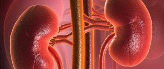
You should quit smoking within a few hours. Also, do not chew gum or suck on hard candies as they may release gastric juice. When diagnosing kidneys, you need to drink 0.5 liters of water half an hour before the procedure.
Following these rules on the eve of the procedure will help identify organ dysfunctions and prescribe effective treatment. You can visit a doctor, get an ultrasound and get tested in Rostov-on-Don at the medical office. The clinic has modern equipment to obtain accurate data.
You can make an appointment by calling the 24-hour hotline! You can call 24/7
Diet before ultrasound examination
You need to prepare for an ultrasound three days in advance. During this time, it is necessary to reduce the load on the mucous membrane and gradually normalize the function of the digestive tract. Diet is the basic preparation, but it is not limited to it. Avoid foods that cause gas and reduce fiber intake.
The number of meals should be at least five times a day in small portions, every 3.5–4 hours. The last dose is no later than 4 hours before bedtime. Liquids should be consumed 1.5–2 liters per day, a little less for children. If the examination is scheduled for dinner, then the last meal taken in the evening is a light dinner. If the procedure is in the afternoon, then a light breakfast, including a couple of eggs and green tea. You can have breakfast depending on the appointed time at 8–10 am (6–8 hours before the procedure). After this, it is advisable not only not to eat, but also not to drink. You should not eat candy or chew gum during this time.
specialist
Our doctors will answer any questions you may have
Chikov Sergey Vladimirovich Ultrasound Doctor
Ask a Question
Prohibited Products
Gas formation in the intestines greatly interferes with ultrasound examination. Therefore, three days before the procedure, you need to exclude from your diet a number of foods that cause flatulence and minimize fiber intake.
Legumes
The fruits of legumes are rich in vegetable protein and complex carbohydrates. This combination leads to intense gas formation. Peas, vetch, beans, lentils, and beans are prohibited when preparing for an ultrasound.
Carbonated drinks
Carbonated drinks contain carbon dioxide, also known as carbon dioxide. Sometimes it is formed as a result of the natural fermentation process, for example, in kvass. Therefore, all these drinks are prohibited.
Flour products and pasta
These foods contain gluten as well as polysaccharides, which when combined cause bloating. This also includes semolina.
Whole grain cereals
These include corn, rice and millet cereals. All of them are rich in fiber and can cause gas formation.
Onion and garlic
These foods contain fructans, a polymer of fructose. Even in small quantities, onions and garlic can cause bloating.
Raw vegetables
It is recommended to exclude cabbage, as well as radishes, radishes, daikon and watercress. These foods irritate the intestinal walls, contain a lot of fiber and can cause gas.
Dairy
Flatulence can be caused by milk. It contains milk sugar - lactose. A lack of the lactase-breaking enzyme can cause gas.
Alcoholic drinks
Alcohol irritates the walls of the stomach and intestines and provokes an inflammatory reaction. Beer also contains fermentation products, including carbon dioxide.
Fruits and berries
First of all, fruits cause bloating. Containing large amounts of simple sugars - fructose. These include grapes, melon, bananas. Berries and fruits with sourness cause gas formation to a lesser extent.
Sweets
Candies, chocolate and other sweets containing simple carbohydrates that promote gas formation are excluded from the menu.
On the eve of the study, you should also refrain from fatty foods, including rich soups with meat broth, fatty fish, lard, and fatty meat.
Authorized Products
Three days before the ultrasound, it is recommended to switch to food with easily digestible proteins and other components that do not cause putrefactive processes in the intestines and gas formation. All dishes should be eaten in moderation.
The following dishes are recommended:
- porridge made from buckwheat and oatmeal cooked in water (rolled oats);
- low-fat broths, soups cooked with vegetables;
- dietary meat with easily digestible protein (turkey, chicken, rabbit);
- boiled or steamed vegetables (carrots, cucumbers, pumpkin, zucchini);
- quail and chicken eggs;
- low-fat fish (any river fish, cod species);
- mashed potatoes.
The following drinks are allowed the day before:
- still water (mineral or artesian);
- lightly brewed tea;
- dried fruits compote;
- fruit drinks;
- herbal infusions;
- fruit juices.
Ultrasound of organs in Volgograd: where to go
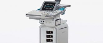
The quality and accuracy of ultrasound diagnostics largely depend on the device and the qualifications of the doctor conducting the examination. Dialine clinics have the most advanced technical equipment and a staff of highly qualified specialists. Our clinics are located in Volgograd, Volzhsky and Mikhailovka. You can choose the most convenient address for yourself and the appropriate time for the examination. With us you can perform an ultrasound of any organ and immediately receive advice from a specialist in the required profile. For ease of use of our services and access to discounts, we recommend registering in your personal account on our website. To clarify your questions, leave a request for a call back or call us. We guarantee high-quality and prompt service to all patients.
Taking medications before the study
Before the procedure, you should refrain from taking medications. The exception is for daily medications. Whether you can take them should be checked with your doctor.
To eliminate any manifestations of gas formation on the eve of the procedure, the doctor prescribes enterosorbents that absorb gas. This improves visualization. It could be Espumisan, Motilium. The drugs are taken the day before the examination in the usual dosage 3 times a day, 80 mg, and once in the morning on the day of the ultrasound, 80 mg. Activated carbon can be a replacement. It is recommended to take 4 tablets 3 times a day. The medicine is taken after meals; before use, it is recommended to finely crush the tablets into powder to increase the area of absorption. For children and adolescents, the dosage may be different.
Enzyme preparations help reduce gas formation. If the patient suffers from constipation, an enema is recommended, but glycerin rectal suppositories can be used. You can take lactulose (Laktulak, Duphalac or an analogue).
Factors influencing ultrasound results?
In addition to diet and cleansing, the following points also affect the correctness of the results:
- Tobacco smoking on the day of screening. You should give up cigarettes a couple of hours before the scan.
- Chewing gum, caramel, lollipops. Avoid using them on the day of your appointment.
- Parallel radiography. The interval between different types of screening should be at least 3 days.
- The degree of fullness of the bladder. When the kidneys are scanned, the urine should be full. In others the situation is devastated.
- Excess body weight. When referring for ultrasound screening, the weight and condition of the patient should be taken into account, since excessive obesity complicates ultrasound penetration.
How is an abdominal ultrasound performed?
Ultrasound is a non-invasive procedure. The abdominal organs are examined transabdominally - through the walls of the abdomen. There is no need to undress completely, just lift the edge of your clothing and move it to your chest.
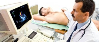
The patient lies on his back on the couch, the doctor applies a hypoallergenic gel to the stomach, which facilitates the movement of the sensor and improves signal conduction into the cavity. This makes it possible to get a clear image on the display. It is important that the patient relax the abdominal muscles as much as possible.
During the examination, the doctor gives certain commands to examine a particular organ in detail. He may ask you to take a deep breath and hold your breath for a few seconds. The procedure does not cause pain or discomfort and usually takes 25 minutes to an hour.
The conclusion of the study is usually issued immediately. Separate printed frames may be attached to it. The protocol indicates the size of the organs, shape, their location, compliance with anatomical standards, integrity, level of intestinal motility, changes found and pathology.
Cleansing
If the patient has a tendency to constipation, it is necessary to take a natural laxative a day before the examination or use suppositories to enhance intestinal motility. If such therapy does not bring results, you can use an enema 12 hours before an ultrasound of the abdominal cavity. It is advisable to do the enema in the evening before the examination. To do this, you need to prepare an Esmarch mug, into which you pour three liters of cool boiled water. After cleansing, it is recommended to take adsorbents or Simethicone. You can replace the enema with herbal laxatives:
- Senade: 1 tablet
- Fortrans: dissolve the sachet in water and drink at least three hours before the examination. It is recommended to take 1 sachet for every 20 kg of weight
- Microclysters: Norgalax or Microlax
Choosing a clinic
Usually, after receiving a referral for an abdominal examination, the patient tries to undergo diagnostics in a good clinic with modern equipment and professional medical staff.
The 36go website has compiled a rating of the best medical institutions where it is possible to get examined. We will guide you through the facility’s equipment, provide a description of the clinic and patient reviews of this study.
Call and our staff will help you make the right choice, advise you on prices for the procedure, and make an appointment. When you contact us through our website, there are discounts, as well as promotions for diagnostic tests.
Advantages of comprehensive ultrasound diagnostics
Ultrasound diagnostics is a simple, painless and quick way to examine all internal organs. The ultrasound diagnostic method has become popular because it is simple and painless and does not require invasive interventions.
- Diagnosis of all organs occurs in a short time during one visit
- A conclusion with a detailed description of all examined organs is issued in person.
- Performing all ultrasound examinations in one visit will help to significantly save money without losing quality
- Carrying out an examination using a modern ultrasound machine allows you to obtain high quality images and provides reliable results.
- A preventive ultrasound of the body will give you peace of mind and confidence in your health and will allow you to identify diseases at an early stage.