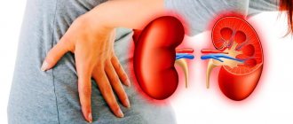
Glomerulonephritis is a common kidney disease. As a rule, the renal glomeruli are affected (to a lesser extent, the interstitial tissue and tubules). If the disease is chronic, the symptoms may be almost invisible or completely absent. But if the disease is acute, then this condition can lead to serious complications that can lead to disability of a person at an early age.
Glomerulonephritis refers to inflammatory diseases of the urinary system. Those, in turn, affect the functioning of not only the kidneys, but also the functioning of the heart. This is explained by the fact that the work of the cardiac and renal systems is connected, therefore, signs such as increased blood pressure or other symptoms of diseases of the cardiovascular system are often concomitant symptoms for diseases of the urinary system. Therefore, it is necessary to start treatment of glomerulonephritis on time to avoid many negative consequences.
At the same time, the success of treatment largely depends on the timeliness of contacting a doctor. It is necessary to pay attention to such signs of pathology as the appearance of edema and blood in the urine.
General information
With glomerulonephritis, damage occurs to the glomeruli - the glomeruli of the kidneys. Most often this occurs due to an infectious or inflammatory process, but other causes may also influence the appearance of pathology.
According to statistics, glomerulonephritis is one of the three most common kidney diseases occurring in adult patients. The danger is that this disease often leads to disability due to the development of chronic renal failure if treatment is not started in time.
At the same time, there is one very interesting pattern. The use of effective antibacterial treatment can reduce the proportion of acute glomerulonephritis, but the number of patients whose disease becomes chronic is steadily growing. This situation is explained by poor quality nutrition, as well as environmental degradation.
Most often, glomerulonephritis is diagnosed in adult patients under 40-50 years of age. But the disease also occurs in childhood. Moreover, men suffer from it much more often than women.
Classification (forms)
In recent years, the classification of chronic glomerulonephritis has been changed. Previously, the disease was divided into types (species) based on symptoms. Today, classifications are based on pathomorphological changes, which are identified by examining a kidney biopsy.
In the CIS countries, the clinical classification , authored by E.M., Tareev. According to this division, there are the following forms of chronic glomerulonephritis:
- Hematuric
- Latent
- Nephrotic
- Hypertensive
- Mixed
According to the classification of Serov V.V. and other authors, based on pathomorphological characteristics the following forms of the disease can be distinguished:
- With half moons
- Diffuse proliferative
- Membranoproliferative
- Mesangioproliferative
- Minimal change nephropathy
- Membranous nephropathy
- Fibroplastic
- Fibrillar-immunotactoid
- Focal segmental glomerulosclerosis
Doctors believe that each of these forms can be acute or chronic.
Main symptoms of glomerulonephritis
The acute course of the disease is characterized by 3 main groups of symptoms:
- urinary – increased red blood cells and protein in the urine;
- edematous – swelling of the arms, legs and face;
- hypertensive – an increase in blood pressure that cannot be reduced with conventional blood pressure medications.
It all starts with an increase in body temperature, a headache begins, and the general condition worsens. Swelling of the eyelids is visible, appetite decreases, and the skin turns pale.
Another telltale sign is a change in urine color caused by blood. In 85% of cases, microhematuria develops, in 15% - macrohematuria (this condition is characterized by a change in the color of urine to a very dark color).
Another symptom of glomerulonephritis in adults is facial swelling. It is very pronounced in the morning, gradually disappearing during the day.
Other symptoms include:
- gagging;
- pain in the lumbar region;
- urge to urinate;
- swelling of the limbs.
Diagnosis of chronic glomerulonephritis
For diagnosis, it is important to identify the leading syndrome: nephrotic, acute nephrotic, isolated urinary, arterial hypertension. Symptoms of chronic renal failure are also important.
Nephrotic syndrome most often occurs with:
- membranous glomerulonephritis
- glomerulonephritis with minimal changes
- kidney amyloidosis
- diabetic glomerulosclerosis, renal amyloidosis
Acute nephritic syndrome is a combination of hematuria, hypertension, edema and often deterioration of the filtration function of the kidneys. It can occur with the following diseases:
- mesangiocapillary glomerulonephritis
- rapidly progressive glomerulonephritis
- exacerbation of lupus nephritis
- mesangioproliferative glomerulonephritis
Arterial hypertension in combination with proteinuria and minimal changes in urinary sediment occurs, in addition to chronic glomerulonephritis, with diabetic nephropathy, kidney damage as part of hypertension.
Urinary syndrome is a combination of proteinuria, hematuria, cylindruria, leukocyturia with lymphocyturia (any combination of the above).
There are the following causes of isolated hematuria :
- infection
- stone
- tumor in the urinary tract
- Berger's disease
- Allport's disease
Proteinuria occurs with inflammatory and non-inflammatory lesions of the glomeruli or with tubulointerstitial lesions of various etiologies. A special variant of massive proteinuria is overflow proteinuria, which is associated with the presence of paraprotein in the blood and myeloma. Benign proteinuria occurs with obstructive sleep apnea syndrome, heart failure; sometimes occurs during emotional stress, hypothermia, or becomes a consequence of a febrile reaction. It is called benign because the prognosis for kidney function is favorable.
Kidney biopsy
A puncture biopsy of the kidney is performed to determine the morphological form of chronic glomerulonephritis, which affects the choice of treatment tactics. Contraindications :
- Hypocoagulation
- The presence of not two, but one functioning kidney
- Suspicion of renal vein thrombosis
- Suspicion of malignancy
- Increased venous pressure in the systemic circulation
- Polycystic kidney disease
- Hydro- and pyonephrosis
- Impaired consciousness
- Renal artery aneurysm
Differential diagnosis
Differential diagnosis is carried out with the following diseases:
- acute glomerulonephritis
- chronic pyelonephritis
- chronic tubulointerstitial nephritis
- nephropathy in pregnancy
- amyloidosis
- alcoholic kidney damage
- kidney damage in systemic connective tissue diseases
- diabetic nephropathy
Characteristic features of chronic pyelonephritis that need to be taken into account when diagnosing:
- neutrophiluria
- bacteriuria
- exacerbations with fever and chills
- changes in the collecting system
- asymmetry of the lesion
In acute glomerulonephritis, a history of connection with a previous streptococcal infection is considered, but, however, unlike IgA nephropathy, the time interval is 10-14 days. Typically acute onset. Among the patients are mainly teenagers and children.
Chronic tubulointerstitial nephritis has the following features (taken into account in differential diagnosis):
- slight proteinuria
- polyuria
- urine acidification disorder
- decreased relative density of urine
If amyloidosis is suspected, an underlying pathology should be looked for, namely chronic inflammation, which occurs, for example, with rheumatoid arthritis. Amyloidosis is suspected when chronic renal failure persists with enlarged or normal kidney size and in the presence of nephrotic syndrome. Tissue biopsy is of decisive importance in the differential diagnosis of such cases.
The diagnosis of diabetic nephropathy is likely (as well as chronic pyelonephritis) if a person has diabetes mellitus or its complications, slowly increasing chronic renal failure, scanty changes in urinary sediment, normal or slightly enlarged kidney sizes.
Nephropathy in pregnant women is said to occur if symptoms of kidney damage appear in the second half of the gestational period, accompanied by high hypertension and other signs of preeclampsia and eclampsia.
Symptoms according to the form of the disease
Each form of glomerulonephritis has its own signs and symptoms. The acute stage occurs in different ways: some patients have severe symptoms, others have them, but are not so intense. The main features include:
- swelling;
- general weakness, feverish state;
- strong thirst;
- increased blood pressure;
- blood in urine.
This stage develops within three weeks from the moment of infection. The peculiarity of acute glomerulonephritis in children is that it often leads to recovery, while in adults, the acute form is more pronounced, becoming chronic (without proper treatment).
The chronic form of glomerulonephritis is characterized by the death of the renal glomeruli, which are replaced by connective tissue.
If drug therapy fails to get rid of the inflammatory process, the situation may worsen due to various complications. Moreover, if you start the course of chronic glomerulonephritis, the kidneys will become wrinkled in the later stages of this process, i.e. There is a real danger to life if therapy is not started.
Publications in the media
Chronic glomerulonephritis (CGN, slowly progressive glomerular disease, chronic nephritic syndrome) is a group concept that includes diseases of the glomeruli of the kidney with a common immune mechanism of damage and a gradual deterioration of renal function with the development of renal failure.
CLASSIFICATION
Clinical (Tareev E.M., Tareeva I.E., 1958, 1972) • By form •• Latent form •• Hematuric form (see Berger’s disease) •• Hypertensive form •• Nephrotic form •• Mixed form • By phases •• Exacerbation (active phase) - increasing changes in urine (proteinuria and/or hematuria), the appearance of acute nephritic or nephrotic syndrome, decreased renal function •• Remission - improvement or normalization of extrarenal manifestations (edema, arterial hypertension), renal function and changes in urine.
Morphological (Serov V.V. et al., 1978, 1983) includes eight forms of CGN • Diffuse proliferative glomerulonephritis (see Acute glomerulonephritis) • Glomerulonephritis with crescents (see Rapidly progressive glomerulonephritis) • Mesangioproliferative glomerulonephritis • Membranous glomerulonephritis • Membrano-pro proliferative ( mesangiocapillary) glomerulonephritis • Focal segmental glomerulosclerosis • Fibroplastic glomerulonephritis.
Statistical data. Incidence is 13–50 cases per 10,000 population. Primary CGN occurs 2 times more often in men than in women, secondary - depending on the underlying disease. It can develop at any age, but is most common in children 3–7 years old and adults 20–40 years old.
Etiology
• The same etiological factor can cause different morphological and clinical variants of nephropathies and, conversely, different causes can cause the same morphological variant of damage.
• Diffuse proliferative - see Acute glomerulonephritis.
• Glomerulonephritis with crescents (see Rapidly progressive glomerulonephritis).
• Mesangioproliferative glomerulonephritis - hemorrhagic vasculitis, chronic viral hepatitis B, Crohn's disease, Sjögren's syndrome, ankylosing spondylitis, adenocarcinomas.
• Membranous glomerulonephritis - carcinomas of the lung, intestines, stomach, mammary glands and kidneys (paraneoplastic glomerulonephritis), non-Hodgken lymphoma, leukemia, SLE (see Lupus nephritis), viral hepatitis B, syphilis, filariasis, malaria, schistosomiasis, exposure to drugs (gold preparations and mercury, as well as trimethadione and penicillamine).
• Membrane-proliferative glomerulonephritis - idiopathic, as well as secondary to SLE, cryoglobulinemia, chronic viral (HCV) or bacterial infections, drugs, toxins.
• CGN with minimal changes - idiopathic, as well as acute respiratory infections, vaccinations, NSAIDs, rifampicin or a-IFN, Fabry disease, diabetes, lymphoproliferative pathology (Hodgken lymphoma).
• Focal segmental glomerulosclerosis - idiopathic, as well as sickle cell anemia, kidney transplant rejection, cyclosporine, surgical excision of part of the renal parenchyma, chronic vesicoureteral reflux, heroin use, congenital pathology (nephron dysgenesis, late stages of Fabry disease), HIV infection.
Pathogenesis
• Immune mechanisms are involved in the development and maintenance of inflammation •• Immunocomplex •• Antibody (autoantigenic) •• Activation of complement, attraction of circulating monocytes, synthesis of cytokines, release of proteolytic enzymes and oxygen radicals, activation of the coagulation cascade, production of pro-inflammatory PGs.
• In addition to immune and non-immune mechanisms, the progression of CGN involves •• Intraglomerular hypertension and hyperfiltration •• Proteinuria (nephrotoxic effects of proteinuria have been proven) •• Hyperlipidemia •• Excessive formation of oxygen free radicals and accumulation of lipid peroxidation products •• Excessive calcium deposition •• Intercurrent recurrent urinary tract infections.
Pathomorphology depends on the morphological form of CGN. In any form, signs of sclerosis of varying degrees are revealed in the glomeruli and interstitium - synechiae, sclerotic glomeruli, tubular atrophy. Proliferation and activation of mesangial cells play a key role in the processes of accumulation and changes in the structure of the extracellular matrix, which ends in sclerosis of the glomerulus. Pathomorphological changes are of exceptional importance for the diagnosis of glomerulonephritis, because Diagnosis almost always requires a biopsy of the kidney tissue.
• Diffuse proliferative - diffuse increase in the number of glomerular cells due to infiltration by neutrophils and monocytes and proliferation of glomerular endothelium and mesangial cells.
• Glomerulonephritis with crescents (rapidly progressive) - see Glomerulonephritis, rapidly progressive.
• Mesangioproliferative - proliferation of mesangial cells and matrix.
• Membrane-proliferative - diffuse proliferation of mesangial cells and infiltration of glomeruli by macrophages; increase in mesangial matrix, thickening and doubling of the basement membrane.
• CGN with minimal changes - light microscopy without pathology, with electron microscopy - disappearance of podocyte feet.
• Focal segmental glomerulosclerosis - segmental collapse of capillaries in less than 50% of glomeruli with deposition of amorphous hyaline material.
• Membranous - diffuse thickening of the glomerular basement membrane with the formation of subepithelial projections surrounding immune complex deposits (a jagged appearance of the basement membrane).
• Fibroplastic glomerulonephritis is the outcome of most glomerulopathies and is characterized by the severity of fibrotic processes.
Clinical manifestations • Symptoms appear 3-7 days after exposure to the provoking factor (latent period), they can also be detected accidentally during a medical examination • Recurrent episodes of hematuria • Edema, urinary syndrome, arterial hypertension in various variants - nephrotic or acute nephritic syndrome (nephrotic form, mixed form - up to 10%, hypertensive form - 20–30%) • Possible combination of manifestations of acute nephritic and nephrotic syndromes • Complaints of headache, darkening of urine, swelling and decreased diuresis • Objectively - pastiness or swelling, increased blood pressure, expansion of boundaries heart to the left • Body temperature is normal or subfebrile.
Clinical manifestations in various clinical forms
• Latent CGN (50–60%) •• No edema or arterial hypertension •• In urine proteinuria no more than 1-3 g/day, microhematuria, leukocyturia, casts (hyaline and erythrocyte) •• Can transform into nephrotic or hypertensive forms • • The development of chronic renal failure occurs over 10–20 years.
• Hypertensive chronic hepatitis •• Clinical manifestations of arterial hypertension syndrome •• There is slight proteinuria in the urine, sometimes microhematuria, cylindruria •• Chronic renal failure develops over 15–25 years.
• Hematuric CGN •• In the urine - recurrent or persistent hematuria and minimal proteinuria (less than 1 g/day) •• There are no extrarenal symptoms •• CGN develops in 20–40% over 5–25 years.
• Nephrotic form - clinical and laboratory manifestations of nephrotic syndrome.
• Mixed form •• Combination of nephrotic syndrome, arterial hypertension and/or hematuria •• It is usually noted with secondary CGN, systemic diseases (SLE, systemic vasculitis) •• CRF develops within 2–3 years.
Clinical picture depending on the morphological form
• Mesangioproliferative CGN •• Isolated urinary syndrome •• Acute nephritic or nephrotic syndrome •• Macro- or microhematuria - Berger's disease •• CKD develops slowly.
• Membranous CGN manifests itself as nephrotic syndrome (80%).
• Membrane-proliferative CGN •• Begins with acute nephritic syndrome, in 50% of patients - nephrotic syndrome •• Isolated urinary syndrome with hematuria •• Arterial hypertension, hypocomplementemia, anemia, cryoglobulinemia are characteristic •• The course is progressive, sometimes rapidly progressive.
• Glomerulonephritis with minimal changes •• Nephrotic syndrome, in 20-30% of cases with microhematuria •• Arterial hypertension and renal failure occur rarely.
• Focal segmental glomerulosclerosis •• Nephrotic syndrome •• Erythrocyturia, leukocyturia in the urine •• Arterial hypertension •• Chronic renal failure is a natural development.
• Fibroplastic glomerulonephritis •• Nephrotic syndrome (up to 50%) •• Chronic renal failure •• Arterial hypertension.
Laboratory data
• In the blood - a moderate increase in ESR (with secondary CGN, a significant increase can be detected, which depends on the primary disease), an increase in the level of CEC, antistreptolysin O, a decrease in the content of complement in the blood (immune complex CGN), with Berger's disease an increase in the IgA content is detected.
• Reduced concentrations of total protein and albumin (significantly in nephrotic syndrome), increased concentrations of a2- and b-globulins, hypogammaglobulinemia in nephrotic syndrome. In secondary CGN caused by systemic connective tissue diseases (lupus nephritis), g-globulins may be increased. Hyper- and dyslipidemia (nephrotic form).
• Decreased GFR, increased levels of urea and creatinine, anemia, metabolic acidosis, hyperphosphatemia, etc. (AKI due to chronic renal failure or chronic renal failure).
• In the urine there is erythrocyturia, proteinuria (massive in nephrotic syndrome), leukocyturia, casts are granular, waxy (in nephrotic syndrome).
Instrumental data • With ultrasound or survey urography, the size of the kidneys is normal or reduced (with chronic renal failure), the contours are smooth, the echogenicity is diffusely increased • X-ray of the chest organs - expansion of the borders of the heart to the left (with arterial hypertension) • ECG - signs of left ventricular hypertrophy • Kidney biopsy ( light, electron microscopy, immunofluorescence study) allows you to clarify the morphological form, activity of CGN, and exclude kidney diseases with similar symptoms.
Diagnostics . If diuresis decreases, dark urine appears, edema or pastiness of the face, or blood pressure increases (may be normal), a set of studies is carried out: measurement of blood pressure, total blood flow, total blood volume, determination of daily proteinuria, total protein concentration and evaluate the proteinogram, lipid content in the blood. An in-depth physical and clinical laboratory examination is aimed at identifying the possible cause of CGN - a general or systemic disease. Ultrasound (x-ray) of the kidneys allows you to clarify the size and density of the kidneys. Assessment of kidney function - Reberg-Tareev test, determination of the concentration of urea and/or creatinine in the blood. The diagnosis is confirmed by a kidney biopsy.
Differential diagnosis: with chronic pyelonephritis, acute glomerulonephritis, nephropathy of pregnancy, chronic tubulo-interstitial nephritis, alcoholic kidney damage, amyloidosis and diabetic nephropathy, as well as kidney damage in diffuse connective tissue diseases (primarily SLE) and systemic vasculitis.
TREATMENT
General tactics • Hospitalization in case of exacerbation of CGN, newly diagnosed CGN, newly diagnosed chronic renal failure • Diet with restriction of table salt (for edema, arterial hypertension), protein (for chronic renal failure, exacerbation of CGN) • Impact on the etiological factor (infection, tumors, drugs ) • Immunosuppressive therapy - GCs and cytostatics - for exacerbation of CGN (indicated also for azotemia, if it is caused by the activity of CGN) • Antihypertensive drugs • Antiplatelet agents, anticoagulants • Antihyperlipidemic drugs • Diuretics.
Immunosuppressive therapy
• GCs are indicated for mesangioproliferative CGN and CGN with minimal glomerular changes. With membranous CGN, the effect is unclear. For membrane-proliferative CGN and focal segmental glomerulosclerosis, GCs are ineffective • Prednisolone is prescribed at 1 mg/kg/day orally for 6–8 weeks, followed by a rapid decrease to 30 mg/day (5 mg/week), and then slowly (2.5–1.25 mg/week) until complete withdrawal •• Pulse therapy with prednisolone is carried out with high CGN activity in the first days of treatment - 1000 mg IV drip 1 r/day for 3 days in a row. After CGN activity decreases, monthly pulse therapy is possible until remission is achieved.
• Cytostatics (cyclophosphamide 2-3 mg/kg/day orally or intramuscularly or intravenously, chlorambucil 0.1–0.2 mg/kg/day orally, as alternative drugs: cyclosporine - 2.5 –3.5 mg/kg/day orally, azathioprine 1.5–3 mg/kg/day orally) are indicated for active forms of CGN with a high risk of progression of renal failure, as well as in the presence of contraindications for the use of GCs, ineffectiveness or complications. when using the latter (in the latter case, combined use is preferred, allowing to reduce the dose of GC). Pulse therapy with cyclophosphamide is indicated for high CGN activity, either in combination with pulse therapy with prednisolone (or against the background of daily oral prednisolone), or alone without additional prescription of prednisolone; in the latter case, the dose of cyclophosphamide should be 15 mg/kg (or 0.6–0.75 g/m2 body surface area) IV monthly.
• The simultaneous use of GC and cytostatics is considered more effective than GC monotherapy. It is generally accepted to prescribe immunosuppressive drugs in combination with antiplatelet agents, anticoagulants - the so-called multicomponent regimens: •• 3-component regimen (without cytostatics) ••• Prednisolone 1–1.5 mg/kg/day orally for 4–6 weeks, then 1 mg/day kg/day every other day, then reduced by 1.25–2.5 mg/week until discontinuation ••• Heparin 5000 IU 4 times a day for 1–2 months with a transition to phenindione or acetylsalicylic acid at a dose of 0.25 –0.125 g/day, or sulodexide at a dose of 250 IU 2 times/day orally ••• Dipyridamole 400 mg/day orally or intravenously •• 4-component Kinkaid-Smith regimen ••• Prednisolone 25–30 mg/day daily orally for 1–2 months, then reduce the dose by 1.25–2.5 mg/week until discontinuation ••• Cyclophosphamide 200 mg IV daily or double dose every other day for 1–2 months, then half dose until remission decreases (cyclophosphamide can be replaced with chlorambucil or azathioprine) ••• Heparin 5000 units 4 times a day for 1–2 months with a transition to phenindione or acetylsalicylic acid, or sulodexide ••• Dipyridamole 400 mg/day orally or IV •• Ponticelli regimen: initiation of therapy with prednisolone - 3 consecutive days at 1000 mg/day, the next 27 days prednisolone 30 mg/day orally, 2nd month - chlorambucil 0.2 mg/kg •• Steinberg regimen • •• Pulse therapy with cyclophosphamide: 1000 mg IV monthly for a year ••• In the next 2 years - once every 3 months ••• In the next 2 years - once every 6 months.
Symptomatic therapy
• Antihypertensive therapy •• ACE inhibitors have an antiproteinuric and nephroprotective effect, because, by reducing intraglomerular hyperfiltration and hypertension, they slow down the rate of progression of chronic renal failure: captopril 50–100 mg/day, enalapril 10–20 mg/day, ramipril 2 .5–10 mg/day •• Non-hydropyridine calcium channel blockers: verapamil at a dose of 120–320 mg/day, diltiazem at a dose of 160–360 mg/day, isradipine, etc.
• Diuretics - hydrochlorothiazide, furosemide, spironolactone.
•Antioxidant therapy (vitamin E), but there is no convincing evidence of its effectiveness.
• Lipid-lowering drugs (nephrotic syndrome): simvastatin, lovastatin, fluvastatin, atorvastatin at a dose of 10–60 mg/day for 4–6 weeks, followed by a dose reduction.
• Anticoagulants (in combination with GC and cytostatics, see above) • Heparin 5000 IU 4 times a day subcutaneously (under the control of the international normalized ratio [INR]) for at least 1–2 months; before discontinuation, the dose is reduced 2–3 days before •• Low molecular weight heparins: calcium nadroparin in a dose of 0.3–0.6 ml 1–2 times/day subcutaneously, sulodexide IM 600 IU 1 time/day for 20 days, then orally 250 IU 2 times a day.
• Antiplatelet agents (in combination with GCs, cytostatics, anticoagulants; see above) •• Dipyridamole 400–600 mg/day •• Pentoxifylline 0.2–0.3 g/day •• Ticlopidine 0.25 g 2 r /day •• Acetylsalicylic acid 0.25–0.5 g/day.
The following therapeutic measures are also used in the treatment of CGN (the effect of which has not been proven in controlled studies).
• NSAIDs (an alternative to prednisolone for low clinical activity of CGN): indomethacin 150 mg/day for 4–6 weeks, then 50 mg/day for 3–4 months (contraindicated in arterial hypertension and renal failure).
• Aminoquinoline derivatives (chloroquine, hydroxychloroquine) are prescribed in the absence of indications for active therapy for sclerosing forms, 0.25–0.2 g orally 2 times a day for 2 weeks, then 1 time a day.
• Plasmapheresis in combination with pulse therapy with prednisone and/or cyclophosphamide is indicated for highly active CGN and there is no effect from treatment with these drugs.
Treatment of individual morphological forms
• Mesangioproliferative CGN •• With slowly progressive forms, incl. with IgA nephritis, there is no need for immunosuppressive therapy •• With a high risk of progression - GCs and/or cytostatics •• 3- and 4-component regimens •• The effect of immunosuppressive therapy on long-term prognosis remains unclear.
• Membranous CGN •• Combined use of GCs and cytostatics •• Pulse therapy with cyclophosphamide 1000 mg IV monthly •• In patients without nephrotic syndrome and normal renal function - ACE inhibitors.
• Membrano-proliferative (mesangiocapillary) CGN •• Treatment of the underlying disease •• ACE inhibitors •• In the presence of nephrotic syndrome and decreased renal function, therapy with GC and cyclophosphamide with the addition of antiplatelet agents and anticoagulants is justified.
• CGN with minimal changes •• Prednisolone 1–1.5 mg/kg for 4 weeks, then 1 mg/kg every other day for another 4 weeks •• Cyclophosphamide or chlorambucil if prednisolone is ineffective or cannot be discontinued due to relapses •• For ongoing relapses of nephrotic syndrome - cyclosporine 3-5 mg/kg/day (children 6 mg/m2) 6-12 months after achieving remission.
• Focal segmental glomerulosclerosis •• Immunosuppressive therapy is not effective enough •• GC is prescribed for a long time - up to 16–24 weeks •• Patients with nephrotic syndrome are prescribed prednisolone 1–1.2 mg/kg daily for 3–4 months, then every other day for another 2 months month, then the dose is reduced until discontinuation •• Cytostatics (cyclophosphamide, cyclosporine) in combination with GC.
• Fibroplastic CGN •• With a focal process, treatment is carried out according to the morphological form that led to its development •• Diffuse form is a contraindication to active immunosuppressive therapy.
Treatment according to clinical forms is carried out if it is impossible to perform a kidney biopsy.
• Latent form •• Active immunosuppressive therapy is not indicated. For proteinuria >1.5 g/day, ACE inhibitors are prescribed.
• Hematuric form •• Inconsistent effect of prednisolone and cytostatics •• Patients with isolated hematuria and/or slight proteinuria - ACE inhibitors and dipyridamole.
• Hypertensive form •• ACE inhibitors; target blood pressure level is 120–125/80 mmHg •• For exacerbations, cytostatics are used as part of a 3-component regimen •• GC (prednisolone 0.5 mg/kg/day) can be prescribed as monotherapy or as part of combined regimens.
• Nephrotic form is an indication for a 3- or 4-component regimen.
• Mixed form - 3- or 4-component treatment regimen.
Surgical treatment • Kidney transplantation is complicated in 50% by relapse in the graft, in 10% by graft rejection.
Features of the course in children • More often than in adults, post-streptococcal nephritis with an outcome in recovery • Nephrotic syndrome up to 80% is caused by CGN with minimal changes.
Features of the course in pregnant women • The effect of pregnancy on the kidneys: function decreases, the frequency of secondary gestosis increases • The effect of CGN on pregnancy - three degrees of risk (Shekhtman M.M. et al., 1989): •• I degree (minimal) - pregnancy is possible allow (latent form) •• II degree (severe) - high risk (nephrotic form) •• III degree (maximum) - pregnancy is contraindicated (hypertensive and mixed forms, active CGN, chronic renal failure).
Complications • Renal failure, left ventricular failure, acute cerebrovascular accident, intercurrent infections, thrombosis.
Course and prognosis. The frequency of progression to chronic renal failure depends on the morphological form of CGN • Diffuse proliferative - 1-2% • Mesangioproliferative - 40% • Rapidly progressive - 90% • Membranous - 40% • Focal and segmental glomerulosclerosis - 50-80% • Membranous-proliferative - 50% • IgA nephropathy - 30–50%.
Reduction. CGN - chronic glomerulonephritis.
ICD-10 • N03 Chronic nephritic syndrome
Features of treatment
Regardless of the form of glomerulonephritis, all treatment is carried out exclusively in an inpatient setting. The length of hospitalization depends on the severity of the individual case.
The treatment regimen is determined by the attending physician. Here is just one of the possible options:
- Diet No. 7.
- Bed rest.
- Antibacterial therapy (erythromycin, oxacillin + ampicillin).
- Taking prednisolone and non-hormonal drugs to correct immunity.
During treatment, various drugs are also used to eliminate associated symptoms. These are diuretics to eliminate swelling, drugs to lower blood pressure, and anti-inflammatory drugs.
Medical recommendations are standard: exclude spicy, salty foods from the diet, refrain from smoking and drinking alcohol. You need to monitor your weight, periodically loading yourself with physical exercise.
Preventive recommendations
Preventive measures come down to timely treatment of infectious and other diseases of any organs and systems.
Care should be taken when treating tonsillitis of bacterial origin. After recovery, be sure to take a general urine test and monitor the condition of your kidneys for several weeks.
After the first episode of glomerulonephritis, the patient must:
- visit a doctor according to your schedule;
- follow nutritional rules;
- treat diseases regardless of genesis, course and etiology, since any pathology can cause a relapse of glomerulonephritis.
Possible complications
As a rule, complications are caused by chronic glomerulonephritis. The consequences can be quite serious:
- Kidney failure.
- Serious heart problems.
- Uremic coma.
- Nephritic encephalopathy.
Glomerulonephritis is a disease that often leads to early disability. It is important to begin conservative treatment when the first signs of the disease are detected.
At the end of therapy, sanatorium treatment is recommended. You need to be regularly monitored by a specialist.
Pathogenesis
The pathogenesis is similar to that of acute glomerulonephritis. The cells of the inflammatory infiltrate and the glomerular cells secrete various mediators. Complement is activated, cytokines, chemokines, and growth factors are produced. Proteolytic enzymes and oxygen radicals are produced, pro-inflammatory prostaglandins are produced, and the coagulation cascade is activated.
In the processes of accumulation and changes in the structure of the extracellular matrix, the activation and proliferation of mesangial cells are of primary importance. Non-immune factors are also important for further progression of the disease. We are mainly talking about changes in blood dynamics (intraglomerular hypertension and hyperfiltration). The progression of glomerulonephritis is also associated with tubulointerstitial changes. Proteinuria is of great importance here. Excessively filtered proteins cause activation and release of vasoactive and inflammatory factors by tubular epithelial cells. They cause an inflammatory interstitial reaction, pronounced accumulation of fibroblasts, etc.
Hyperlipidemia, which is essential for nephrotic syndrome, affects the development of glomerulosclerosis. Intercurrent recurrent urinary tract infections can greatly influence the deterioration of kidney function. In recent years, the importance of such factors as obesity in the pathogenesis of chronic glomerulonephritis has been considered. It is also an independent etiological factor for kidney damage.
At the beginning of the development of obesity, a state of relative oligonephronia develops, resulting in an increased filtration load of the glomeruli. Hyperfiltration is initiated and maintained by metabolites and hormones from the adipose tissue itself.
Pathogenesis and pathomorphology of fibroplastic glomerulonephritis
The severity of fibrotic processes is significant: synechiae or fusion of vascular lobules with the capsule are formed, and the capillary loops of the glomerulus are sclerosed. The progressive accumulation of extracellular matrix in the mesangium and beyond causes sclerosis of the glomerular capillaries. When the integrity of the capillary walls is disrupted, plasma components penetrate into the extracapillary space, and the resulting fibrin provokes the development of sclerotic changes.
Prevention
Glomerulonephritis can be prevented by following several effective preventive measures. Here are the main ones:
- Engage in an active hobby (especially if your job involves sitting for long periods of time).
- Stop or significantly limit alcohol consumption and smoking.
- Minimize the consumption of salty foods.
- Avoid hypothermia.
- Constant control of blood sugar (diabetes).
Glomerulonephritis is a progressive disease, the course of which can be stopped if the cause of the pathology is determined in time. Moreover, the overall picture is very favorable: most cases end in complete recovery (in about 30% of cases the disease progresses to the chronic stage).
Etiology
Mesangioproliferative glomerulonephritis can be caused by the following reasons:
- Sjögren's syndrome
- Crohn's disease
- chronic viral hepatitis B
- Henoch-Schönlein hemorrhagic vasculitis
- adenocarcinoma of the gastrointestinal tract
- ankylosing spondylitis, etc.
Causes of membranous glomerulonephritis:
- stomach cancer
- bowel cancer
- lung cancer
- systemic lupus erythematosus
- leukemia
- non-Hodgkin's lymphoma
- so kidneys
- mammary cancer
- exposure to medications, etc.
Glomerulonephritis with minimal changes can be triggered by vaccination or acute respiratory infections. It also occurs in some cases when taking NSAIDs, interferon-a and rifampicin. Other causes: Hodgkin's lymphoma, diabetes mellitus, Fabry disease, etc. The cause of chronic glomerulonephritis with minimal changes in a significant proportion of cases is not established.
The last stage is decompensation
Over time, the chronic disease leads to uremia, the last stage of decompensation. Significant amounts of urea and creatinine accumulate in human blood, which cannot be eliminated due to impaired renal function. Intoxication occurs. It is expressed by the following signs:
- headache;
- general weakness;
- diarrhea;
- vomiting;
- thirst.
The tongue and skin are dry, the patient quickly loses weight, which leads to cachexia, an extreme form of exhaustion of the body. Dystrophy of internal organs develops. Since the body cannot eliminate toxins naturally, it tries to get rid of them in other ways: through the skin and intestines. A person develops a strong ammonia odor from sweat and breath. Intoxication leads to increasing hypoxia (low oxygen content) of all tissues, and uremic coma occurs.
Advantages of JSC "SZTsDM"
Our divisions are different:
- availability of the latest equipment;
- qualified, experienced laboratory technicians;
- friendly employees;
- quick availability of results and convenient ways to obtain them.
Medical centers and laboratory terminals are located in St. Petersburg, Leningrad region, Veliky Novgorod, Novgorod region, Kaliningrad, Baltiysk, Pskov. Call us to find out the location of the center closest to you and make an appointment for testing or a doctor's appointment.
Diagnostic methods
The main methods for diagnosing acute glomerulonephritis are laboratory tests of the composition of urine and blood, including:
- general blood analysis;
- biochemical blood test;
- clinical examination of urine;
- analysis of total protein and individual fractions;
- blood coagulogram;
- linked immunosorbent assay;
- urine analysis using the Zimnitsky method;
- Reberg's test;
- urine analysis using the Nechiporenko method.
In addition, to make a diagnosis, it may be necessary to conduct an ultrasound of the kidneys and other organs located in the abdominal cavity, ultrasound of the renal vessels, ECG and EchoECG, CT or MRI of the kidneys, as well as a biopsy of renal tissue for histological examination.


