Indications
The irrigoscopy method is used to diagnose the following diseases:
- Fistulas;
- Tumors in the large intestine;
- Megacolon;
- Spikes;
- Scarring;
- Diverticula;
- Inflammation in the colon (chronicle).
Irrigoscopy is prescribed when:
- The functioning of the intestines (large) is impaired;
- There are difficulties with defecation;
- There are abdominal pains, but the causes are unclear;
- An inflammatory process in the intestinal area is suspected;
- There are substances of a different color in the feces;
- There are recurrent formations;
- They want to assess the consistency of anastomoses in the intestines.
Irrigoscopy
Irrigoscopy (Latin irrigare - to irrigate + other Greek σκοπέω - I observe, I examine) is an X-ray method for examining the colon with the retrograde introduction of a radiocontrast agent into it. The length of the human large intestine is about 1.5 m, and a detailed examination of all sections presents certain difficulties. Irrigoscopy, in this regard, is a very valuable research method, since it is not too burdensome for the patient and is very economical. In addition, irrigoscopy gives an idea not only of the geometry of the intestine, but also allows you to get an idea of its motor function, which is very important for selecting therapy for various bowel disorders. The latter circumstance must be taken into account especially if we remember that constipation occurs in almost 20% of the population of developed countries. Let me remind you in passing that we speak of constipation in cases where the intervals between stool excretion exceed 48 hours, stool elimination is difficult, accompanied by a feeling of incomplete bowel movement, and stool is of increased hardness and is passed in small quantities (less than 100 g).
Indications for irrigoscopy
- Stomach ache.
- Pain in the anus.
- Discharge from the anus of mucus or pus.
- Stool disorders (constipation, diarrhea).
- Suspicion of any intestinal disease (malformations, tumors, chronic colitis, diverticula, fistulas, cicatricial narrowings, etc.).
- Decreased blood hemoglobin levels (anemia).
- “Small signs” syndrome: loss of appetite, unmotivated weakness, weight loss.
Contraindications to irrigoscopy
There are no absolute contraindications for the procedure. The main contraindication is suspicion of perforation, since free barium is a strong irritant to the mediastinum and peritoneum; water-soluble contrast agent is less irritating and can be used if perforation is suspected.
The following contraindications are considered relative:
- pregnancy;
- severe general condition of the patient;
- inability to hold the enema, which is often found in older people;
Preparation for irrigoscopy begins on the eve of the study and consists of eliminating foods that contribute to gas formation from the diet:
- sweets;
- baking;
- wholemeal bread;
- vegetables rich in plant fiber (cabbage, zucchini, peas, carrots, beets, fresh herbs);
- fruits rich in plant fiber (plums, oranges, bananas, apricots, peaches, apples);
- whole milk.
Here is a list of foods that most often interfere with the examination.
Each person can have his own. In any case, be guided by your own reaction. 2 hours before the examination it is necessary to give two cleansing enemas. If the procedure is carried out after lunch, and there is no opportunity to prepare during the day, then a cleansing enema can be given after the morning bowel movement. When examining the esophagus and stomach, the examination is performed on an empty stomach. If your area of interest is the intestines, pelvic organs, eat to your health, but only those foods that do not cause you discomfort. And, of course, food portions should be moderate. You can arrange a holiday for yourself the next day
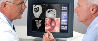
The fluoroscopy techniques of the esophagus, stomach and intestines have been worked out in great detail, are safe and not burdensome for the patient. Its diagnostic results are very effective. For millions of patients, doctors annually conduct fluoroscopic examinations of the gastrointestinal tract both to establish a diagnosis of the disease and to monitor the results of treatment.
Before the contrast agent is administered, the patient lies down on the X-ray machine table. A freshly prepared barium suspension, heated to 33-35 °C, is introduced into the colon using a Bobrov apparatus through a rubber tube without a rigid tip. The contrast enema is introduced gradually under low pressure with visual control on the fluoroscope screen. At the same time, they try to use the minimum amount of contrast possible and are always guided by the patient’s feelings. If unpleasant sensations appear, the administration of the contrast enema is suspended and continued only after they disappear.
When contrast is administered, the doctor observes the straightening of the intestine and the condition of its contours. They are also interested in the speed and uniformity of the advancement of the contrast mass and the places where it is delayed. Pathological changes found during fluoroscopy are subject to radiography (photographs are taken). The examination continues until the contrast mass reaches the cecum (the initial part of the large intestine) and fills it. Once filling of the entire colon has been achieved, one panoramic image is taken. The patient is then asked to have a bowel movement, after which he is again subjected to fluoroscopy. At the same time, attention is paid to how much and in what parts of the colon the contrast mass is retained. In certain diseases, the affected parts of the colon are emptied faster and more completely than healthy parts. If there is a narrowing in any area, then the contrast mass is retained above this place, while the section of the intestine located below is free of contents. After emptying the colon, the relief of the mucous membrane is clearly determined.
In addition to what has been described, there is a method for examining the colon using a combined injection of contrast mass and air. In this case, a contrast suspension is first introduced, then the patient is asked to partially empty the intestines, after which, under the control of the fluoroscope screen, the colon is slowly inflated with air at short intervals. With this method of examination, the relief of the intestinal mucosa is better visualized. This option is used if a tumor is suspected.
The duration of the procedure is about half an hour.
Irrigoscopy does not cause serious complications. With overstretching of the intestine and an overdose of tannin, abdominal pain and painful urge to stool may occur, which disappear within 2-3 hours and do not require special treatment. In such a situation, particularly sensitive patients can take 1 tablet of baralgin. The drug contains analgin, antispasmodic and sedative components. It is contraindicated in cases of low blood pressure, heart failure, heart rhythm disturbances, severe angina, benign prostatic hyperplasia (adenoma).
You should not plan any serious business on the day of irrigoscopy. After the procedure, it is best to go home and rest for the rest of the day.
You may pass some barium residue in your stool for a few days after the test, so you shouldn't be surprised if your stool is lighter than usual. Some patients may experience short-term constipation. In this regard, it makes sense to use castor oil 15 ml in the morning on an empty stomach.
Registration and preparation for complex studies (stomach x-ray, urography, irrigoscopy, etc.) must be discussed with the x-ray laboratory assistant.
Contraindications for surgery
The study is contraindicated in patients who have:
- Acute ulcerative colitis;
- Perforation (formation of a through hole) of the colon;
- Serious condition;
- Suspicion of perforation of the colon;
- Megacolon is toxic;
- Ischemic colitis;
- Arterial hypertension with the presence of tachycardia;
- Lactation;
- Pregnancy;
- Early childhood.
Biopsy
Irrigoscopy should be performed carefully if:
- There is diverticulitis;
- Bloody diarrhea;
- The chronicle was diagnosed with ulcerative colitis;
- There is cystic pneumatosis.
When is a test ordered and what does it show?
Irrigoscopy is prescribed to a patient who consults a doctor with complaints:
- for the presence of pus, blood or mucus in the stool;
- for pain in the anal area;
- for pain in the lower abdomen;
- for alternating diarrhea or constipation.
The study is also indicated if there is a suspicion of a malignant neoplasm of the large intestine or acute obstruction.
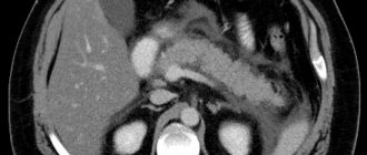
In the absence of pathology, the shape and location of the intestine corresponds to the age norm of the patient. Extensibility and elasticity are uniform along the entire length of the intestine, the lumen is of equal diameter (there are no sharp narrowings or adhesions), the relief of the mucous tissue is without pathological protrusions or depressions, and the contrast does not penetrate above the large intestine.
If ulcers or diverticula have formed in the intestinal mucosa, they are filled with a contrast agent and appear as depressions in the photographs. Polyps, on the contrary, have the appearance of protrusions similar to a mushroom. Inflammation and infiltration can be determined by the presence of thickening of the intestinal wall.
A cancerous tumor has the appearance of a round formation, which can have a smooth surface or be covered with erosions. With intestinal obstruction, a certain section narrows, and the contrast agent does not reach the bauhinium valve, which separates the small and large intestines.
With increased organ tone, the image shows a noticeably narrowed intestinal lumen and increased folding, and with atony, the lumen is expanded and the folds are smoothed
Thus, from the image, the doctor can judge changes in the structure and contraction of the organ, which makes it possible to make or clarify the diagnosis. For example, irrigoscopy makes it possible to see:
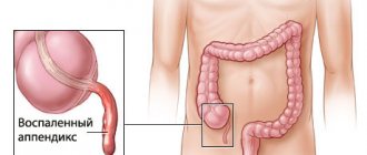
Intestinal diverticulitis
- irritable bowel syndrome (the haustra folds in one place are very relaxed, but in another, on the contrary, they are tense, the lumen is narrowed, after defecation the contrast agent is not completely removed);
- non-ulcerative colitis (the lumen in the colon is of unequal diameter, which indicates uneven contraction of the muscles of the organ, foci of inflammation are present, in areas where spasm of the folds is pronounced, the distance between them is too small);
- nonspecific ulcerative colitis (ulcers are filled with contrast, which is why the mucous membrane looks like a mesh or spotty, formations of “pseudopolyps” are noticeable, part of the intestine is relaxed, and part is spasmodic);
- diverticulosis (contrast penetrates into diverticula (depressions ranging in size from a millimeter to several centimeters) due to which the intestinal wall has an uneven contour; next to the inflamed diverticulum, thickening of the wall due to infiltration is visible; if the protrusion ruptures, the contrast flows into the abdominal cavity);
- colon cancer (at the initial stage, round formations with smooth edges are noticeable; around the tumor, as a result of edema and atony, the relief of the intestine changes; physiological folds may be absent; accumulations of barium near the formation or on it indicate the emergence and disintegration of the tumor);
- presence of polyps.
Kinds
There are two forms of irrigoscopy.
Procedure with a simple contrast method
This form of irrigoscopy involves the use of barium alone.
This research method should be done for older people and patients whose health has deteriorated in the postoperative period. It is done to determine intestinal motility.
With such filling of the intestine, this method allows you to obtain the clearest contours.
Double contrast procedure
This method involves the use of barium suspension and air.
It reveals tumors, ulcerative defects of the intestine, polyps in the intestinal lumen, intestinal obstructions and diverticula.
Irrigoscopy in Moscow
Experienced diagnosticians of the Russian State Scientific Center perform irrigoscopy with minimal risk to the health of patients. Thanks to high-tech equipment, specialists obtain high-quality images. The results are deciphered on the day of the examination. Further treatment of identified pathologies is possible in the outpatient department or in the hospital.
The center is located in Moscow at the address: 1st Leonova Street, building 16. To clarify prices for services and make an appointment for treatment, call the phone numbers listed on the website or use the online form.
Preparation
It is very important to properly and well prepare the patient for the procedure. If the intestines are not completely cleansed, particles from other procedures remain, there are difficulties with barium retention in the body, or there is a psychological factor, then the result of the study may be distorted.
You need to prepare for the procedure like this:
- For a week, it is recommended to go on a diet with a minimized amount of protein and fiber, as well as foods that promote gas formation;
- Four days in advance, it is necessary to discontinue medications that increase or decrease intestinal motility;
- The day before irrigoscopy, you can fast for medicinal purposes and drink water heavily so that you can drink about three liters per day;
- From six o'clock in the evening the night before irrigoscopy you do not need to eat or drink;
- Requires bowel cleansing with an enema and laxatives.
If you are taking medications that affect blood clotting or anti-inflammatory drugs, your doctor should indicate their use. The day before surgery, all medications are discontinued, with the exception of insulin.
Differences from colonoscopy
Irrigoscopy or colonoscopy - which is better for the intestines? Before answering this question, it is necessary to highlight their main distinguishing features. This:
- Execution method
. Colonoscopy does not require the use of contrast or x-rays. Unlike competitive medical manipulation, it only uses a flexible probe with a camera. - Purpose
. Irrigoscopic examination is used exclusively for diagnostic purposes (except for intussusception in children), and during colonoscopy, polyps can be removed, bleeding can be stopped, or a biopsy can be taken.
Carrying out the operation
The patient is placed on one side. Using an enema, the intestines are filled with barium, which is ready for the procedure. X-ray images are taken at the same time.
X-ray images show the location of the intestine, its length, shape, how elastic and stretchable it is.
When emptying the intestine from barium, an image is taken with air to understand whether there are functional or organic disorders in the intestinal wall.
This procedure usually takes half an hour if double contrast is done.
The results take from thirty minutes to three days to interpret (it all depends on the place where the procedure was performed).
Irrigoscopy is done without painkillers or anesthesia. It is painless, but may cause some discomfort.
Technique
The duration of the examination is 30-40 minutes
. Before it starts, an x-ray is taken of the organs in a supine position, after which:
- The patient lies on his side, raises his legs to his stomach and puts his arms behind his back.
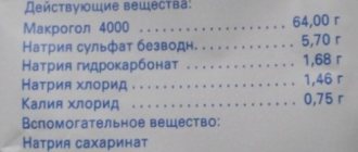
- The X-ray contrast agent is barium sulfate: which is first diluted with water and heated.
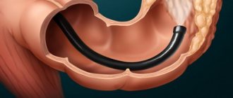
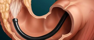
- The endoscopist inserts it into the rectum through a tube (in rare cases, the contrast is given orally). For barium to spread throughout all intestinal sections, you must wait about 2 hours. For uniform distribution, the patient will need to remain in a motionless lying position.
- Air is pumped into the intestines, straightening the folds, and several photographs are taken.
- Air is removed through the equipped valve, and the endoscope is removed.
Using double contrast, it is possible to diagnose colon cancer with high accuracy.
What complications
Typically, this type of research is not considered dangerous. Complications during irrigoscopy occur extremely rarely. It happens that a perforation of the intestine occurs and the contrast agent enters the peritoneal cavity. In this case, urgent surgery will be needed.
If suddenly after the operation signs such as nausea, vomiting, fever, pain in the abdominal area, dizziness and weakness up to loss of consciousness appear, then you need to be wary.
A feature of this type of research is the ease of execution, the ability to carry out the procedure with little equipment, and the procedure’s low invasiveness. Further, X-ray images will become visual material for subsequent consultations with doctors to assess the patient’s health status.
The Onco.Rehab clinic guarantees painless and high-quality diagnostics, which will help you in treating the disease.
What is irrigoscopy?
The human intestine is a hollow organ. It is not visible on a regular X-ray because the rays pass through the intestinal walls. During irrigoscopy, visualization is ensured by the introduction of a contrast agent - barium suspension.
After filling the large intestine with a contrast agent, the doctor takes pictures in different projections. Conventional irrigoscopy shows only large abnormalities. To detect minor pathological changes, including tumors, irrigoscopy with double contrast is performed: after taking images with a barium mixture, additional images of the intestine, previously filled with air, are taken. This allows you to examine the mucous membrane in more detail.
Structure of the apparatus
Irrigoscopy - procedure
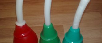
The irrigoscopic device, based on the examination of various areas of the intestine, is designed quite simply. The basis of this device is a small container with two tubes attached to it. The existing container can hold from one to three liters of a special substance. A special contrast agent necessary for the examination is poured into the bottom of the container. By the way, without this substance it is simply impossible to study the structure of the intestine.
Next, a special bulb is attached to one end of the tube to allow air to be pumped. At the second end of the other tube there is a special tip, which is inserted directly into the intestine when examining the intestines. The material from which the tubes are made is silicone. The choice of this composition by the designers is not accidental; the tubes can be used repeatedly, cleaning them from various microorganisms through conventional sterilization. At the same time, this material is absolutely safe for the human body.
The metal parts of the device are made of high quality stainless steel. The tips of this device are made in different dimensional units. This condition helps to use the device for both adults and children. This simple structure of this device makes it possible to detect quite serious malfunctions in the intestines. Along with the device, the kit includes detailed operating instructions and relevant certificates that guarantee the quality of the manufactured device.
Patient preparation
Before the patient is examined using an irrigoscopic device, he needs to prepare a little for this procedure. To do this, he must be on a special diet for about three days before the examination. This diet provides for the complete exclusion from the human diet of rye bread, potato and spicy dishes, the consumption of apples and mushrooms, legumes, chocolate products, and cabbage. During the diet, the patient is prohibited from consuming all types of gas-forming drinks.
The diet should not contain fresh herbs, raw vegetable and fruit dishes, as well as berries. The patient is recommended to eat more cereals during the diet, especially semolina, fermented milk products, soups, boiled meat or fish and eggs. Drink more tea, mineral water or juices.
What is the essence of irrigoscopy?
A regular x-ray does not show internal organs. X-ray beams pass right through them. But if you introduce a contrast agent into the required area and take pictures, you will be able to see all the details of a previously invisible organ and examine all the changes that are not possible with other methods.
When choosing where to have irrigoscopy, remember: you should undergo the examination with experienced diagnosticians, using high-tech equipment, so that the ratio of the effectiveness of the procedure and your comfort is maximum. In Clinic No. 1, the X-ray room is equipped with modern digital equipment, which provides minimal exposure to X-ray radiation, excellent visualization, and accurate interpretation of images. Our radiologists have extensive experience.
Irrigoscopy analysis
In the absence of pathological changes, the radiologist will see in the picture a swollen intestine with clear physiological bends and a smooth mucosal surface.
Irrigoscopy makes it possible to detect even small malignant formations, diagnose cancer at an early stage and, thereby, giving the patient a chance for recovery. The photographs show damage, mucosal ulcers, scars, diverticula, etc. The border between healthy and damaged areas of the mucosa is clearly visible.
The data allows the doctor to make the correct diagnosis and prescribe adequate treatment.
Contraindications
Irrigoscopy is not prescribed to pregnant women, since the patient must receive a dose of radiation, and this presumably can negatively affect the development of the fetus. Also, the study is not carried out if the patient has problems with the myocardium and coronary vessels or a high pulse rate, since fear associated with the procedure can worsen the functioning of the cardiovascular system.
Irrigoscopy is contraindicated if there is a suspicion of a violation of the integrity of the intestinal wall (as a result of perforation of an ulcer, rupture of a diverticulum or tumor disintegration), since the contrast agent can leak into the abdominal cavity and cause a number of pathologies. At the discretion of the doctor, the examination is carried out, but not barium sulfate is used as a contrast, but an aqueous solution of radiopaque substances.
In case of diverticulitis and ulcerative colitis, irrigoscopy should be carried out with extreme caution, since an organ overfilled with air or contrast agent may rupture at the site of the lesion. In order not to provoke complications, the barium suspension and air are supplied under low pressure.