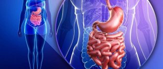
Malabsorption
In case of malabsorption syndrome, the main signs are identified by laboratory testing of blood, feces, and urine. A general blood test may show signs of anemia (iron deficiency and B12 deficiency); vitamin K deficiency affects the prothrombin time (extension occurs). During biochemical analysis, the level of albumin in the blood, calcium and alkaline phosphatase are noted. A study of the amount of vitamins is being carried out.
Stool examination shows an increase in its daily volume (with fasting - a decrease). The coprogram reveals the presence of muscle fibers and starch in the feces. With some enzyme deficiencies, the pH of the stool may change. If fatty acid malabsorption is suspected, a test for steatorrhea is performed.
Before collecting stool for research, it is necessary for the patient to consume about 100 grams of fat per day for several days. Then daily feces are collected and the amount of fat in it is determined. Normally, it should not be more than 7 grams. If the fat content in the stool exceeds this value, malabsorption may be suspected. If the amount of fat is more than 14 grams, functional disturbances in the functioning of the pancreas are likely. In cases of severe malabsorption and celiac disease, half or more of the dietary fat is excreted in the feces.
Functional tests to detect absorption disorders in the small intestine - D-xylose test and Schilling test (B12 absorption assessment). As an additional diagnostic measure, a bacteriological examination of stool is performed. X-ray examination can reveal signs of disease of the small intestine: blind loops, horizontal levels of liquid or gas may form in some loops, interintestinal anastomoses, diverticula, strictures, and ulcerations are visible.
When performing abdominal ultrasound, MSCT and magnetic resonance imaging, the abdominal organs are visualized and their pathologies are diagnosed, which may be the root cause of the developed malabsorption syndrome. Endoscopy of the small intestine reveals Whipple's disease, amyloidosis and intestinal lymphangiectasia, allows you to collect material for histological examination, and aspirate intestinal contents for bacteriological examination (for excessive contamination of the small intestine with microorganisms and the presence of pathological flora).
As additional diagnostic measures, the state of the exocrine pancreas is assessed (secretin-cerulein, bentriamine, LUND and PABA tests, determination of the level of immunoreactive trypsin); Bacterial overgrowth syndrome is detected (hydrogen and carbon dioxide breath tests); lactase deficiency is diagnosed (lactose test).
Publications in the media
Malabsorption syndrome can be caused by a defect in any stage of digestion or pathology of any of the gastrointestinal tract organs.
Etiology • Hypervitaminosis and hypovitaminosis E • Syndrome of impaired absorption of glucose and galactose (*182380, 22q13.1, transmembrane sodium glucose transporter gene SLC5A1, r).
Clinical manifestations vary, because malabsorption may involve one or more nutrients • Changed foul-smelling stool, films of fat or drops of oil can be observed on the surface of the toilet water • Loss of body weight • Edema and ascites secondary to hypoalbuminemia • Secondary anemia due to impaired absorption of iron, vitamin B12, folic acid or a combination thereof • Bone pain or fractures due to vitamin D deficiency • Paresthesia or tetany due to calcium deficiency • Bleeding due to vitamin K deficiency • Neuropathies and other manifestations of hypovitaminosis E • Muscle weakness due to potassium deficiency.
The diagnosis is based on clinical findings and confirmed by laboratory tests.
• Analysis of fat content in feces. A positive Sudan stain indicates fecal excretion of more than 15 g of fat per day. Fat content is determined in 72-hour stool samples. Normally, 93–95% of dietary fats are absorbed. Pancreatic disease is often associated with fat excretion in excess of 20–30 g/day.
• The d-xylose absorption test, which does not require enzymatic degradation or micelle formation for absorption, is used to assess the integrity of the intestinal mucosa. Take 25 g orally. Urine collected over a 5-hour period should contain at least 4–5 g of d-xylose.
• Test for unabsorbed carbohydrates. The entry of unabsorbed carbohydrates into the large intestine and their fermentation by bacteria reduces the pH of the stool; The condition is especially common in lactase deficiency, as well as in celiac disease and short bowel syndrome.
• Study of pancreatic functions. Bicarbonate content and total fluid secretion from the duodenum after secretin stimulation followed by the use of pancreatic chymotrypsin (bentyramide test) to release para-aminobenzoic acid excreted in the urine; urinary excretion of less than 60% suggests pancreatic insufficiency.
• Measuring the level of carotene in the blood. Vitamin A is fat soluble, and carotene levels are determined by vitamin metabolism; A low serum carotene level with normal vitamin A intake may be helpful in assessing fat malabsorption.
• Bacterial growth •• Culture of jejunal aspirates. Deviation from the norm: >104 microorganisms/ml. It is safe to assume dysbiosis in the presence of obligate anaerobes (clostridium and bacteroides), facultative anaerobes (lactobacillus and enterococci) or coliform bacteria •• Breath tests for the presence of bile salts are based on the ability of bacteria to deconjugate 14C-labeled glycine cholate before it can suck in. 14C-Glycine is then transformed into 14CO2 and measured in the exhaled breath of patients to detect bacterial overgrowth •• Urinary indican and 5-hydroxyindoleacetic acid test: elevated levels due to increased tryptophan metabolism; the test is nonspecific, because changes in the levels of tryptophan metabolites are also detected in carcinoid syndrome and Whipple's disease.
• X-ray of the small intestine is an informative diagnostic method, especially with air contrast. Sequestration or flocculation of barium suggests celiac disease. Thickened folds are seen in Whipple's disease, lymphoma, amyloidosis, radiation enteritis, Zollinger-Ellison syndrome, and eosinophilic enteritis.
• Schilling excretory test. Used to diagnose vitamin B12 malabsorption. The first stage of the Schilling test is the intake of 57Co-labeled vitamin B12, followed by determination of urine radioactivity. The second stage is to repeat the test after introducing the internal factor. Incomplete urine collection is a common cause of underestimated results, so radioisotopes in the blood plasma are analyzed at the same time. Low levels in blood plasma and urine indicate decreased absorption. Normalization of the results after the second stage of the test indicates the presence of B12-deficiency anemia and intrinsic factor deficiency. Low second-stage excretory test scores are often due to severe celiac disease, bacterial overgrowth, resection, or inflammation of the terminal ileum where absorption occurs. The presence of a high titer of antibodies to intrinsic factor in the gastric juice can also cause an underestimated result in the test with the intake of intrinsic factor.
• Biopsy of the small intestine. Flattening of the villi with infiltration of inflammatory cells is characteristic of celiac disease; isolated flattening of the vills is observed in infectious enteritis, giardiasis, lymphoma and bacterial overgrowth.
Treatment tactics for specific causes of malabsorption • Pancreatic insufficiency occurs in chronic pancreatitis, pancreatic carcinoma, cystic fibrosis. Changes in tests with bentiramide or secretin (pancreatic function test) are possible. Pancreatic enzyme replacement therapy is performed • Bacterial overgrowth accompanies changes in peristalsis (eg, diabetes and amyloidosis), small intestinal diverticula, strictures (eg, lymphoma and Crohn's disease) or the formation of blind loops after Billroth-II gastrectomy. Antibacterial therapy with ampicillin or tetracycline is often successful, and surgical correction of anatomical changes is possible • Celiac disease - see Celiac disease • Whipple's disease - see Whipple's disease.
ICD-10 • K90 Intestinal malabsorption
MALABSORPTION SYNDROME IN CHILDREN (PART 1)
Malabsorption syndrome is a difficult diagnostic problem not only for a pediatrician, but also for a gastroenterologist. This is due both to the variety of causes of intestinal absorption disorders, the polymorphism of their clinical manifestations, and to difficulties in diagnosis, which often requires the use of labor-intensive, expensive, invasive and fairly specific methods
A significant limitation of diagnostic capabilities, combined with a lack of modern reliable information about diseases accompanied by malabsorption syndrome in our country, have become the causes of all sorts of “distortions” in the diagnosis and treatment of this pathology. Thus, in our country, primary lactase deficiency is unjustifiably widely diagnosed in children in the first year of life - that is, at an age when it cannot exist by definition. Late forms of celiac disease in older children, on the contrary, are not sufficiently detected. We do not diagnose the majority of diseases associated with malabsorption syndrome at all, because we have neither the capabilities nor the appropriate knowledge for this.
We have planned a series of articles in which we will try to highlight the problem of malabsorption syndrome in childhood, as widely as possible, but at the same time mainly in the applied aspect. The first part will be devoted to general diagnostic issues, and the subsequent ones will be devoted to specific diseases and pathological conditions.
Definition and classification
The syndrome of impaired intestinal absorption or malabsorption syndrome (MS) can accompany a number of diseases and is not an independent diagnosis. The term “malabsorption” is used to refer to conditions in which there is a decrease in the absorption of one or more food nutrients in the intestine. Malabsorption may result from impaired breakdown (digestion) of nutrients in the intestinal lumen or defects in mucosal absorption. All diseases accompanied by SM can be divided into 2 groups: diseases with generalized damage to the intestinal mucosa, in which the absorption of many nutrients is usually impaired (Table 1), and diseases in which there is a predominant malabsorption of individual nutrients - proteins, fats , carbohydrates, vitamins or microelements (Table 2). Almost all diseases with SM are accompanied by chronic diarrhea, which aggravates malabsorption.
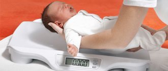
Clinical features of malabsorption syndrome depend on the nature and degree of malabsorption of nutrients. General symptoms typically include diarrhea, bloating, decreased weight gain, or even weight loss, which appears as flattening and stagnation on body mass index charts. Physical examination reveals muscle atrophy and the disappearance of the subcutaneous fat layer with the formation of hanging folds of skin. The consequences of malnutrition are most pronounced in young children, since their energy reserves are limited and their caloric needs are relatively higher due to increased linear and volumetric growth in this age period. In older children, SM may result in a slower rate of linear growth, as is often seen in children with late-onset celiac disease. In the absence of adequate treatment for SM, long-term malnutrition can be fatal, although today this is observed mainly in developing countries.
Some diseases accompanied by SM are characterized by certain specific symptoms. Thus, edema, as a rule, is associated with exudative enteropathy, drumstick-type nail changes are associated with cystic fibrosis and celiac disease, perianal excoriation and severe flatulence are detected with carbohydrate malabsorption, perianal and perioral rashes with enteropathic acrodermatitis; Menkes syndrome is characterized by abnormal hair, and Johanson-Blizzard syndrome is characterized by typical facial features.
Children with SM often have an increased appetite, which helps them compensate for the loss of protein and energy in their feces. Thus, with exocrine pancreatic insufficiency, signs of decreased nutrition appear only when the level of loss of consumed protein and energy begins to exceed 40%, if these losses are compensated by increased appetite.
In other situations, with diseases associated with villous atrophy or inflammation of the intestinal mucosa (celiac disease, post-infectious enteropathy), on the contrary, anorexia develops, so even with small losses of protein and energy, signs of malnutrition appear early.
Thus, assessment of nutrition (weight gain, changes in body mass index) is the most important part of the diagnosis of malabsorption syndrome.
In addition, it is important to identify signs of deficiency of certain nutrients. Long-term malabsorption of calcium and vitamin D can lead to decreased bone mineral density and the development of metabolic bone diseases with an increased risk of bone fractures.
Vitamin K malabsorption, regardless of the cause (impaired fat absorption, mucosal atrophy), can lead to coagulopathy. When iron absorption is impaired, microcytic hypochromic hyporegenerative anemia develops. Anemia may also occur when serum folic acid levels are reduced due to atrophy of the gastrointestinal mucosa. Low serum concentrations of vitamin A and vitamin E are usually due to fat malabsorption.
A carefully collected medical history can help you choose a more structured and rational approach to further examination. Thus, indications of the onset of diarrhea in very early infancy suggest the presence of a congenital defect (Table 3). However, secretory diarrhea can be a manifestation of such disorders as congenital chloride diarrhea and microvilli inclusion disease. The stool in these diseases is so copious and watery that it can be mistaken for urine. The appearance of symptoms after the introduction of certain foods into a child’s diet can also have diagnostic significance, as, for example, after the introduction of sugar-containing foods in case of sucrase-isomaltase deficiency. The nature of diarrhea also matters: watery diarrhea with an “explosive” character of the stool indicates a malabsorption of carbohydrates, voluminous unformed feces are observed in celiac disease, pasty, fatty, yellowish and with an unpleasant odor are observed in exocrine pancreatic insufficiency.
But the color of stool usually doesn’t mean anything. For example, green feces with undigested pieces of vegetables or other food only indicate accelerated intestinal transit in a child with diarrhea of almost any origin.
The scope and nature of additional diagnostic tests depend on the history and physical examination. In children with chronic or recurrent diarrhea, the initial examination should include stool culture, coprological examination (including determination of the presence of helminth and parasite eggs, leukocyte content), and stool occult blood test.
Impaired absorption of carbohydrates is indicated by a decrease in stool pH and an increase in the level of reducing substances in the stool. Quantitative determination of fat and fecal a1-antitrypsin levels is indicated to confirm fat and protein malabsorption, respectively. To diagnose exocrine pancreatic insufficiency, the content of elastase-1 in the feces is determined.
A complete blood count can reveal microcytic anemia (iron deficiency), lymphopenia (with primary or secondary intestinal lymphangiectasia), neutropenia (Shwachman syndrome), acanthocytosis (with abetalipoprteinemia). If celiac disease is suspected, the level of IgA antibodies to tissue transglutaminase (TG2) in the blood serum is determined. Further, depending on the results of the initial tests, more specific studies may be carried out.
Studies for carbohydrate malabsorption
A screening test for carbohydrate malabsorption is the determination of reducing substances in the stool.
This method is based on a reaction that allows one to detect the presence of sugars that have reducing activity (the ability to reduce copper from the Cu2+ state to Cu1+). These include glucose, galactose, lactose, fructose, and maltose. Sucrose (like starch) does not have this ability.
Normally, the content of sugars with reducing activity in feces is insignificant. Exceeding the reference values (more than 0.25%) in combination with acidic stool pH values indicates the presence of disturbances in the breakdown and absorption of sugars.
Hydrogen breath tests are used to diagnose specific types of carbohydrate malabsorption. After an overnight fast, the suspected sugar (lactose, sucrose, fructose or glucose) is administered orally in the form of a solution (at a rate of 1–2 g/kg, not more than 50 g of carbohydrates).
If this carbohydrate is not broken down or absorbed in the small intestine, it enters the large intestine, where it is metabolized by normal microflora to produce hydrogen. Hydrogen is absorbed through the mucous membrane of the colon and excreted through the lungs.
An increase in the concentration of hydrogen in the exhaled air after a carbohydrate load confirms the presence of malabsorption of this carbohydrate. An increase in hydrogen concentration by 20 ppm (ppm –1´10-6) compared to the initial one is considered diagnostically significant.
Before and during the study, the child should not receive antibiotics, as they suppress the activity of intestinal microflora necessary for the fermentation of sugars.
Using a biopsy of the small intestinal mucosa, the concentrations of disaccharidases (lactase, sucrase, maltase, palatinase) in it can be measured directly.
With primary enzyme defects, low levels of disaccharidases are combined with normal morphology of the intestinal mucosa. Partial or complete villous atrophy, caused, for example, by celiac disease or rotavirus gastroenteritis, can lead to secondary disaccharidase deficiency. In this case, the levels of disaccharidases are normalized as the mucous membrane of the small intestine is restored.
Studies on fat malabsorption
The presence of globules or droplets of fat in the stool indicates a malabsorption of fats. The ability to metabolize fats depends on age. Thus, the intestines of a premature newborn absorb only 65–75% of dietary fat consumed, in a full-term newborn this figure is almost 90%, and in an older child, about 95% of the fat received from food is absorbed. The gold standard for confirming fat malabsorption is to measure the amount of fat absorbed. To do this, feces are collected for 72 hours (provided that the patient consumes at least 100 g of fat per day) and the fat absorption coefficient is calculated based on the fat content in it using the formula:
fat absorption coefficient, % = (fat consumed (g) – amount of fat in feces (g)/fat consumed (g)) ´ 100%.
However, such a study is quite labor-intensive, expensive, and unpleasant for analysis, so it is used only in some specialized centers. And for clinical practice it is more convenient to use such a simple method as steatocrit.
Steatocrit is a gravimetric method for estimating the percentage of fat in an individual stool sample; it has high sensitivity and specificity (when compared with the standard - a study of fat loss in 72 hours). Take 0.5 g of feces, mix it with 0.05 g of sand and 2 ml of water. The resulting mass is filled into a microhematocrit tube, centrifuged, and the percentage of the fat layer is measured (similar to hematocrit measurement). For children older than 6 months, a steatocrit of less than 10% is considered normal, 10–20% is considered a borderline result, and more than 20% is considered steatorrhea.
If a deficiency of bile acids is suspected as the cause of malabsorption of fats, then a useful test may be to determine their level in the duodenal contents.
With fat malabsorption, including that caused by exocrine pancreatic insufficiency, the absorption of fat-soluble vitamins A, D, E and K is usually impaired, which is accompanied by a decrease in their concentrations in the blood serum. An increase in prothrombin time may indirectly confirm the presence of vitamin K deficiency.
Studies for exocrine pancreatic insufficiency
Cystic fibrosis is the most common cause of pancreatic insufficiency in children, so if its presence is suspected, determination of the level of chlorides in sweat should be carried out first (even if the results of neonatal screening are negative).
Fecal elastase-1 is a sensitive test for assessing exocrine pancreatic function in cystic fibrosis and chronic pancreatitis. Elastase-1 is a stable endoprotease that is not destroyed by exogenous pancreatic enzymes.
The only drawback of the method is the lack of a clear distinction between primary pancreatic insufficiency and secondary exocrine pancreatic dysfunction due to intestinal villi atrophy. The proximal small intestine produces pancreozymin/cholecystokinin, a hormone that stimulates the secretion of pancreatic enzymes. With atrophy of the mucous membrane, a decrease in the production of pancreozymin/cholecystokinin leads to disruption of the exocrine activity of the pancreas. In addition, fecal elastase-1 levels may be low during episodes of acute diarrhea.
A decrease in the concentration of elastase-1 in feces to 100 mg/g or less indicates the presence of exocrine pancreatic insufficiency and the need for enzyme replacement therapy. At borderline values (100–200 mg/g), repeated determination of elastase-1 is recommended (in three independent stool samples) and then, depending on the clinical situation and the results obtained, either further examination (imaging methods, endoscopy) or dynamic observation with study of fecal elastase concentration once a year.
Serum trypsinogen concentration can be used as a screening test for the presence of exocrine pancreatic insufficiency. In cystic fibrosis, its level is significantly increased in the early period of life.
Subsequently, trypsinogen levels gradually decrease, so that by the age of 5–7 years, in most patients with cystic fibrosis with pancreatic insufficiency, its concentrations correspond to subnormal values. Cystic fibrosis without pancreatic insufficiency usually has normal or elevated serum trypsinogen levels. In such patients, monitoring serum trypsinogen concentrations can be used to monitor exocrine pancreatic function. In Shwachman syndrome, another disease associated with exocrine pancreatic insufficiency, serum trypsinogen levels are low.
Other studies (NBT-PABA (nitroblue tetrazolium-para-aminobenzoic acid) test and pancreolauril test) involve measuring in urine or exhaled air the concentrations of substances released or absorbed in the intestine under the influence of pancreatic enzymes. These tests are low specific and are rarely used in clinical practice.
The “gold” standard for studying the exocrine function of the pancreas is direct analysis of duodenal contents with assessment of its volume, concentration of bicarbonates, trypsin and lipase after stimulation with secretin and pancreozymin/cholecystokinin. This test requires duodenal intubation and the presence of specially equipped laboratories, and is therefore performed only in some specialized centers.
Studies for exudative enteropathy (protein-losing enteropathy)
Dietary proteins and endogenous proteins secreted into the intestinal lumen are almost completely absorbed: less than 1 g of protein from these sources reaches the colon. Most of the nitrogen in stool comes from proteins from intestinal bacteria. Significant protein losses through the intestines lead to hypoalbuminemia with corresponding clinical manifestations (edema).
When identifying hypoalbuminemia in children, the level of protein excretion in the urine is first determined, since kidney disease is the most common cause of hypoalbuminemia. Other potential causes of low blood albumin levels include liver disease (decreased production) and insufficient dietary protein intake. Very rarely, hypoalbuminemia develops due to massive protein loss through the skin.
A screening test for intestinal protein loss is the level of a1-antitrypsin in the stool. This whey protein has a molecular weight close to that of albumin, but, unlike the latter, a1-antitrypsin is resistant to the action of proteolytic enzymes in the gastrointestinal tract. If the excretion of a1-antitrypsin in feces increases, further research is required to clarify the cause of exudative enteropathy (we wrote more about the causes, differential diagnosis and algorithm for examining children with exudative enteropathy in No. 2 (52) for 2015).
Research in diseases of the intestinal mucosa
Diagnosis of a number of diseases accompanied by malabsorption syndrome requires histological examination of biopsy samples of the small intestinal mucosa. A biopsy is performed during an endoscopic examination; the general rule is to take several samples from different parts of the intestine, since damage to the mucous membrane can be expressed to varying degrees, especially in celiac disease. To diagnose congenital microvilli atrophy, the PAS reaction (or PAS (Periodic Acid Schiff) reaction) is used, followed by electron microscopy.
With intestinal lymphangiectasia, damage to the mucous membrane can be segmental. In this case, a series of x-rays or repeated ultrasound examinations of the small intestine can help identify the thickened area of the intestine through which protein loss occurs.
In addition, the activity of mucosal disaccharidases can be determined in biopsy samples of the small intestinal mucosa.
Duodenal aspirate obtained by endoscopy may be examined for pancreatic enzyme concentrations, bacterial cultures, and the presence of other pathogens (eg, some helminths and protozoa).
In the following issues, we will consider approaches to the diagnosis and treatment of specific diseases manifested by malabsorption syndrome.
The main causes of spasms in children
The diseases and causes that can lead to this problem are quite extensive. It is worth noting that in children, spasms and pain are most often observed in the gastrointestinal tract and accompany various organic and functional diseases of the stomach and intestines. The difference between organic and functional diseases lies in the presence of structural or biochemical changes in organs and tissues. Organic diseases have them, but functional ones do not. Functional pathology is caused by changes in the regulation of these organs, which can also cause symptoms similar to organic ones. In young children, spasms are more often observed against the background of functional pathology. With age, the incidence of organic pathology increases. The most common causes of excessive contraction and abdominal pain in children are:
- Intestinal colic.
- Functional diarrhea and constipation.
- Acetonemic syndrome.
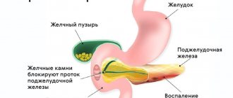
- Biliary dyskinesia, inflammation of the gallbladder.
- Gastritis, peptic ulcer.
- Diseases accompanied by malabsorption syndrome. Malabsorption is a malabsorption of substances in the small intestine.
- Inflammatory bowel diseases.
- Chronic colitis, intestinal infections, parasitic infestations.
There are also many other diseases that are accompanied by excessive contraction of muscle cells and pain. We list only the most common of them. Often spasms are accompanied by inflammatory diseases of the urinary tract (cystitis, pyelonephritis). In this case, they occur in the urinary tract. Often, during the formation of the menstrual cycle, teenage girls may experience severe cramps and pain in the lower abdomen. This problem is called dysmenorrhea. Vascular spasms often develop when the temperature rises in children, which is characterized by the appearance of “white fever”. In this article we will briefly talk about the main causes of spasms in children, as well as the diseases that lead to them. Let us explain the basic principles of use and the mechanism of action of drugs used for this pathology.
There are many other diseases that are accompanied by excessive contraction of muscle cells and pain.
Symptoms of spasms in children
As mentioned above, the most common cause of spasms in children is pathology of the gastrointestinal tract.
Below we consider the main symptoms of gastrointestinal diseases accompanied by spasms in children:
- Intestinal colic. This pathology is characterized by sudden and severe bouts of crying in the baby. Colic appears during the neonatal period. Intestinal colic is associated with immaturity of the nervous regulation of intestinal activity and increased gas formation against the background of intestinal dysbiosis.
- Functional diarrhea and constipation. Spasms in these pathologies are not the main symptom. They can only appear periodically. The diseases are functional disorders of young children, which are accompanied by diarrhea or constipation, respectively. At the same time, an in-depth examination does not reveal any other pathology.
- Acetonemic syndrome. Often occurs in children, especially during early childhood. As a rule, the cause of its development is an error in diet. Other reasons include ARVI, harmless excitement (birthdays, etc.), stress. In this case, the pain is no longer caused by spasm, but by irritation of the mucous membrane with ketone bodies (those substances that cause acetone syndrome). Characteristic symptoms of this disease are the smell of acetone in the exhaled air, nausea, vomiting, lethargy, and drowsiness.
- Biliary dyskinesia, inflammation of the gallbladder . Dyskinesia is a functional disorder of the gallbladder and bile ducts, which may be accompanied by spasms in these organs. As a result, children experience pain, often in the right hypochondrium. In addition, children feel heaviness in the right hypochondrium, bitterness in the mouth, and yellowness of the skin and eyes appears.
- Gastritis and peptic ulcer. The causes are often Helicobacter pylori. Children experience: nausea, often vomiting, abdominal pain, heartburn, tendency to constipation, bad breath.
With gastritis, children often experience nausea, vomiting, and abdominal pain.
- Malabsorption syndrome . Among the pathologies in this group, it is worth highlighting celiac disease and lactase deficiency. Celiac disease is a gluten intolerance that is accompanied by a variety of symptoms, including abdominal pain. Lactase deficiency is an intolerance to dairy products, after consuming which occurs bloating, abdominal pain and diarrhea.
- Inflammatory bowel diseases. These include Crohn's disease and ulcerative colitis. It is most often characterized by the appearance of blood in the stool and abdominal pain. Subsequently, symptoms intensify and other signs appear.
- Intestinal infections. Acute intestinal infections are manifested by fever, often vomiting and diarrhea, which is characterized by persistent progression. As a rule, it can develop after eating bad foods or after contact with a sick person.
In addition to pathology of the gastrointestinal tract, other diseases can lead to the development of spasms and pain. Urinary tract infections are quite common, especially in girls. This is due to the short and wide urethra, through which pathogenic bacteria can quickly penetrate. The development of cystitis is accompanied by the appearance of symptoms such as pain when urinating, pain, frequent urination, burning and pain in the urethra and bladder. Teenage girls often develop dysmenorrhea, which is characterized by pain and heaviness in the lower abdomen.