Causes of inflammation of the papillae on the tongue
If you have an inflammatory process in your mouth, contact your dentist immediately. Family dentistry in St. Petersburg is a clinic where every patient will be provided with assistance. The doctor will determine the causes of the disease and prescribe treatment. Problems may arise:
- When the patient’s diet includes excessive amounts of sour, sweet, spicy foods;
- If chronic stress develops in the body;
- When the patient received a tongue injury, burn, cuts, punctures;
- If the organ is affected by an allergic reaction;
- For some diseases of internal organs;
The surface of the muscular organ changes significantly under aggressive influence. The provoking factor is xerostomia, disease of the salivary glands, Sjogren's syndrome, diabetes mellitus. With glossitis, the sensitivity of the papillae increases. During the examination, the doctor must exclude HPV, leukoplakia, aphthous stomatitis, syphilis, and oral fibroma. Children are examined for scarlet fever.
Sometimes the disease can be triggered by acid reflux, gastritis, throat infections, systemic inflammation of the gingival tissues when the pathogenic microorganism develops rapidly. Taking some medications can cause pain. High concentrations of alkali or acid have a negative effect on the condition of the muscular organ. Injuries on the tongue can occur from a hard brush, dentures, or tartar.
Symptoms
When the papillae on the tongue become inflamed, tissue sensitivity increases significantly and taste changes. Patients complain of unpleasant discomfort in the mouth. The taste spectrum is distorted and becomes unnatural. In addition, a number of other symptoms appear.
- Noticeable tissue swelling.
- Marked increase in size of the papillae.
- Tingling and itching.
- Burning, mild soreness.
If you do not consult a dentist in time, complications may develop: lingual eruptive papillitis. This disease occurs over a long period of time with an increase in body temperature. Most often the disease affects children. An alarming symptom is the appearance on the surface of the vocal organ of bubbles (pustules) filled with a transparent liquid. When the first signs of illness appear, you should immediately visit the dentist. Timely treatment of the disease minimizes complications and speeds up recovery.
Diagnosis and treatment methods for glossitis
In most cases, the basis for diagnosis is the patient’s complaints and visual examination of the oral cavity.
When visiting a doctor, be sure to indicate when the pathological manifestations appeared and what they are associated with. If any dental treatment was performed, be sure to mention it. It is better to seek medical help at the first signs of illness. Avoid taking any medications, especially antibiotics. This may make diagnosis difficult and reduce the effectiveness of subsequent therapy.
To determine the cause of glossitis, additional diagnostic methods are used: examination of scrapings from the mucous membrane under a microscope, ELISA, PCR. These methods make it possible to determine what pathogen caused the disease, as well as to confirm or exclude syphilitic glossitis.
Determining the causative agent of the disease is the basis for successful treatment. The emphasis is on etiotropic therapy, that is, eliminating the cause of the disease. If glossitis is bacterial in nature, properly selected antibiotics will give an excellent result, but these drugs are completely useless for viral or candidal glossitis. For fungal infections, antimycotic drugs are prescribed; for viral infections, antiviral and immunomodulators are prescribed. If the cause of glossitis is anemia, treatment of the underlying disease is necessary. Special tactics are necessary for the pathological process caused by Treponema pallidum. A dermatovenerologist must be involved in the treatment of syphilitic glossitis.
An integral part of glossitis treatment is diet. In the acute period, eating food causes serious difficulties for patients, so porridges, purees, and pureed soups are recommended. Food should not be cold or hot. Solid foods, spicy, salty, sour are excluded. This avoids irritation of the mucous membrane, which minimizes discomfort and speeds up the healing process.
In some patients, the pain syndrome is significant. In such a situation, unpleasant sensations can be relieved with the help of applications with anesthetics. To moisturize excessively dry mucous membranes, products containing glycerin are used.
Local therapy includes removing plaque from the tongue and treating with antiseptic solutions. Substances such as chlorhexidine, furacilin, and potassium permanganate are widely used. To speed up the recovery of the mucous membrane, vitamin A is used topically.
Systemic antibiotics and antiviral drugs are prescribed according to indications. This tactic is used in severe cases. With a mild clinical course of glossitis, a positive result can be achieved with local treatment.
In severe cases, swelling of the tongue can be significant and make breathing difficult. In such situations, it is necessary to take glucocorticosteroids. More often they are used in the form of ointments.
During treatment you need to give up smoking and alcohol. These substances irritate the mucous membrane and prevent its regeneration.
Some patients with glossitis have keratinized areas on the tongue. Such changes cannot be treated conservatively and can only be removed surgically.
If you consult a dentist in a timely manner and follow all his recommendations, the outcome of the disease will be favorable. Glossitis responds well to treatment if its cause is determined and targeted. If the disease is not treated, it can be complicated by an abscess or phlegmon. These conditions require surgical treatment and long-term antibiotic therapy, so it is better to start treatment at the initial stage.
Localization
If the papilla on your tongue is inflamed, visit the dentist. Only he will be able to correctly determine the causes of the disease, find out what caused the damage to the mucous membrane of the mouth and the body of the tongue. The area where the disease is localized plays an important role in making the correct diagnosis. Pathology can cover the entire organ, part of it, or the sides. Based on this factor and the clinical picture, we can assume what exactly caused the inflammation.
In all languages
When the mucous layer of an organ is affected over its entire surface, the most common cause is a thermal or chemical burn. One can assume the infectious nature of the pathology. If the damage is severe, there may be no taste at all. Patients complain of severe burning.
On the root
Most often, the papillae on the tongue at the base become inflamed due to the action of allergens. The affected root of the muscular organ causes difficulties such as the perception of bitterness, because the inflammatory process has affected the circumvallate papillae. They are the ones responsible for this function. The patient's salivation increases, and the tissues in the oral cavity swell. This condition can also be caused by problems with the digestive system, too high or low acidity of gastric juice. It is on the back of the speech organ that bacterial or fungal glossitis is localized.
On the tip
The disease can affect the tip. This often occurs due to mechanical injuries or damage. The edge of the tongue is the first to come into contact with excessively hot food or aggressive drinks, which burns this area. Soft tissues often rub against sharp chips of crowns and are subject to accidental biting.
Side
The delicate surface on the sides often suffers from various anomalies. Leaf-shaped, conical, filamentous, mushroom-shaped structures are exposed by the teeth. This leads to an increase in the papillae, their hyperemia due to mechanical damage from fangs and molars. Also provoking factors are painful microorganisms and burns of chemical origin.
Taste organ and taste perception
The formation of a taste sensation occurs due to the perception of taste stimuli by the receptors of the papillae of the tongue. Certain areas of the tongue are responsible for each type of stimulus. Sweet is perceived by the receptors of the tip of the tongue, sour by its lateral zones, bitter by the root of the tongue, salty by the tip of the tongue and its lateral surfaces [1, 9].
On the back and lateral surfaces of the tongue there are various groups of papillae (see figure).
Figure 1. Papillae of the tongue (diagram) according to H. Fenish (cited from [1]).
Filiform and cone-shaped papillae perceive tactile and temperature stimuli, and also perform a mechanical function; mushroom-shaped, grooved (grooved) and leaf-shaped - flavoring. Additionally, lentiform papillae (papillae lentiformes) are also distinguished.
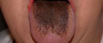
The filiform papillae are the most numerous, located on the dorsal surface of the tongue in the transverse direction in parallel rows, tightly adjacent to each other, so the surface of the tongue has a velvety appearance. In the region of the root of the tongue, these rows of filiform papillae repeat the pattern of the V-shaped groove separating the body of the tongue from its root. As has been established by a number of researchers [1, 6], the sensory function of the papillae is provided by neurofilament protein and protein S-100. The filiform papillae include the lamina propria and epithelium. Each papilla consists of a primary papilla formed by the lamina propria, from which smaller secondary papillae of the lamina propria arise. The primary papilla is covered by epithelium, which also covers each secondary papilla. The name “filiform papillae” is due to the fact that the areas of the epithelium covering the secondary papillae resemble filaments in structure [1]. At the top of the papilla, the threads are split in the form of a “comb”. These areas consist of a substance similar to horny tissue. The epithelium covering the filiform papillae becomes keratinized. The process of desquamation of the surface layer of the epithelium of the filiform papillae is an expression of physiological regeneration. If the function of the digestive organs is impaired, or with general inflammatory or infectious diseases, the rejection of the surface layer of the epithelium of the filiform papillae slows down, the tongue becomes “coated.” The same is observed with tongue inactivity [1, 6, 9].
Cone-shaped papillae are a special type of filiform papillae, differing in the anatomical structure of the apex. In contrast to the “comb” of filiform papillae, their unsplit conical apex is curved backwards at the tip.
Fungiform papillae are located at the tip of the tongue, leaf-shaped papillae are located on the sides, and groove-shaped papillae are located on the root [1, 3, 9]. The latter are surrounded by a roller into which the secretion of small protein glands opens. This is necessary, firstly, to remove food particles from the surface of the papilla, and secondly, in this case the dissolved substance is better perceived by the receptor. This is why poor tongue hygiene reduces its taste sensitivity.
There are fewer fungiform papillae than filiform papillae [1]. These papillae are few in number and randomly scattered among the filiform ones. They are observed more on the tip of the tongue than in other parts of it. They have a narrow base and a widened top. In the region of the epithelium of the apex of the papilla there are taste buds. The largest number of them is located in the area of the tip of the tongue. Each fungiform papilla is formed by its own plate and is called the primary papilla, from which the secondary papilla projects into the integumentary epithelium. However, the surface of their epithelium does not follow the contours of the secondary papillae in the lamina propria, as is observed in filiform papillae. Therefore, the secondary papillae of the lamina propria bring capillaries close to the surface of the epithelium. The covering epithelium of these papillae is relatively transparent, since it does not keratinize [1]. Due to this, the blood in the vessels passing through the high secondary papillae becomes translucent, giving them a red color during life. Normally, the fungiform papillae are pale pink in color and do not stand out above the level of the filiform papillae. In pathology - bright red, hyperplastic; may be the initial stage in the development of erosions and ulcers on the tongue. In case of systemic disorders due to damage to these papillae, loss of taste sensitivity is often observed [1, 2, 9]. Perhaps they are interconnected with the endocrine system. In the thickness of the papillae themselves, a large number of Merkel bodies responsible for sensory function are determined.
They are very similar to mushroom-shaped lentiform papillae, which are wide flat caps on a very short stalk [1]. Their function has not been studied; they suggest a role in the formation of taste sensations. The circumvallate papillae are also organs of taste, numbering 8-15, they are located on the border between the root and body of the tongue, close to each other in a V-shaped terminal groove, and do not protrude above the surface of the mucous membrane. The ridge of mucous membrane surrounding each papilla is separated from it by a deep groove into which the excretory ducts of the small serous salivary glands of Ebner open. This “ditch” is filled with liquid and is cleared of various particles due to the activity of glands located deeper than the papilla, which open through ducts at the bottom of the “ditch”. Each circumvallate papilla consists of a centrally located primary papilla (formed by the lamina propria). The secondary papillae of the lamina propria protrude from it, protruding into the stratified squamous epithelium that covers the entire surface of the papilla [1]. The circumvallate papillae in the areas of their attachment are narrower than on the free surface and resemble mushroom-shaped papillae in shape. The epithelium of the papilla contains a taste bud containing a large amount of acetylcholinesterase and acid phosphatase, which indicates their importance in the conduction of taste stimulation. Leaf-shaped papillae in the form of 3-8 parallel folds 2-5 mm long are located at the base of the lateral surface of the tongue. They are separated by slits into which the ducts of the serous salivary glands open. Fibrous connective tissue forms the basis of the leaf-shaped papillae of the tongue. It forms secondary protrusions that penetrate the epithelium.
In the epithelium of the papillae, oval-shaped taste buds are revealed, their base adjacent to the basement membrane and reaching their apices to the surface of the epithelium, separated from it by 2-3 rows of flat epithelial cells. In the space separating the leaf-shaped papillae, the ducts of the protein glands open. In the area of the tip of the tongue there are mixed mucous glands, which open through several excretory ducts on the lower surface of the tongue [1].
Scheme of perception of taste sensation
Taste bud - taste bud - microvilli membrane - receptor proteins - ion composition changes - signal of irritation to the cerebral cortex - taste perception.
Determination of the taste sensitivity of the tongue
Gustometry - determination of the threshold of taste sensitivity of the tongue. Gustometry is carried out to determine the state of the organ (taste buds of the tongue and the central link in the perception of taste sensations). Gustometry is also a criterion for assessing the quality of tongue cleaning, since it has been found that after high-quality tongue hygiene, taste sensitivity increases. Gustometry is carried out using various substances - taste irritants [9].
The minimum concentration of a chemical substance that causes a taste sensation when applied to the entire surface of the tongue is called the taste threshold.
Normal threshold sensitivity values are:
- for sweets - 1-2% glucose solution;
- for sour - 0.1-0.2% solution of tartaric acid;
- for salty food - 0.1-0.2% solution of table salt;
- for bitters - 0.001-0.002% solution of quinine hydrochloride.
Reagents used for density testing: glucose solution 0.5, 1, 2, 10, 20%; table salt solution 0.15, 0.1, 0.2, 0.5, 1%; tartaric acid solution 0.1, 0.2, 0.5, 1%; solution of quinine hydrochloride 0.00025, 0.001, 0.002, 0.01%.
When carrying out density testing, the application of irritants is carried out in the following sequence: sweet, salty, sour, bitter, starting from the minimum concentration in increasing order. Patients are asked to rinse their mouth with distilled water and stick out their tongue as much as possible. Then the back of the tongue is dried with filter paper. Using a pipette, apply a drop of irritant solutions to the lateral surfaces and tip of the tongue. Simultaneously with the application of the stimulus, the stopwatch is started with the left hand. The stopwatch is turned off as soon as the subject begins to answer about the quality of the stimulus.
The sensation of taste may vary depending on the mass fraction of the substance. For example, a solution of table salt below a threshold concentration is perceived as sweet. The threshold of taste sensitivity also depends on the temperature of the solution and is maximum at 37°C.
For high-quality density testing, patients are not recommended to take alcoholic beverages or foods with persistent aromatic characteristics 2-3 days before the start of the study.
Changes in taste sensitivity
Reduced taste sensitivity to all or individual substances is called hypogeusia,
and unusually high sensitivity -
hypergeusia.
The perverted ability to perceive a taste that is unusual for a given substance or group of substances is designated by the term
parageusia.
The complete loss of all sense of taste is called
ageusia.
These disorders concern all types of taste sensitivity (sweet, salty, sour, bitter) or only some of them [14, 15].
Parageusia
— Congenital — taste color blindness (in hereditary diseases).
— Acquired: 1) post-traumatic; 2) for various somatic diseases.
— Temporary: 1) during pregnancy; 2) when taking certain medications; 3) when using certain oral antiseptics (chlorhexidine, listerine); 4) after eating certain foods.
An example of temporary parageusia: after eating pineapples and artichokes, a temporary disturbance in taste perception occurs.
Artichokes, as scientists have discovered, contain the chemical cynarin .
which in most people disrupts the sense of taste and also increases the sensitivity of the tongue papillae to sweets. Disorders of taste sensitivity can occur with lesions of the oral mucosa, diseases of the gastrointestinal tract, peripheral nerves, and central nervous system.
In any process accompanied by increased desquamation or temporary loss of epithelium on the tongue (erythema multiforme exudative, leukoplakia, lichen planus, stomatitis, xerostomia, etc.), a weakening or loss of taste may occur. Atrophic processes in the tongue due to gastrointestinal diseases and vitamin deficiencies also lead to a decrease in taste. In this case, taste sensitivity is restored only with the regeneration of the papillae of the tongue.
A sour taste in the mouth may appear in patients with galvanism. Various ulcerative-necrotic and purulent processes in the oral cavity with ulcerative-necrotic gingivitis, periodontitis, tonsillitis can also lead to an unpleasant taste in the mouth. In this case, taste sensitivity does not change, but only a source of unpleasant taste appears.
With hepatocholecystitis and gastritis, patients may experience a feeling of bitter and sour taste in the oral cavity. If a patient has a unilateral taste disturbance in the anterior 2/3 of the tongue, this indicates damage to the peripheral nerve - the chorda tympani, lingual nerve, facial nerve, solitary fasciculus [12, 14]. Simultaneously with damage to the peripheral taste nerves, other types of innervation are usually disrupted, which makes it possible to determine the location of the lesion (for example, when the lingual nerve is damaged, all types of superficial sensitivity of the tongue are disrupted) [13]. This is due to the fact that taste fibers are located next to other fibers almost throughout their entire path.
In chronic otitis and epitympanitis, isolated damage to only the chorda tympani is sometimes observed in the form of a decrease or distortion of the sensitivity of the tongue. Taste disturbance in the posterior third of the tongue, the soft palate, indicates damage to the glossopharyngeal nerve. Bilateral, less often unilateral, complete loss of taste occurs with organic lesions of the central nervous system (brain tumors, neurosyphilis) [5]. Perversion of taste or loss of taste is not uncommon in hysteria.
Changes in taste during pregnancy
In a pregnant woman, after the attachment of the egg to the mucous membrane of the uterus, a so-called dominant pregnancy arises - a focus of excitation in the brain, caused by the constant flow of signals from the uterus to the brain. At the hormonal level, the dominant is supported by increased production of progesterone [17, 19].
Progesterone is synthesized by the ovaries, placenta and adrenal cortex. From the moment the fertilized egg attaches to the wall of the uterus, increased production of progesterone begins, which helps maintain pregnancy (suppresses the activity of the smooth muscles of the uterus; acting on the central nervous system, supports the formed dominant of pregnancy; stimulates the preparation of the mammary glands and the growth of the uterus; suppresses the reaction of rejection of the fertilized egg). The content of progesterone in the mother’s blood increases unevenly, doubling by the 7-8th week, and then gradually increasing until 37-38 weeks [18].
With a pathological decrease in progesterone levels, replacement therapy is required. An increase in the level of the hormone indicates the presence of renal failure (impaired excretion). The production of progesterone stops completely only with advanced changes in the placenta, for example, with frozen pregnancy [16, 20].
Elevated levels of progesterone trigger a cascade of biochemical changes in the body.
In this regard, the body “automatically” determines the lack of certain substances, vitamins and microelements. As a result, the expectant mother’s body is given a “command” to satisfy the deficit, causing the “necessary” need. For example, with a lack of calcium there is a desire to eat chalk, with a deficiency of ascorbic and folic acid - green vegetables, with a lack of B vitamins - a desire to drink beer [4]. At the same time, a woman should not eat anything that will be harmful to her or the child [16, 21]. To achieve this, changes occur in the gastrointestinal tract that facilitate the process of rejection of currently unsuitable food and prolong the process of processing suitable food [7]. Some scientists [18, 21] have found that nausea, vomiting and altered taste preferences during pregnancy perform a beneficial function: it is a natural mechanism for protecting the expectant mother and fetus from painful conditions caused by food, as well as protecting the fetus from harmful substances that can negatively affect the formation of its organs and tissues. Their findings explain why many pregnant women develop an aversion to meat, certain vegetables and caffeinated drinks, and a preference for mildly flavored foods. Thus, with the help of aversion to food, protection against toxins produced by microorganisms and other teratogenic (causing defects in fetal development) substances is also formed [11].
At the same time, during the first 3 months of pregnancy, embryonic cells differentiate and begin to form structures that, during the critical stage of new life development, can be adversely affected by teratogenic substances contained in some plants [6, 8]. Their components can also have a negative effect on the course of pregnancy: for example, parsley can cause uterine contractions and provoke the threat of miscarriage. It is no coincidence that a pregnant woman’s body often rejects spices.
Parageusia during pregnancy may be caused by central discordination of taste sensations [3, 6]. Its mechanism is not fully understood. With hypergeusia in pregnant women, the body seems to be “programmed” to perceive only certain taste sensations associated with the intake of necessary substances into the body. Hypogeusia in pregnancy may be associated with a dense layer of viscous, protein-rich residual saliva on the dorsum of the tongue and poor oral hygiene [4]. A decrease in taste sensitivity is often associated with a strong coating of the tongue and extensive lesions of its leukoplakia, since this makes it difficult for taste substances to reach the taste buds [6]. Hypogeusia in pregnancy has not been well studied, but researchers increasingly associate it with poor tongue hygiene [10, 12].
Diagnostics
Basic diagnostic methods in dentistry allow a thorough examination to be made in order to make the correct diagnosis. During the initial examination, the dentist determines the size of the pathological zones, appearance, color, and shape of the organ. The doctor finds out whether there is swelling of the tongue, ascertains plaque and its localization, abrasions, punctures, eczema, ulcers. The health of gum tissue and teeth is important.
In order to determine the type of pathogen and the form of the disease, a number of tests are prescribed:
- Histological smear;
- Sensitivity is determined using a special test;
- A general blood test is informative;
Systemic pathologists are also excluded: HIV, syphilis, hepatitis, AIDS. If necessary, the patient is referred for consultation to a gastroenterologist, dermatologist, endocrinologist, otolaryngologist, or immunologist.
Glossitis
Glossitis
is an inflammation of the tissues of the tongue, manifested in its soreness, changes in color and structure, and the appearance of a dense coating. There is a burning sensation, hyperemia (overflow of blood vessels), food loses its taste, and salivation increases.
If the doctor has diagnosed the patient with glossitis, then the next step is to find out whether this indicates a pathology of other organs. This could be a herpes virus, poisoning with heavy metal salts, etc. Treatment is prescribed depending on the initiating cause, trying to exclude it. It usually includes local antiseptic therapy.
Treatment
The diagnostic result allows you to prescribe treatment. Therapeutic dentistry has new methods and technologies that will return the patient to health.
- The patient is advised to adjust his diet and reduce spicy and sour foods in his diet. Dishes should not be hot.
- Fillings and dentures that injure the mucous membranes need to be replaced.
- You should have your oral cavity professionally sanitized.
- Remove soft and hard deposits on crowns.
- Cure caries, gum inflammation.
The therapeutic course may include taking antibiotics, anti-inflammatory drugs, and a course of treatment for diseases of internal organs. Regular visits to the dentist are a way to prevent oral problems and complications of existing diseases.
An injured, inflamed muscle organ requires tissue regeneration. For this purpose, drugs containing carotene are prescribed: Aekol, Retinol, Chlorophyllipt, Lugol. Ointment applications are made from Solcoseryl and Cholisal. For pain, anesthetics are used: Trimecaine solution, Anastezin emulsion. Patients are recommended medications to stimulate the body's defenses and vitamins.
If the disease is infectious, a scraping is first done. A pathogenic microorganism is identified in the laboratory. Then therapy is carried out according to the obtained strains. Candidiasis is eliminated with fluconazole. Acyclovir is prescribed against herpes. Metrogil-denta treats protozoal infections.
Inflammation of the papillae on a child's tongue
If the child’s body is weakened and he complains of discomfort in the mouth and pain in the muscle organ, parents should immediately show him to the pediatrician and pediatric dentist. The doctor will determine whether his papillae are red and whether there is an inflammatory process in the mouth. Once the diagnosis is made, therapy is prescribed. As a preventive measure, parents should ensure that the baby regularly brushes his teeth and rinses his mouth thoroughly. Children's immunity is not strong enough, so the body cannot cope with the disease on its own. The situation is complicated by the fact that children are curious; they put most objects in their mouths. To avoid serious complications, take your child to the dentist regularly.
Tongue injuries
Another reason why the tongue hurts on the side lies in mechanical injuries to the organ. They occur when eating food with sharp parts (seeds, nuts, undercooked cereals), hitting, or biting. Damage can occur due to a seizure, sports, falls, or various accidents.
If you are absolutely sure that the soreness is the result of an injury, then you just need to give the tongue time to recover. Nevertheless, examination by a specialist to clarify the diagnosis and treatment will not be superfluous.