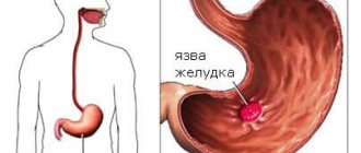
Bacterial vaginosis - first symptoms and treatment regimen
Bacterial vaginosis (gardnerellosis, vaginal dysbiosis, vaginal dysbiosis) is a common disease in women associated with a disruption of the normal microflora of the vagina and an increase in the number of other microbes, including gardnerella.
The nature of the disease depends on many factors, so when your health improves, the symptoms completely disappear. The disease is not sexually transmitted and does not affect men. Unprotected sexual intercourse has a certain role in the occurrence of the disease. Frequent changes of partners contribute to changes in the vaginal microflora.
Causes
To date, science does not fully have information about what actually provokes the development of non-inflammatory syndrome. However, the relevance of this problem is increasing every year.
Factors that provoke the development of the disease include:
- weakening of local and general immunity;
- poor nutrition;
- long-term antibacterial and hormonal therapy;
- frequent douching;
- the use of local contraceptives (condoms, creams and suppositories) that contain 9-nonoxynol;
- frequent change of sexual partners;
- wearing synthetic underwear;
- endocrine and gynecological pathologies;
- failure to comply with basic rules of personal hygiene;
- intestinal diseases.
Currently, bacterial vaginosis is one of the most common diseases among women of active reproductive age (from 23 to 33 years). According to statistics, about 30-35% of women suffer from vaginosis, but only half of the total number of patients know about their problem due to the presence of a characteristic odor. The rest, as a rule, don’t even know about it.
Causes of vaginal dysbiosis
Vaginal dysbiosis can develop in a woman at any age, regardless of the presence or absence of sexual activity. However, women who often change sexual partners are at risk. The causes of dysbiosis are internal and external.
Internal reasons:
- hormonal changes;
- long-term use of antibiotics;
- avitaminosis;
- endocrine system disorders;
- indiscriminate use of medications;
- recent surgery or injury;
- diseases in the field of gynecology;
- hypothermia of the body;
- gastrointestinal diseases.
External reasons:
- improper vaginal hygiene;
- climate change;
- stressful situations and emotional tension;
- bad ecology;
- strong physical or intellectual stress;
- unhealthy diet (excessive consumption of sweets, fatty foods, salty foods).
Symptoms
Often the only symptom of bacterial vaginosis is the presence of copious vaginal discharge with an unpleasant smell of stale fish, which can bother you for a long time. At the beginning of the disease, the discharge is thin, white or grayish.
The general symptoms of bacterial vaginosis are as follows:
- discharge with an unpleasant odor (fishy), which occurs as a result of the breakdown of amines produced by anaerobic bacteria.
- copious, homogeneous, creamy vaginal discharge of a grayish-white color, adhering to the walls of the vagina.
- sometimes vulvovaginal irritation appears in the form of itching and burning, discomfort during sexual intercourse.
- signs of vaginal inflammation (addition of vaginitis) are observed in half of the patients.
- rarely - urination disorders and pain in the perineum.
If the disease continues for a long time, more than 2 years, then the following symptoms occur:
- the color of the discharge becomes dark green;
- leucorrhoea changes its consistency, becomes more viscous or resembles a curdled mass;
- Also, the following signs are characteristic of discharge due to vaginal dysbacteriosis: over time they become thick and sticky, and their distribution along the vaginal walls is even. Leucorrhoea is easily removed from the walls with a cotton swab;
- with a long-term process, a number of patients complain of minor or moderate itching/burning in the vulva area (see vaginal itching);
- pain during sexual intercourse (see pain during intercourse);
- the volume of vaginal discharge reaches 0.02 liters per day (taking into account that the normal amount of leucorrhoea is no more than 2 - 4 ml);
- in a number of situations, pathogenic flora joins the described infectious process, which contributes to the development of vaginitis;
- Sometimes urinary disorders occur (frequent and painful urination in women).
A distinctive feature of the disease is the absence of visible signs of inflammation. That is, upon visual examination, a physiological pink color of the vaginal mucosa is observed. Only in some cases, single reddish dots are observed in menopausal women.
Bacterial vaginosis: new perspectives in treatment
A.A. KHRYANIN
, Doctor of Medical Sciences, Professor,
Novosibirsk State Medical University of the Ministry of Health of Russia; Vice-President of the ROO "Association of Obstetricians-Gynecologists and Dermatovenerologists", Novosibirsk, O.V.
RESHETNIKOV , MD, Senior Researcher,
Research Institute of Therapy and Preventive Medicine, Novosibirsk
Bacterial vaginosis is an infectious non-inflammatory syndrome characterized by the replacement of normal microflora (mainly lactobacilli) with polymicrobial associations of anaerobes and Gardnerella vaginalis. In recent years, the use of molecular biology techniques has shown that there is a much greater diversity of microorganisms associated with bacterial vaginosis than previously thought. Clindamycin has established itself as an effective and safe drug in the treatment of bacterial vaginosis in modern conditions.
The vaginal flora is a multicomponent microecological system that provides protection to all reproductive organs of women both under normal conditions and in pathology. The main representatives of the vaginal microflora are normally lactobacilli of various species (Lactobacillus spp.) and, to a lesser extent, bifidobacteria and corynebacteria, as well as anaerobic gram-negative bacilli of the genus Fusobacterium
and gram-negative cocci of the genus
Veillonella
. In healthy women of reproductive age, the leading place in the vaginal microcenosis is occupied by lactobacilli (anaerobic and aerobic origin), united under the general name “Dederlein's rods,” which make up more than 95% of the total vaginal microflora. Bifidobacteria, like lactobacilli, protect the vaginal mucosa from the effects of not only pathogenic, but also opportunistic microorganisms and their toxins, prevent the breakdown of secretory IgA, stimulate the formation of interferon and the production of lysozyme. In healthy women, anaerobic microflora prevails over aerobic microflora in a ratio of 10: 1 [1, 2].
Lactobacilli convert glycogen, which is contained in large quantities in the vaginal epithelial cells of women of reproductive age, into lactic acid, increasing the acidity of the vagina. In addition, lactobacilli produce hydrogen peroxide. As a result, the acidic environment of the vagina and hydrogen peroxide inhibit the growth of opportunistic microbes (staphylococci, streptococci, E. coli, anaerobic bacteria, Gardnerella vaginalis, Mobiluncus spp.
), which are found in small quantities in the vagina of the vast majority of women. If the proportion of lactobacilli decreases, their place in the ecosystem is taken by opportunistic microbes (primarily Gardnerella vaginalis). Thus, the acidic environment of vaginal contents, lactobacilli and the protective factors they produce form a powerful natural barrier to the penetration of pathogenic bacteria, protecting the upper parts of the woman’s reproductive tract.
A feature of the vaginal microflora is its variability under the influence of both exogenous (use of tampons, frequent vaginal showers and douching, change of sexual partner) and endogenous factors (neuroendocrine diseases, diabetes, hypothyroidism). Microcenosis is influenced by physiological and hormonal changes (puberty, pregnancy, menopause), phases of the menstrual cycle, and various disorders of menstrual function [3]. The use of certain medications (antibiotics, hormones) and surgical interventions also plays a role.
Bacterial vaginosis (BV) (formerly known as vaginal dysbiosis) is a general infectious non-inflammatory syndrome associated with vaginal dysbiosis and accompanied by an excessively high concentration of obligate and facultative anaerobic opportunistic microorganisms in combination with a sharp decrease in the number or absence of lactic acid bacteria in the vaginal discharge ( table 1
).
| Table 1. Vaginal ecosystem | ||
| Microorganism | Healthy women | Women with BV |
| Total number of microorganisms | <107 microorganisms/g | >109 microorganisms/g |
| Aerobic:anaerobic ratio | From 1:2 to 1:10 | Reaches 1:100 |
| Lactobacilli | Prevail | Minor amount |
| Gardnerella vaginalis | Availability 5-25% | Availability 71-92% |
| Mycoplasma hominis | Availability 15-30% | Availability in 63% |
| Mobiluncus spp. (facultative anaerobe) | Availability 0-5% | Availability 50-70% |
| Bacteroides spp. (anaerobe) | Availability in 52% | Availability up to 100% |
| Peptococcus spp . (anaerobe) | Availability in 26% | Availability up to 100% |
With bacterial vaginosis, elimination of lactobacilli occurs, accompanied by colonization of the vagina by anaerobes: Fusobacterium, Mobiluncus, Peptostreptococcus, Gardnerella vaginalis
. Despite the fact that BV is characterized by its polymicrobial nature, the main microorganism that triggers the process is Gardnerella vaginalis, a facultative anaerobic gram-negative rod; It is this that determines the main symptoms of BV.
The fact is that G. vaginalis
has the unique ability to form a so-called biofilm on the surface of the urogenital mucosa.
Biofilm a
conglomerate of microorganisms located on any surface, the cells of which are attached to each other. Typically, cells are immersed in an extracellular polymeric substance (extracellular matrix) they secrete - mucus. It is believed that 95-99% of all microorganisms in the natural environment exist in the form of biofilm. Microorganisms form a biofilm under the influence of a number of factors, including cellular recognition of sites of attachment to the surface and the presence of nutrients or aggressive substances, oxygen, etc. In the biofilm formation mode, the cell changes its behavior, which is determined by the regulation of gene expression.
It is this biofilm, like cement or glue, that attracts other microorganisms to itself, forming a conglomerate of bacteria, most of which have a pathogenic, or at least dangerous, effect for humans. Biofilms have been found to consist primarily of Gardnerella vaginalis
, while
Atopobium vaginae
was present in 80% of cases and constituted 40% of the biofilm mass.
Other bacteria are much less common, including bacteria belonging to the genera Bacteroides, Corynebacterium, Lactobacillus, Veillonella, Ruminococcus and Streptococcus
[4].
Factors contributing to the development of BV include:
— Immunodeficiency states of the body (chronic stress, diseases, massive treatment with antibiotics and cytostatics, radiation therapy, diabetes, vitamin deficiency). — Hormonal dysfunction of the ovaries, including age-related hormonal changes, hormone therapy. — Inhibition of local immunity factors and lactobacilli (vaginal douching, foreign bodies, intrauterine contraceptives, use of spermicides, contraceptive suppositories and creams containing 9-nonoxynol (Patentex Oval, Nonoxynol) — Massive infection of the vagina, promiscuous relationships.
Prevalence of BV
It is not possible to determine the true incidence of BV due to the fact that in 1/3 of women this disease is asymptomatic. The few studies have found the prevalence of BV to range from 3.14% in asymptomatic women aged 18 to 72 years (screened in the Netherlands) to 49% in women aged 13 to 65 years in a colposcopy office in the United States. The wide variation in reported prevalence rates may be due to the inclusion of different patient groups, demographic variations, and different diagnostic criteria. Overall, based on the results of 21 studies, the overall prevalence of BV was 27.1%, with no significant difference between developed (28.0%) and developing (23.5%) countries [5].
During the Human Microbiome Project, molecular biology techniques revealed that there is a much greater diversity of microorganisms associated with BV than was apparent using culture methods. As an example, here is a list of microorganisms previously unknown in BV: Atopobium vaginae, BV-associated bacteria (BVAB-1, BVAB-2 and BVAB-3) from the order Clostridiales, Megasphaera spp, Leptotrichia spp, Dialister spp, Chloroflexi spp, Olsenella spp, Streptobacillus spp, Shuttleworthia spp, Porphyromonas asaccharolytica
[6].
These diverse organisms accumulate to form distinct communities or profiles, which suggest that BV is not a single entity but a syndrome of variable composition, causing a variety of symptoms, different phenotypic outcomes, and resulting in variable responses to different antibiotic regimens. Some organisms or combinations of organisms are highly specific for BV, so in the future the use of molecular quantitative assays will allow better diagnosis of each BV subtype and individualized therapy. A woman's race and geographic region, as well as different racial groups within the same geographic region, have significant differences in which microorganism is dominant in the vaginal environment. In most populations, L. crispatus is the dominant isolate, and in white women, L. crispatus and/or L. jensenii are more common than any other Lactobacillus
[6].
Among African American women in the United States, the prevalence of gram-negative bacteria BVAB1, which was previously mistakenly perceived as Mobiluncus spp,
. [7].
A recent meta-analysis involving more than 10,000 women demonstrated an association between BV and precancerous conditions, namely cervical intraepithelial neoplasia/dysplasia [5]. Since only a minority of patients infected with HPV develop cervical dysplasia, studies of cervical carcinogenesis should include the presence of an additional contributing factor. This factor is BV. Biochemical changes in the vaginal secretions of women with BV include the formation of metabolic products such as propionate and butyrate, which can damage epithelial cells. In addition, BV-associated anaerobes release volatile amines (especially putrescine, trimethylamine and cadaverine), which appear in the vaginal environment after the conversion of amino acids derived from the abundance of anaerobes, and form, in combination with nitrites (derived from bacterial nitrates), nitrosamines. These carcinogenic compounds are capable of forming DNA adducts and therefore mutagenic events. Local accumulation of nitrosamines during episodes of BV may promote cellular transformation in the cervical epithelium in combination with other oncogenic agents such as HPV infection. In addition, patients with BV and dysplasia showed an altered profile of local cervical immunity, namely nitric oxide (NO) and cytokine concentrations (IL-6, IL-8 and IL-10). Finally, another important additional cofactor in cervical carcinogenesis may be the relative absence of hydrogen peroxide (H2O2), normally produced by lactobacilli. This prevents the selective induction of apoptosis, which is a key element of lactobacilli-stimulated antitumor defense [5].
In non-pregnant women, the presence of BV is associated with an increased risk of infection of the upper genital tract with non-sexual infections and STIs, as well as HIV infection. During pregnancy, BV increases the risk of post-abortion sepsis, early miscarriage, recurrent miscarriage, late miscarriage, premature rupture of membranes, spontaneous preterm contractions and preterm birth, histological chorioamnionitis and postpartum endometritis. As a result, abnormal vaginal flora may predispose to increased colonization of the genital tract, infiltration of membranes, microbial invasion of the amniotic cavity, and fetal damage [6].
Careful observation of 49 women (vaginal samples taken weekly during pregnancy and monthly after childbirth) showed that a relatively greater diversity of microorganisms present in the birth canal was associated with the risk of preterm birth, and the highest risk was found in women whose vaginal secretions contained few lactobacilli , as well as microorganisms of the species Gardnerella spp
and
Ureaplasma spp.
Most women have postpartum disturbances in the vaginal microbiota with a decrease in
Lactobacillus spp
and an increase in various anaerobes, such as
Peptoniphilus, Prevotella and Anaerococcus
species. This impairment was not associated with gestational age at delivery and persisted for up to 1 year postpartum. The findings have important implications for predicting preterm birth and for understanding the potential impact of persistent changes in the postpartum microbiota on maternal health, including outcomes in subsequent short-term pregnancies [8].
Laboratory diagnosis of BV
BV can be diagnosed clinically or using a set of clinical criteria, microscopic, enzymological, chromatographic methods, as well as using qualitative or semi-quantitative cultural methods [6].
In world medical practice, clinical and laboratory criteria proposed by Amsel R. (1983) are used, listed in Table 2 [9]. The diagnosis of bacterial vaginosis is considered confirmed if three or four of the following criteria are present:
| Table 2. Clinical and laboratory criteria for bacterial vaginosis [9] | |||
| Criteria | № | Definition | Sign of BV |
| Clinical | I | Examination of the vagina with a speculum, colposcopy | Copious, homogeneous, white-gray discharge with an unpleasant odor |
| Clinical and laboratory | II | Determination of vaginal pH with an indicator strip | pH > 4.5 |
| III | KOH test (whiff test) - adding 10% KOH to vaginal discharge in a test tube | The appearance of a specific odor | |
| Laboratory | IV | Microscopy of a smear from vaginal discharge as a native specimen or Gram-stained | Detection of “key cells”* |
| Note: *“Key cells” are mature epithelial cells with microorganisms adhered to them (gardnerella, mobiluncus, gram-positive cocci). You can get false positive results by identifying epithelial cells with lactobacilli adherent to them; in this case, it is necessary to perform microscopy of vaginal smears stained with Gram. | |||
The presence of at least 3 positive signs out of 4 is considered diagnostically significant:
1. Clinical manifestations. 2. Increased pH of vaginal discharge > 4.5. 3. Positive amine test (increased smell of rotten fish when reacting with 10% KOH). 4. Gram smear microscopy criteria: desquamated epithelium in large quantities, “key cells” make up 20% of all epithelial cells, leukocytes are rare.
The culture method has the highest sensitivity and specificity in the diagnosis of bacterial vaginosis.
Its high information content is due to qualitative and quantitative indicators of the composition of the vaginal microbiocenosis. Accordingly, with bacterial vaginosis, there is a decrease in the number of lactobacilli and an increase in the content of opportunistic flora. Disadvantages of the method: relative high cost and duration of implementation. The study of Gardnerella DNA in scrapings from lesion sites using PCR is an important additional criterion for BV. Expanded criteria for diagnosing BV
1. Predominance of epithelial cells over leukocytes (no more than 30 per field of view). 2. No visual signs of inflammation. 3. The presence of at least 20% key cells. 4. Detection of less than 5 lactobacilli per field of view by immersion microscopy. 5. Polymicrobial picture of the smear (abundant polymicrobial coccal and rod G-/G+ flora. 6. Increased bacterial contamination in the cytological preparation.
Nugent criteria
The low sensitivity of the Amsel criteria and the presence of asymptomatic forms of bacterial vaginosis forced us to look for other methods and criteria for confirming the diagnosis. At the end of the 80s. Spiegel proposed using a scoring system for diagnosing bacterial vaginosis, taking into account the ratio of morphotypes of lactobacilli and vaginal gardnerella during microscopy of a Gram-stained vaginal smear. However, the system did not take root, and only in 1991 Nugent RP et al. proposed their laboratory criteria for diagnosing bacterial vaginosis (Nugent's Diagnostic Criteria for Bacterial Vaginosis), which are still widely used in world medicine [10]. It is based on a system of points (points) from 0 to 7 and their combination for diagnosing and assessing the degree of bacterial vaginosis by assessing three bacterial morphotypes of the vagina ( Table 3
):
A - Lactobacilli - large gram-positive rods ( Lactobacillus acidophilus: large gram-positive rods)
B - Gardnerella vaginalis and Bacteroides species:small gram-variable or
gram-negative
C -
Mobiluncus species:curved gram-variable
rods
| Table 3. A vaginal smear is stained with Gram and the number of identified morphotypes is counted separately under an immersion microscope system | |||
| Points | A Lactobacilli | B Gardnerella | C Mobiluncus |
| 0 | more than 30 morphotypes | no morphotypes | no morphotypes |
| 1 | 5–30 morphotypes | one morphotype | one morphotype |
| 2 | 1-4 morphotypes | 1-4 morphotypes | 1-4 morphotypes |
| 3 | one morphotype | 5–30 morphotypes | 5–30 morphotypes |
| 4 | no morphotypes | more than 30 morphotypes | more than 30 morphotypes |
| The number of points received is summed up (A + B + C). 0-3 points: normal microflora; 4-6 points: intermediate microflora; => 7 points: bacterial vaginosis [10]. | |||
A recent study compared the Amsel and Nugent criteria; as a result, it turned out that the Amsel criteria are somewhat less informative, but can be used in the absence of a specialized laboratory [11].
In recent years, the global scientific community has developed criteria for the differential clinical and laboratory diagnosis of BV and other similar or associated conditions (diseases). There are nonspecific manifestations that can be recorded by a gynecologist, followed by more accurate laboratory analysis (Table 4) [12].
| Table 4. Differential diagnosis of vaginal discharge syndrome (bacterial vaginosis, vulvovaginal candidiasis, trichomoniasis) [12] | |||
| Indicators | BV | IN VK | Trichomoniasis |
| Discharge | White-gray, abundant | Curdled or creamy white | Foamy, yellow-green, abundant |
| Smell | Yes | No | Yes |
| Itching, burning, irritation | No | Yes | Yes |
| Edema, hyperemia | No | Yes | Yes |
| Dyspareunia | No | Yes | Yes |
| pH | > 4,5 | ≤ 4,5 | > 4,5 |
| White blood cell count | Norm | Increased | Increased |
| Gram smear microscopy | Key cells | Mushrooms | Trichomonas |
| Culture method | Not carried out | Fungi of the genus Candida | Trichomonas |
| Note. BV - bacterial vaginosis, VVC - vulvovaginal candidiasis. | |||
Treatment
Recognition of the importance of BV and its association with STIs and poor reproductive prognosis has led to the search for better and more comprehensive treatment options. There is a wide range of differential diagnosis for vaginal discharge, and the success of treatment often depends on the correct diagnosis, however, a large percentage of patients are treated without additional specific tests.
The presence of a wide range of therapeutic options diagnosing the main causes of vaginitis, and the lack of a clear diagnosis in 30% of patients, even after additional expensive examination, explains why many gynecologists use these drugs. Misdiagnosis or failure to diagnose other infections, associated mainly in cases of BV and T. vaginalis
, can lead to inadequate treatment, a new exacerbation and re-infection.
In non-pregnant women, treatment will not only eliminate vaginal discharge, but will also reduce the likelihood of infectious complications after an abortion and/or hysterectomy is possible for each woman. In addition, treatment of BV by restoring the acidic pH in the vagina reduces the risk of infection with the immunodeficiency virus and other sexually transmitted diseases.
In pregnant women, treatment with BV, along with the above-mentioned effects, helps reduce the risk of developing pregnancy complications, namely premature rupture of amniotic fluid, the onset of labor (contractions) and childbirth itself, as well as postpartum inflammation of the inner surface of the uterus (endometritis). Pregnant women with asymptomatic BV should also be treated, especially if there is a threat of premature birth.
The drug of choice for the treatment of BV is clindamycin.
This is an antibiotic of the lincosamide group for topical use in gynecology. The mechanism of action of the drug is associated with a disruption of intracellular protein synthesis in the microbial cell at the level of the 50S ribosomal subunit. It has a bacteriostatic effect, and in higher concentrations it has a bactericidal effect against some microorganisms. Has a wide spectrum of action. Active against microorganisms that cause bacterial vaginosis:
•
Gardnerella vaginalis. • Mobiluncus spp. • Bacteroides spp. • Mycoplasma hominis. • Peptostreptococcus spp.
Clindamycin (Dalacin)
.
Release form: vaginal cream and vaginal suppositories
. 5 g of cream (1 dose) vaginal 2% and one vaginal suppository contain: clindamycin phosphate 100 mg.
Pharmacokinetics.
After a single intravaginal administration of 100 mg of clindamycin, an average of 4% of the administered dose is systemically absorbed. Cmax in blood plasma averages 20 ng/ml.
Clinical studies on the use of clindamycin in women in the first trimester of pregnancy have not been conducted, therefore the use of Clindacin in the first trimester of pregnancy is possible only when the expected benefit to the mother outweighs the risk to the fetus. Use in the second and third trimesters of pregnancy is possible if the expected effect of therapy outweighs the potential risk to the fetus (adequate and strictly controlled studies have not been conducted in pregnant women; clindamycin passes through the placenta and can concentrate in the fetal liver, but no complications have been reported in humans). Studies have not determined whether treating bacterial vaginosis reduces the risk of adverse pregnancy outcomes such as premature rupture of membranes, preterm labor, or preterm delivery. FDA category of effect on the fetus is B.
A study conducted in Switzerland examined 5,377 pregnant women with symptoms of potential obstetric complications at 25–37 weeks' gestation. pregnancy. Symptomatic women were tested by culture for Mycoplasma hominis and Ureaplasma spp. and treated with clindamycin if positive. As a result of treatment, the percentage of premature births and respiratory complications in newborns significantly decreased [13].
Our colleagues from Belgium searched the PubMed and Web of Science databases to find new approaches in the prevention, treatment and prevention of relapses of BV. As a result, it turned out that clindamycin and metronidazole remain the main drugs in the treatment of BV. Other medications such as tinidazole, rifaximin, nitrofuran, decalinum chloride, ascorbic acid (vitamin C) and lactic acid are being intensively studied. It is believed that the use of a combination regimen, alternating and long-term administration to prevent relapses is promising. The benefits of parallel administration of probiotics are also undoubted [14].
A multicenter, randomized, double-blind study conducted in Germany, Austria, and Switzerland compared the effectiveness and tolerability of 2% vaginal clindamycin cream (5 g at night for 7 days) and oral metronidazole (500 mg orally for 7 days) in the management of BV. Patients were observed 5-10 days and 25-39 days after completion of treatment. As a result, cure or improvement was noted after 1 month in 83% of patients in the clindamycin group versus 73% in the metronidazole group, side effects were noted with equal frequency (12%) in both groups [15].
Recently, the concept of aerobic vaginitis has appeared in the world literature. Aerobic vaginitis is an inflammatory disease of the vagina caused by aerobic microflora with a sharp decrease or absence of normal vaginal lactoflora. Previously, the term “aerobic vaginitis” meant bacterial vaginitis. The basis of aerobic vaginitis, as with bacterial vaginosis, is the reduction or absence of normal vaginal lactoflora and its replacement with aerobic bacteria. The exact causes and mechanism of development of aerobic vaginitis are still unknown. It is also unknown why in some cases there is a proliferation of anaerobic microflora and the development of bacterial vaginosis, and in others the colonization of the vagina by aerobic microorganisms and the development of aerobic vaginitis. The most common etiological agents of aerobic vaginitis ( Escherichia coli, Enterococcus sp.
, group A beta-hemolytic streptococcus, Staphylococcus aureus).
In resolving this controversy, it turned out that vaginal suppositories containing kanamycin or clindamycin showed high effectiveness in relieving aerobic vaginitis in non-pregnant women. Additionally, clindamycin (vaginal suppositories) in combination with probiotics was found to be a better choice for pregnant women with aerobic vaginitis than metronidazole [16]. Conclusion
Bacterial vaginosis is a long-known pathological condition of the female genital area with well-developed clinical diagnostic criteria (Amsela and Nugenta).
New molecular diagnostic capabilities are constantly expanding our understanding of the various types of native and foreign microflora, indicating the diversity of the vaginal microbiota in each individual woman. In this case, the choice of the optimal drug provides clinical effectiveness against most pathogenic microorganisms. Such a drug can be clindamycin, the significance of which in the treatment of bacterial vaginosis is beyond doubt in recent scientific publications. References
1. Anderson MR, Klink K, Cohrssen A. Evaluation of vaginal complaints. JAMA 2004, 291:11:1368–1379. 2. Mitchell H. Vaginal discharge – causes, diagnosis, and treatment. BMJ 2004, 328:7451:1306–1308. 3. Khryanin A.A., Reshetnikov O.V. Bacterial vaginosis: new ideas about the microbial biosocium and treatment possibilities. Medical Council, 2014, 17: 128-133. 4. Verstraelen H, Swidsinski A. The biofilm in bacterial vaginosis: implications for epidemiology, diagnosis and treatment. Curr Opin Infect Dis 2013, 26: 86–89. 5. Gillet E, Meys JFA, Verstraelen H et al. Association between bacterial vaginosis and cervical intraepithelial neoplasia: Systematic review and meta-analysis. Plos One, 2012, 7, Issue 10 e45201. 6. Lamont RF, Sobel JD, Akins RA et al. The vaginal microbiome: New information about genital tract flora using molecular based techniques BJOG, 2011, 118(5): 533–549. 7. Muzny CA, Sunesara IR, Griswold ME et al. Association between BVAB1 and high Nugent scores among women with bacterial vaginosis. Diagn Microbiol Infect Dis., 2014 December, 80(4): 321–323. 8. Di Giulio DB, Callahan BJ, McMurdie PJ et al. Temporal and spatial variation of the human microbiota during pregnancy Proceedings of the National Academy of Sciences 2015/ www.pnas.org/cgi/doi/10.1073/pnas.1502875112. 9. Amsel R, Totten PA, Spiegel CA, et al. Nonspecific vaginitis. Diagnostic criteria and microbial and epidemiologic associations. Am J Med 1983, 74(1): 14–22. 10. Nugent RP, Krohn MA, Hillier SL. Reliability of diagnosing bacterial vaginosis is improved by a standardized method of gram stain interpretation. J Clin Microbiol, 1991, 29(2): 297–301. 11. Mohammadzadeh F, Dolatian M, Jorjani M, Alavi Majd H. Diagnostic value of Amsel's clinical criteria for diagnosis of bacterial vaginosis. Glob J Health Sci., 2014 Oct 29, 7(3): 8-14. 12. Workowski K.A. Sexually transmitted diseases treatment guidelines, 2010. MMWR Recommend Rep 2010; 59:61–63. 13. Vouga M, Greub G, Prodhom G, et al. Treatment of genital mycoplasma in colonized pregnant women in late pregnancy is associated with a lower rate of premature labor and neonatal complications. Clin Microbiol Infect, 2014 Oct, 20(10): 1074-9. 14. Donders GG, Zodzika J, Rezeberga D. Treatment of bacterial vaginosis: what we have and what we miss. Expert Opin Pharmacother, 2014 Apr, 15(5): 645-57. 15. Fischbach F, Petersen EE, Weissenbacher ER, et al. Efficacy of clindamycin vaginal cream versus oral metronidazole in the treatment of bacterial vaginosis. Obstet Gynecol, 1993 Sep, 82(3): 405-10. 16. Han C, Wu W, Fan A, Wang Y, Zhang H, Chu Z, Wang C, Xue F. Diagnostic and therapeutic advancements for aerobic vaginitis. Arch Gynecol Obstet, 2015 Feb, 291(2): 251-7.
Severity
According to the severity of vaginal dysbiosis, there are:
| Compensated or 1st degree | there is no microflora in the smear, epithelial cells are present without changes and the possibility of infection with other pathogenic microorganisms remains. |
| Subcompensated or 2nd degree | the content of Doderlein bacilli decreases, gram-negative and gram-positive flora increases, there are from 1 to 5 “key” cells, a slight increase in leukocytes - up to 15 - 25. |
| Decompensated or 3rd degree | there are no lactic acid bacteria, there is a clinical picture of the disease, “key” cells entirely, various pathogenic and facultative or conditionally pathogenic microorganisms. |
According to the flow, acute, torpid or erased and asymptomatic vaginal dysbiosis is distinguished.
Diagnosis of bacterial vaginosis at the Miracle Doctor clinic
- A study to determine the acidity of vaginal discharge, which normally should not exceed 4.5. Anything greater than this number indicates the presence of pathology;
- Amine test using a substance such as potassium hydroxide. Discharge from bacterial vaginosis is mixed with it, and the resulting odor is assessed. If it resembles fish, then this indicates the disease being described;
- A smear of the vaginal mucosa and studying its contents using microscopy. The reliability of the latter is highly assessed. A decrease in the number of lactobacilli or their absence, an increase in the number of other microorganisms indicates bacterial vaginosis.
Diagnostic measures are not limited to the above list. Doctors also resort to procedures with a higher level of complexity. These, for example, include identifying the volume of lactic and succinic acid in the vaginal discharge and other studies.
Diagnostics
A preliminary diagnosis of bacterial vaginosis can be made already during a gynecological examination. After the examination, discharge is taken from the posterior inferior vaginal vault.
The diagnosis can be made if 3 of the 4 listed signs are present:
- specific nature of the discharge;
- acidity >4.5 (normal 3.8-4.5);
- positive amino test;
- presence of “key” cells. The so-called “key cells” are mature epithelial cells (the superficial layer of the vaginal epithelium), on the entire surface of which microbes are densely and in large numbers attached.
Completing one of the 4 tests is not sufficient to make a diagnosis.
Prevention of vaginal dysbiosis
Follow simple rules to prevent the formation of dysbiosis:
- maintain daily intimate hygiene;
- give up alcoholic drinks;
- temper your body;
- Avoid regular use of panty liners;
- Maintain proper nutrition and consume enough dairy products.
When the first signs appear, immediately contact a gynecologist to prevent the development of the disease in time. Do not make diagnoses yourself and do not self-medicate.
How to treat bacterial vaginosis?
Initially, a woman is prescribed antibiotics to treat bacterial vaginosis: they have a detrimental effect on nonspecific bacteria and clear the vaginal mucosa of them.
The drugs of choice are Metronidazole, Tinidazole, Clindamycin, as they are active against anaerobes. Local use of antibiotics is preferable to avoid systemic side effects, but in some cases the gynecologist is forced to resort to tablet forms.
The treatment regimen is selected individually:
- Tinidazole 2.0 in tablet form is taken orally 1 time per day for 3 days;
- Metronidazole in the form of a 0.75% gel is administered into the vagina once a day for 5 days;
- Clindamycin suppositories 100 mg are administered into the vagina once a day for 3 days;
- A cream containing 2% Clindamycin is injected into the vagina once a day for 7 days;
- Metronidazole 2.0 tablets are taken orally once.
During antibacterial therapy and a day after its completion, it is necessary to avoid drinking alcohol, even in minimal doses. The drugs disrupt the metabolism of ethyl alcohol in the body, which causes the accumulation of toxic metabolites and severe intoxication. In its course, it resembles a severe hangover: the woman experiences severe weakness, limbs shake, blood pressure rises, a severe throbbing headache occurs, and painful nausea and vomiting develop.
Clindamycin cream contains fat, so it can damage the condom or latex contraceptive membrane. All local forms of drugs are administered immediately before bedtime to prevent them from flowing down the vaginal walls.
If antibiotics are intolerant or there are contraindications to their use, the first stage of treatment is carried out with local antiseptics:
- Hexicon 1 suppository is administered 2 times a day for 7-10 days;
- Miramistin in the form of a solution is irrigated into the vagina once a day for 7 days.
Preparations for bacterial vaginosis, used in the second stage of treatment, contain lactobacilli and create favorable conditions for restoring the vaginal microflora. They are used 2-3 days after completion of antibacterial therapy:
- Acylact 1 suppository 2 times a day is inserted into the vagina for 5-10 days;
- Bifiliz 5 doses are taken orally 2 times a day for 5-10 days.
Antifungal suppositories for bacterial vaginosis are usually not prescribed. The need for them arises if candidiasis, a fungal infection, joins the opportunistic microflora. In this case, Clotrimazole suppositories are prescribed intravaginally once a day for 6 days.
Treatment
Treatment of this pathology is carried out in stages:
- Stage I – reducing the level of pathogenic microorganisms. Antibacterial therapy is used for these purposes;
- Stage II – restoration of the vaginal microflora. In this case, probiotics, suppositories with lactobacilli, and immunomodulators are used.
A control smear is prescribed after one and a half to two months. Until then, it is recommended to use condoms during sexual intercourse.
Possible treatments:
- The “Gineton” device is a low-frequency ultrasound, the effect of which is the basis of this hardware technique, which eliminates the source of infection and improves the penetration of drugs into tissues.
Treatment of pregnant women
Therapeutic therapy during pregnancy differs from the standard regimen in that:
- Antibiotics are not prescribed;
- Treatment is carried out after the first trimester.
Treatment during pregnancy
How to treat bacterial vaginosis in case of pregnancy? In the first trimester of gestation, systemic therapy for the disease is not performed (metronidazole and other drugs are toxic to the embryo). Local administration of etiotropic drugs in the early stages is used with caution.
Taking metronidazole or clindamycin begins in the second trimester and is carried out in short courses. Metronidazole 0.5 g. (2 tablets) twice a day for 3–5 days, and clindamycin is prescribed in a dosage of 0.3 g. 2 times a day for 5 days.
Pregnancy complications that may occur due to the disease include:
- miscarriage - loss of a fetus during the first 23 weeks;
- premature birth - when a baby is born before the 37th week of pregnancy;
- chorioamnionitis - infection of the chorion and amnion membranes (the membranes that make up the fetal sac) and amniotic fluid (the fluid surrounding the fetus);
- premature rupture of the amniotic sac - a bladder containing fluid in which the fetus develops;
- postpartum endometritis - infection and inflammation of the uterine tissue after childbirth.
If you are pregnant and experience symptoms of vaginosis, contact your gynecologist as soon as possible. Although the risk of complications is low, treatment will help reduce it further.
Treatment of gardnerellosis
Treatment in men requires complete elimination of Gardnerella from the genital tract. For this purpose, antibiotics of the 5-nitroimidazole group (Tinidazole, Clindamycin, etc.) are prescribed. If no other infection (except Gardnerella) is detected, the treatment regimen includes only one drug, and the course lasts 3/5/7 days.
You need to understand that contact with a bacterium is not accompanied by the development of immunity against it. Upon completion of the therapeutic course, an episode of gardnerellosis may recur. To prevent reactivation of Gardnerella, both partners should undergo treatment, and the woman should pay attention to measures to prevent relapses of bacterial vaginosis.
Protection against complications developing against the background of diseases of the male genital area requires an annual visit to a urologist. Highly qualified specialists conduct consultations at the medical clinic. At the clinic you can be tested for gardnerellosis and receive recommendations regarding the prevention and treatment of sexually transmitted infections.
Prevention
The recommendations are as follows:
- using barrier methods of contraception, wearing underwear only made from natural fabrics;
- regular examination by a gynecologist and timely treatment of diseases;
- treatment of chronic pathologies of internal organs;
- strengthening the immune system in natural ways: physical activity, hardening, etc.;
- avoiding douching and other similar procedures.
Bacterial vaginosis is a pathology that reflects a decrease in the body’s level of defense at the moment. Often occurring asymptomatically, gardnerellosis is always detected during examination by a gynecologist. Only a doctor can prescribe the most effective tablets for bacterial vaginosis, suppositories or other forms. Don't delay treatment!
Diagnosis of gardnerellosis in men
If you have signs of the disease, you should contact a urologist. Diagnostics includes the following steps:
- Survey. The doctor clarifies complaints, collects anamnesis, clarifies the features of a man’s sexual life (presence of a regular partner, contraceptive methods used, etc.)
- Laboratory diagnostics. The urologist takes a swab from the urethra. A bacterioscopic examination allows you to identify signs of concomitant inflammation and exclude the most common sexually transmitted infections. Gardnerella in men is detected by PCR.
- Instrumental diagnostics. The doctor prescribes an ultrasound if he suspects the involvement of the pelvic organs in the pathological process.
If necessary, the urologist may recommend consultations with other specialists (therapist, endocrinologist, etc.)