What are leukocytes, their norm in urine
Leukocytes are white blood cells that provide the body's immune system with a response to invading pathogenic agents. They are produced in the bone marrow and have a lifespan of about 7 days. Leukocytes travel with the blood and participate in the absorption of foreign elements. Having done their work, the cells die. They are removed from the body along with urine. The urine of a healthy person contains no more than 3-5 units in the field of view.
In men, the norm of leukocytes in the urine is 0-3, in women - 0-5, in girls 0-7, in boys 0-5 units in the field of view. The absence of cells is a variant of the norm.
If a large number of white blood cells (leukocyturia) are found in the urine, then an inflammatory or infectious process is occurring in the body. Any pathological conditions activate the production of leukocytes, and, as a result, an increase in their concentration.
The body signals a health problem by increasing temperature and fever, deterioration in overall health, and changes in the color and transparency of urine.
The pathological condition is detected using OAM (general analysis of morning urine). To confirm/refute the diagnosis, the doctor will additionally prescribe a Nechiporenko test and a general blood test with a leukocyte formula. A nephrologist, urologist, pediatrician or general practitioner, gynecologist or oncologist deciphers the test results.
When to determine the number of leukocytes in urine
General indications for taking a urine test are:
- examination for infectious diseases and inflammatory processes;
- the need to establish an accurate diagnosis if diseases of the urinary system are suspected;
- examination of pregnant women before registration at the antenatal clinic, routine examinations;
- the occurrence of complications during pregnancy, suspicion of gestosis;
- the need to evaluate the effectiveness of prescribed treatment, including antibiotic therapy;
- undergoing routine medical examinations;
- diagnosing diseases in children;
- monitoring the condition of patients with cancer.
White blood cells in urine should be checked if the following symptoms occur:
- acute pain in the lower abdomen, under the right rib or in the lower back;
- frequent urination;
- a feeling of incomplete emptying of the bladder;
- discomfort, pain, burning and itching when urinating;
- darkening of urine, appearance of an unpleasant odor;
- the appearance of foreign inclusions in urine (blood clots, mucus, flakes);
- increased temperature and fever of unknown origin;
- increased blood pressure.
Sluggish or chronic disease of the urinary organs can be suspected when yellowish spots are found on underwear.
What amount is considered elevated?
It can be stated that leukocytes in the urine are elevated if up to 20 copies are detected. If the number reaches 85-100, then we can talk about an acute disease of the genitourinary system and the presence of pus in the urine. The pathology is called pyuria. In this case, the liquid becomes dark in color, becomes cloudy and has a purulent odor. Upon examination, impurities are detected.
In order not to distort the test result, on the eve of urine collection it is important to exclude the influence of the following factors:
- abuse of fatty, protein foods;
- diaper rash in babies;
- poor genital hygiene;
- severe dehydration;
- pathological vaginal dryness during menopause.
High white blood cells can be caused by the presence of a bladder catheter, as well as the use of certain pharmaceuticals: diuretics, NSAIDs, antibiotics and drugs that suppress the immune system.
Preparing for a urine test in women
Preparation for OAM in women includes a number of hygienic procedures, as well as general restrictions.
- On the day before taking biomaterial, it is advisable to refrain from physical activity, drinking alcohol, and not eat vegetables and fruits (beets, carrots, citrus fruits, watermelons), red wine, and multivitamins, which can change the color of urine.
- Avoid taking diuretics for 48 hours before urine collection (in consultation with your doctor).
- It is not advisable to collect biomaterial earlier than 5-7 days after cystoscopy.
- Women should not collect urine during menstruation, and it is also not recommended to take a urine test 2 days before and 2 days after menstruation. If a general analysis is urgently required during menstruation, the woman is advised to place a tampon in the vagina before the test.
- Carry out a thorough toileting of the external genitalia under the shower with soap (it is prohibited to use hygiene products with antiseptics).
Types of leukocyturia
Depending on the number of white blood cells, three types of leukocyturia are distinguished:
- insignificant (from 7 to 40 units);
- moderate (up to 100 specimens in the field of view);
- pronounced (over 100 units).
A high concentration of white blood cells in the urine may be:
- caused by diseases of the urinary system (true) or arising against the background of an inflammatory process in the genital organs (false);
- infectious or non-infectious origin. In the first case, the presence of infection is confirmed by urine culture or PCR diagnostics. Non-infectious leukocyturia is not associated with bacterial activity; it is caused by an inflammatory process due to allergic cystitis, autoimmune glomerulonephritis, and medications.
Depending on the predominant type of leukocytes, they are distinguished:
- lymphocytic leukocyturia, giving reason to suspect lupus erythematosus or rheumatoid arthritis;
- mononuclear, arising from renal pathologies;
- neutrophilic, which is provoked by tuberculosis, tonsillitis, malaria or pyelonephritis;
- eosinophilic, accompanies allergies.
Urine collection rules
In order for the results obtained to correspond to reality and correctly reflect the picture of the functioning of the body, it is necessary to properly prepare and collect fluid for a general urine test (UCA). As mentioned earlier, the composition and amount of urine excreted is influenced by the food consumed and other external factors, so experts recommend:
- consult a doctor about the possibility of reducing the intake of medications - antipyretics, painkillers, diuretics, and other drugs containing ethyl alcohol, caffeine, glycerol trinitrate, which lead to an increase in the concentration of glucose and creatinine in urine;
- refrain from drinking coffee, tea, drinks containing ethyl alcohol, various artificial and natural dyes in large quantities;
- a few days before collecting urine, avoid excessively salty, fatty, sweet, spicy foods, and smoked foods;
- do not visit the day before, or better yet for several days, a sauna or bathhouse, which help remove fluid from the body, which leads to a decrease in the daily volume of urine;
- do not eat fruits and berries with bright, saturated colors, otherwise the color of the biofluid may change, which is one of the indicators of OAM analysis;
- women should refrain from donating urine during the menstrual period, or be very careful in collecting it so that particles of menstrual blood do not fall into the urine collection container.
There are also restrictions on certain medical procedures. For example, a week or later before urine collection, cystoscopy cannot be performed.
There is also a time limit between the moment of collection and delivery of the collected material to the laboratory. This period should not exceed 2 hours. If an unexpected delay occurs, the container must be placed in the refrigerator. To conduct the study, the laboratory assistant will need no more than 50 ml of biofluid.
To conduct OAM and obtain an accurate, reliable result, morning urine obtained during the first morning urination is given for testing. The urine secreted by the body is absolutely sterile, so that foreign substances and foreign inclusions do not get into it, it is necessary to use sterile containers for analysis. Today they are sold in all pharmacies. As a last resort, if it is not possible to purchase a special container for biosamples, you can prepare a small glass jar with a tightly (hermetically) closing lid yourself. The dishes must be washed very thoroughly and rinsed with boiling water.
After waking up, before going to the toilet, you need to treat the external genitourinary organs with soap and carefully blot off all the liquid. If desired, you can use sterile wipes. Women should place a special tampon or a small piece of cotton wool in the vagina to prevent mucus from entering the urine.
Depending on what pathologies are expected to be identified and where they may be located (in the lower or upper parts of the urinary tract), two options are possible for collecting fluid for research:
- medium urine - after the start of urination, the first portion is flushed into the toilet, then the process is delayed and the remaining urine is collected in a prepared container;
- collection of general urine, for this you will need a 200 ml container for urine or two vessels, after urination is completed, all the liquid is mixed and the excess is poured out, leaving the required amount.
The study of biomaterial is a fairly simple and quick process. Despite the large number of substances being determined, the results of the analysis can be collected from the laboratory the next day.
What diseases cause an increase in leukocytes in the urine?
A large number of diseases can cause an increase in the concentration of leukocytes in the urine. If the analysis shows deviations from the norm, then it is necessary to undergo further examination to exclude serious diseases, and if they are confirmed, begin timely treatment.
The main diseases in which leukocyturia is detected:
- pathologies of the urinary tract: cystitis, urethritis, pyelonephritis, glomerulonephritis, stones in the bladder or kidneys, cystic formations, abscess of kidney tissue, damage to the renal vessels in diabetes mellitus;
- pathologies of the female genital organs: vaginitis, thrush, inflammatory processes in the uterine appendages, bartholinitis, endometritis;
- pathologies of the male genital organs: adenoma, inflammatory process in the prostate gland, balanoposthitis, infectious diseases;
- other diseases: autoimmune origin, cancer, gallstones, pneumonia, diabetes, various allergies, heart and vascular diseases, appendicitis.
Leukocyturia in the structure of urinary tract infection
Urinary tract infection (UTI) is one of the most common diseases of childhood in general and primarily in the structure of diseases of the urinary system in particular.
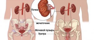
It is necessary to distinguish between urinary tract infection and pyelonephritis. The term UTI implies the presence of infection in the urinary tract (tubule, pelvis, ureter, bladder, urethra) [1–5], while the term “pyelonephritis” means bacterial infection, primarily of interstitial tissue, which should always be accompanied by a violation of its function [1 , 6–8]. In this case, the infection can simultaneously affect the urinary tract, in particular the pelvis and bladder [2]. The presence of an inflammatory process in the pelvis is almost always accompanied by damage to the interstitium of the kidney, so at present pyelitis is not considered as an independent disease. The main criterion for the diagnosis of UTI is the presence of bacteriuria, but the detection of bacteriuria does not always indicate inflammation, which is typical for asymptomatic bacteriuria [1–6]. Bacteriuria can be transient, when colonization of the microbe does not occur, and therefore no inflammatory process occurs. The presence of an inflammatory process is determined by clinical signs (intoxication, pain), paraclinical indicators - accelerated ESR, leukocytosis with neutrophyllosis, increased concentration of acute phase proteins (CRP). These listed signs are characteristic of any acute inflammatory process. An indicator of the presence of an inflammatory process in the kidneys and urinary tract is leukocyturia.
Leukocyturia is the most common symptom found in a urine test. When should the presence of leukocytes in centrifuged urine sediment be interpreted as pathological leukocyturia? The answer is simple: when their number exceeds the norm. However, there is no consensus regarding this norm [9, 10]. It is advisable to take as the norm 0-1-2 leukocytes per field of view (FOV) in boys, 1-2-3 in FOV for girls, and in the presence of signs of exudative-catarrhal diathesis - up to 5-7 in FOV sp. This is usually combined with an increase in the number of epithelial cells. Undoubtedly, a larger number of leukocytes in urine sediment may not be a sign of pathology, but this must be proven by implementing a certain algorithm of actions (Fig.). An increase in the number of leukocytes in the urine should be considered a pathological phenomenon. The presence of a very large number of leukocytes, when they cover all fields of vision, is characterized as pyuria. If you do not pay attention to slightly elevated white blood cell counts in a timely manner, this can lead to a number of complications. It’s worth thinking about why cystitis mainly occurs in girls and often takes a chronic course? Yes, of course, this is facilitated by the anatomical features of the external genitalia. But a short and wide urethra in girls will facilitate the penetration of infection into the bladder if external pathology is not detected in a timely manner, which may not manifest itself clearly for some time. In boys, the presence of phimosis and synechiae in the absence of pain can also be manifested by only slight leukocyturia, which can be mistakenly taken as normal, especially if it returns to normal upon repeated urine testing. It must be borne in mind that children may for some time not pay attention to short-term unpleasant sensations during or after urination, to frequent urges, and only the appearance of pain during urination will cause a reaction in the child and attract the attention of parents. However, at a time when, with slight irritation of the mucous membrane of the external genitalia, there is still no bright clinic, the number of leukocytes in the urine sediment is already increasing. That is why the detection of even 3-5 leukocytes in the visual field requires that this not be considered the norm at first.
| Rice. Algorithm for a doctor’s actions when detecting leukocyturia |
Leukocyturia cannot always be detected by the usual method of examining the urinary sediment of a single portion of urine if it is insignificant and occurs periodically during the day. It is more reliable to evaluate leukocyturia in urine collected over a certain time and taking into account its quantity. To identify unstable latent leukocyturia, there are quantitative methods for its determination. These include the Addis-Kakovsky test and the Amburger test. As for the Nechiporenko test (more precisely, the analysis of urine sediment according to Nechiporenko), which is often used in everyday practice to detect latent leukocyturia, it is unsuitable for this. Since this analysis uses a single portion of urine, as for a conventional analysis, it is impossible to judge the presence or absence of latent leukocyturia (as well as erythrocyturia). However, urine sediment examined using Nechiporenko’s method makes it possible to more reliably quantify the content of formed elements, since the chamber method of counting them makes it possible to estimate urine sediment in one figure. This is especially important when treating urinary tract infections, as it allows for a more reliable assessment of the effectiveness of the therapy.
At one time, great importance was attached to the identification of a special type of leukocytes, named after the authors who first described them, Sternheimer–Malbin cells. They are also called active leukocytes. When stained accordingly, ordinary inactive leukocytes have a pale pink protoplasm filled with dark granules and a purplish-red nucleus. Other, so-called active leukocytes have almost colorless protoplasm, filled with grayish granules that perform Brownian motion, their nuclei are pale violet. The sizes of these cells are usually increased. It was believed that their appearance is characteristic of an acute inflammatory process in the urinary system, in particular, pyelonephritis. Currently, the appearance of such cells is associated with hypoosmolality of urine, and therefore they are not given specific clinical significance. However, the detection of Sternheimer–Malbin cells in a significant number (more than 10–15%) in the absence of urine hypoosmia gives reason to consider them as a sign of an inflammatory process in the urinary system.
Causes of leukocyturia. Leukocyturia occurs in various pathologies and is not always a sign of a bacterial infection, especially when there are no extrarenal manifestations in isolated urinary syndrome manifested by leukocyturia (Table).
In acute pyelonephritis and exacerbation of chronic leukocyturia, as a rule, it is significant and is usually accompanied by moderate proteinuria, often occurring against the background of intoxication and often pain. With cystitis, urethritis, vulvitis and balanoposthitis, leukocyturia is often accompanied by dysuria. Persistent leukocyturia is observed with damage to the urinary tract by mycoplasma, chlamydial, fungal infections, as well as with tuberculosis of the kidneys and urinary tract. With abacterial interstitial nephritis, leukocyturia is usually combined with micro- or macrohematuria and moderate proteinuria. Leukocyturia is observed in the first days of acute glomerulonephritis, as well as with exacerbation of chronic glomerulonephritis. It is represented largely by lymphocytes, reflecting the body's reaction to the deposition of immune complexes in the structures of the glomeruli. If the course of the disease is favorable, leukocyturia disappears after 5–7 days. Its persistence in subsequent days, and even more so its increase, should be regarded as an unfavorable factor in the course of the disease.
In everyday pediatric practice, the appearance of leukocyturia is most often associated with UTI. This often leads to unjustified prescription of uroseptics. The criterion for infection in the urinary system is only the detection of the pathogen by urine culture or taking a smear from the urethra for a latent infection. To confirm or exclude the infectious nature of leukocyturia, an evaluation of the urocytogram is necessary. With the bacterial nature of leukocyturia, leukocytes are represented mainly by neutrophil cells (more than 70–80%). Non-infectious leukocyturia is characterized by the presence of a significant number of cells of the lymphomonocyte series, up to their predominance over neutrophils. The presence of eosinophils in the urinary sediment indicates the allergic nature of the pathology. Eosinophiluria is characteristic of acute interstitial nephritis, the establishment of which can promptly help in differential diagnosis with acute glomerulonephritis, since the clinical and laboratory data of these diseases may be similar.
Currently, the diagnosis of UTI is made quite widely and, as practice shows, not always justified. Isolated urinary syndrome, manifested by leukocyturia, is not a sufficient basis for making this diagnosis. The main criterion for making this diagnosis can only be the detection of bacteriuria in the diagnostic titer. However, practice shows that today in clinics this study is either not carried out, or urine is sent for culture when antibacterial therapy has been started. Leukocyturia itself is not a reason for using a uroseptic or antibiotic in treatment if there are no symptoms of intoxication or pain. It is necessary to first establish the cause of its occurrence and only then decide on the advisability of this or that therapy.
Tactics for managing children with leukocyturia. First of all, you need to know why the urine test was taken, and if this is not the debut of leukocyturia, then when and under what circumstances an increased number of leukocytes in the urine was noted before. You should also rule out the current presence of diaper rash in the perineal area or inflammation of the genitals, and also find out whether it has happened in the past. It is important to know whether there were or are now phenomena of dysuria, which can be painless and manifest only by frequent urination, which children often do not pay attention to. It is also necessary to find out whether there has ever been a rise in temperature without catarrhal phenomena, which could be regarded as a sign of teething or overheating. When starting to examine a child, especially a girl, the doctor must first determine whether the leukocyturia is external or whether it is caused by damage to the urinary tract or pyelonephritis. To do this, it is necessary to conduct a 2-glass test (Fig.), which requires a certain skill and is carried out by the mother after appropriate instructions. If an increased number of leukocytes is detected only in the first portion, smears from the vulva and vagina should be taken for microscopy, as well as a smear from the urethra for hidden infections. In case of a burdened allergic history, it is necessary to submit urine for a urocytogram, and if there are symptoms of vulvitis, a vulvocytogram. This will eliminate urinary allergosis. In addition, all children with leukocyturia are advised to undergo an ultrasound of the kidneys and bladder, as well as a urine test for bacteriuria. To do this, it is advisable to first conduct a screening test for bacteriuria (nitrite test), which will allow you to get an answer after 3 hours and, if the result is positive, submit urine for culture to determine the microbial number and sensitivity of the pathogen to antibacterial drugs.
So, the detection of leukocyturia in isolated urinary syndrome makes it possible to reasonably diagnose a UTI only in the presence of bacteriuria, as determined by a screening test or urine culture. Timely detection of pathology in the external genitalia and its rational treatment will help prevent the occurrence of cystitis in girls and associated neurogenic bladder dysfunctions.
Literature
- Papayan A.V., Erman M.V., Anichkova I.V. et al. Infection of the urinary system in children (etiopathogenesis, diagnosis and treatment). A manual for doctors and senior students. St. Petersburg, 2001. 56 p.
- Shulutko B.I. Urinary tract infection // Nephrology. Current state of the problem. St. Petersburg, 2002, p. 447–458.
- Letifov G. M. Treatment and medical examination of children with nonspecific infectious and inflammatory diseases of the urinary system. Rostov-on-Don, 2004. 64 p.
- Vyalkova A. A. Urinary system infections in children: modern principles of treatment / International Nephrological School of the European Association of Pediatric Nephrologists. St. Petersburg 2004, p. 149–161.
- Malkoch A.V. Urinary tract infection. In the book: Practical guide to childhood diseases. T. 6: Nephrology of childhood. Ed. Tabolina V. A., Belmera S. V., Osmanova I. M. M., 2005, p. 248–250.
- Korovina N. A., Zakharova I. N., Mumladze E. B., Gavryushova L. P. Diagnosis and treatment of pyelonephritis in children. M., 2003. 72 p.
- Magomedova M.N., Rusnak F.I., Klyuchnikov S.O. Pyelonephritis in children. In the book: Lectures on pediatrics. T. 6. Nephrology. Ed. Demina V.F., Klyuchnikova S.O., Rusnaka F.I. and Osmanova I.M.M., 2006, p. 87–107.
- Malkoch A.V., Kovalenko A.A. Pyelonephritis. In the book: Practical guide to childhood diseases. T. 6: Nephrology of childhood. Ed. Tabolina V. A., Belmera S. V., Osmanova I. M. M., 2005, p. 250–282.
- Rivkin A. M., Papayan A. V. Urinary syndrome. In the book: Clinical nephrology of childhood. Ed. Papayana A.V. and Savenkova N.D. St. Petersburg, 2008, p. 66–76.
- Franz M., Horl U. The most common errors in the diagnosis and management of urinary tract infection (UTI) // Nephrology and Dialysis. 2000, vol. 2, no. 4.
A. M. Rivkin, Candidate of Medical Sciences, Associate Professor
St. Petersburg State Pediatric Medical Academy, St. Petersburg
Contact information about the author for correspondence: 194100, St. Petersburg, st. Litovskaya, 2
* For a girl, the test is carried out as follows: in the morning the girl does not wash herself, the child stands over a pre-prepared basin, legs spread wide apart, the mother sits down in front of the child, holding 2 jars in her hands. The girl begins to urinate in the first jar, into which urine will begin to flow, washing the external passages. After 1–2 seconds, the stream evens out, and the mother substitutes the second can. This can is removed before the pressure of the jet begins to weaken and washing of the external genitalia begins. The child finishes urinating in the basin. The process of urination should be continuous, and therefore the mother and child on the eve of the day of urine collection should practice 2-3 times to adapt to each other. The first portion of urine is sent to determine only the sediment. In this case, the laboratory assistant must indicate the amount of urine delivered, the number of leukocytes and erythrocytes in the urine. The second portion is sent for a general urine test. In this case, it is important that the 1st portion contains no more than 5–7 ml of urine, because the formed elements contained in urine sediment can be diluted with urine.
Why do leukocytes increase in pregnant women?
OAM is a mandatory test prescribed for pregnant women. In the third trimester, indicators must be monitored weekly.
Due to the fact that the antigenic load on the body increases, a slight excess of the leukocyte norm is allowed in the tests.
A sharp increase in concentration gives the doctor reason to suspect:
- diabetes mellitus in pregnancy;
- diseases of the urinary system (cystitis or pyelonephritis);
- complication in the form of severe kidney damage;
- thrush;
- disturbance of bladder tone.