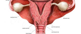
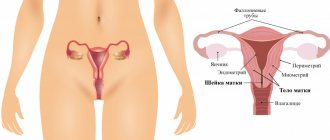
A biopsy and histological examination of a tissue sample obtained with its help is one of the most accurate methods for diagnosing tumors in the human body. In diagnostics, to monitor the condition and identify pathology of the uterine mucosa, gentle aspiration technology is used - endometrial biopsy pipe. Its results, obtained by examining tissue under a microscope, are necessary to identify the causes:
- hyperplastic growth and thickening of the mucous membrane;
- endometrial carcinomas;
- pathological uterine bleeding;
- miscarriage;
- infertility caused by hormonal and hyperplastic disruptions;
- oncological diseases.
Pipel endometrial biopsy is performed before IVF, during bacteriological examination, including before planned conception, as well as to monitor the results of hormone therapy and its effect on the female reproductive system.
Unlike a conventional biopsy, a painful procedure that requires dilation of the cervical canal and produces a high percentage of complications, this technology is painless and non-traumatic
.
What is the research?
Pipelle endometrial biopsy is the collection of elements of the inner layer of the uterus for diagnostic and research purposes. The procedure is carried out using a flexible probe of small diameter, inside of which a piston is placed.
Due to the piston, negative pressure is created in the probe, and the biomaterial enters the device through the hole. A centimeter scale is applied to the surface of the device to control the depth of insertion of the probe. Each device is placed in individual sterile packaging.
The undoubted advantages of the method include:
- atraumatic. When collecting biomaterial, damage to the walls of the uterus is excluded. The method does not require expansion of the cervical canal, therefore it is suitable for nulliparous patients. The study is carried out on an outpatient basis. Complications after the procedure are extremely rare;
- information content. During the diagnostic procedure, it is possible to obtain biomaterial from hard-to-reach areas. Subsequent examination of the samples taken under a microscope allows us to determine the exact cellular composition of the endometrium. The method is able to identify atypical cells characteristic of malignant processes, inflammatory diseases, and hormonal disorders. In addition, the study allows us to identify some of the causes of infertility associated with endometrial pathology and evaluate the results of hormone therapy;
- painlessness. During diagnosis, the woman does not feel pain. The examination is carried out under local anesthesia.
Methodology
Medicine does not stand still, and today a new approach is being used to study the mucous membrane of the female organ, which allows us to solve many problems that are characteristic of the traditional approach.
Conducting a traditional study is impossible without expanding the cervical canal of the organ. The procedure is performed under hysteroscopic control using intravenous anesthesia. This is not only painful, but can also lead to a number of unpleasant consequences. This is why contraindications arise.
Using a new technique, the doctor gets new opportunities. The procedure itself does not require intravenous anesthesia, and dilation of the cervical canal is also not required.
PET/CT examination
Since the instrument is disposable, it is kept in sterile conditions (packaging) before use, so the issue of possible infection is eliminated.
And another important point: the procedure is more economical than the traditional one.
When is a diagnostic procedure prescribed?
The study can be carried out for various diagnostic purposes. Let's consider for what pathologies and disorders it is necessary.
Infertility, failed IVF.
The condition of the endometrium plays a primary role in the onset of pregnancy. The process of successful implantation of the embryo in the uterus depends on the receptivity of the endometrium - structural and functional characteristics.
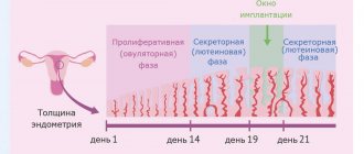
Depending on the phase of the menstrual cycle, the structure and thickness of the endometrium changes. It reaches its greatest maturity during the implantation window, creating favorable conditions for fixation of the embryo in the uterus.
Taking a sample of the endometrium at the expected time of the implantation window and its further immunohistochemical study allows us to assess the receptivity of the endometrium, a decrease in which often leads to reproductive losses, the inability to get pregnant, and unsuccessful IVF programs.
In addition, a pipel biopsy is performed if there is suspicion of:
- anovulatory cycles, in which there is no ovulation and no phase of development of the corpus luteum;
- inflammatory and hyperplastic processes, which can be a source of infertility.
Bleeding of unknown origin, disruption of the menstrual cycle.
The study may be prescribed for bleeding of unknown origin: between menstruation, during menopause.
Also, the diagnostic procedure is performed for women with rare and frequent menstruation, changes in the volume of menstrual blood loss in the direction of increase/decrease. The indication for diagnosis is amenorrhea—the absence of menstruation for six months or more.
The cause of an unstable menstrual cycle and strange bleeding can be inflammatory and hyperplastic processes, endometriosis, and hormonal disorders. Pipelle biopsy of the endometrium allows you to accurately determine the cause of pathological symptoms.
Suspicion of endometrial cancer or other malignant processes in the uterus.
Endometrial cancer is one of the most common malignant diseases in women. Most often, pathology occurs during menopause. Histological analysis of a biopsy (material taken by biopsy) allows not only to detect a cancerous tumor, but also to determine the degree of its malignancy and sensitivity to hormone therapy.
Suspicion of endometriosis.
The disease is characterized by the penetration of cells of the inner layer of the uterus outside the organ. As a rule, the pathology occurs in women of reproductive age. There are many methods for diagnosing endometriosis, and pipel biopsy is one of them.
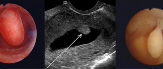
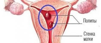
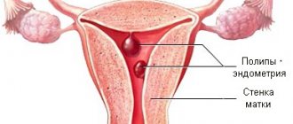
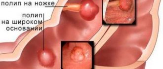
Endometrial polyps.
A polyp is a benign formation. However, any benign tumor under certain conditions can degenerate into malignant. For example, an adenomatous endometrial polyp is considered a precancer due to the fact that its cells are able to divide rapidly.
A biopsy is performed if differential diagnosis of a polyp is needed. Examination of the biopsy allows one to exclude the possibility of malignant degeneration of the tumor.
Suspicion of endometrial hyperplasia.
With pathology, thickening of the endometrium and certain changes in its structure are observed. You can suspect a problem during an ultrasound, but to make an accurate diagnosis, a core biopsy is required. In addition, the study allows us to understand whether it is atypical hyperplasia or not. Atypical hyperplasia is accompanied by changes in the structure and shape of cells and is a precancerous condition.
Suspicion of chronic endometritis.
The disease is an inflammatory process occurring in the endometrium. In the chronic form, the pathology leads to irregular menstruation, infertility, miscarriage and other consequences.
If endometritis is suspected, the biopsy specimen is analyzed for CD138 and CD56 markers, which appear in this pathology.
Monitoring the condition of the endometrium after surgical interventions.
Collection of endometrial elements may be necessary after curettage and other surgical interventions on the uterus. A biopsy allows you to evaluate how well the inner layer of the uterus has recovered after surgery.
What does a Pipel endometrial biopsy show?
The results will be ready within 1-2 weeks. If no pathological changes are found in the aspirate, then the conclusion will indicate that the endometrium corresponds to the age and physiological norm. The specialist will describe the changes in detail.
Histology of the aspirate may indicate the following conditions:
- inflammatory process;
- endometrial atrophy;
- oncological pathologies, precancerous tissue conditions;
- proliferation disorder. In this case, intracellular structures or new cells are formed;
- benign changes in the uterine cavity, including hyperplasia.
On what day of the cycle is the study carried out?
Depending on the diagnostic purposes, the study can be carried out on different days of the menstrual cycle. Detailed information is presented in table ⇓⇓⇓
| Purpose of diagnosis | Recommended period of the menstrual cycle for collecting biomaterial |
| Suspicion of oncological processes in the uterus | Any days except the period of menstruation |
| Polyps, endometritis | The period after the end of menstruation |
| Bleeding of unknown origin | During bleeding of unknown origin. It is important to collect biomaterial during the period when pathological signs are observed |
| Heavy menstruation | Days 5–10 of the cycle |
| Search for causes of infertility, unsuccessful IVF programs | Estimated implantation window period |
| Suspicion of an anovulatory cycle | On the eve of menstrual bleeding |
| Diagnosis of the condition of the endometrium in women during menopause, not related to identifying the causes of unknown bleeding | Any day |
| Tracking the results of hormone therapy | Second phase of the cycle |
Decoding the results
A laboratory assistant will examine the biopsy obtained during the procedure under a microscope. The results of a pipel biopsy can be obtained in person in one or two weeks (in private clinics, as a rule, this happens faster than in public medical institutions). You should contact your gynecologist for a transcript.
This type of biopsy causes minimal trauma to the uterine cavity, so the risk of complications is very low. The advantages of pipel biopsy include:
- Carrying out the procedure in a gynecologist’s office, no need to go to the hospital;
- minimal discomfort;
- the procedure lasts several minutes;
- the growth of the endometrium is not disrupted, since in the process it receives minimal damage, unlike the curettage procedure or other biopsy methods;
- good information content of studies of biopsy obtained by this method, since the sampling is made from different parts of the mucosa;
- low cost.
You can perform a pipel biopsy in the medical institutions presented on the website; the price of the service ranges from 2-5 thousand rubles.
Preparatory stage
Before the study, the woman undergoes a thorough examination, which may include:
- clinical blood test;
- blood test for HIV infection, RW, hepatitis B and C;
- coagulogram;
- cytological and microscopic examination of gynecological smears.
During the period of the cycle in which a biopsy is planned, you need to avoid hormone therapy, which can distort the results of the study. It is also important to stop taking blood thinning medications a week before the test. Such medications may cause bleeding during the procedure.
If the study is carried out in the second phase of the cycle, then after the end of menstruation you need to protect yourself during intimacy. Contraception is necessary to exclude pregnancy at the time of biopsy.
A few days before the procedure, it is recommended to refrain from sexual intercourse, the use of vaginal suppositories, and douching.
The study is carried out on a full bladder.
Material collection
In most cases, aspiration biopsy is performed on an outpatient basis without the use of anesthesia.
The Peipel catheter, which is used to collect biomaterial, is a thin (2-4 mm in diameter) plastic tube with a piston, which is then removed, causing fragments of endometrial tissue to be drawn inward.
After receiving the biomaterial, it is immediately sent for histological examination.
It usually takes about 1 minute to aspirate a piece of endometrial tissue.
Similar methods
Pipelle biopsy is deservedly considered one of the most modern methods of collecting material for studying the uterine cavity. In terms of its effectiveness, it can be compared with aspiration biopsy, but at the same time its advantage is its gentle action and, subsequently, the absence of mechanical damage. Aspiration biopsy takes a sample of mucous membrane in a similar manner, but the procedure does not use a flexible tube. This method cannot be indicated in the presence of cervical cancer.
Best materials of the month
- Coronaviruses: SARS-CoV-2 (COVID-19)
- Antibiotics for the prevention and treatment of COVID-19: how effective are they?
- The most common "office" diseases
- Does vodka kill coronavirus?
- How to stay alive on our roads?
