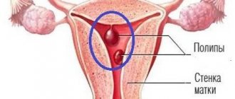
An endometrial polyp is a growth on the inner lining of the uterus into its cavity. It is formed due to excessive growth of cells in the inner lining of the uterus.
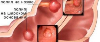
If the polyp is attached to the surface by a narrow stalk, then it is called “pedunculated”; if there is no stem - “on a broad base.”
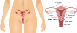
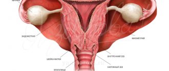
The very concept of “polyp” means an abnormal growth of a certain type of tissue that protrudes above the membrane into the cavity of the organ. Polyps can be found in the nose, stomach, and bladder. An endometrial polyp is a growth specifically from the mucous membrane of the uterus, but this neoplasm is also found in the cervix.
Thus, there are two types of uterine polyps based on their location:
- endometrial polyp of the uterus;
- cervical polyp.
As a rule, such abnormal proliferation of endometrial cells occurs in 10% of women. Most often, an endometrial polyp occurs on an elongated stem. The size of such a neoplasm can vary from 1-2 mm to 3-4 cm, sometimes more. A pedunculated polyp can even penetrate through the cervix into the vaginal cavity. If the polyp reaches a significant size (from 8-10 mm), then it may have thin blood supply vessels.
An endometrial polyp is a benign neoplasm. In some cases, its cells can degenerate into cancerous ones, and then it becomes a malignant tumor. According to scientific research, about 0.7% of endometrial polyps contain adenocarcinoma cells.
Reasons for appearance
Endometrial hyperplasia and polyps, that is, an increase in the number of cells in the inner lining of the uterus or cervix, can occur for several reasons.
Until now, scientists and doctors have not clearly established what the main reason for the appearance is. Hormonal disorders are currently considered the main risk factor for endometrial hyperplasia. In particular, an excess of estrogen and a lack of progesterone in a woman’s body affects her.
Risk factors for education are:
- anovulation;
- persistent follicle syndrome;
- hyperglycemia (with diabetes);
- presence of follicular cysts;
- granulosa cell tumors;
- menopausal syndrome;
- obesity;
- trauma to the uterine cavity (long-term wearing of an intrauterine device);
- abortions or miscarriages;
- complications during or after childbirth;
- chronic inflammatory diseases of the internal genital organs, endometritis;
- immunodeficiency states;
- thyroiditis, thyroidosis and other diseases of the thyroid gland.
Causes of formation of vaginal polyps
There are several reasons that provoke the growth of polyps:
- Human papillomavirus.
Papillomavirus has more than a hundred varieties, each of which forms a priority location for localization. This type of papilloma, such as genital warts, quite often affects the mucous tissues of the genital organs, including the vagina. They usually visually resemble lobed cauliflower, being reddish-pink in color. Polyps caused by the human papillomavirus usually show multiple growths and are localized close to the vaginal opening. - Mechanical damage to the mucous membrane.
Injuries to the vagina during medical procedures and childbirth, accompanied by ruptures of the mucous membrane, inevitably lead to the formation of a scar. Over time, individual polyps may appear in an area of rough connective tissue. - Chlamydia and other sexually transmitted infections.
The infectious-inflammatory process is one of the leading factors stimulating the formation of vaginal polyps. - Hormonal disorders.
Hormonal changes, as well as diseases accompanied by endocrine imbalance, often contribute to the formation of polyps. Sometimes the process of polyp growth itself occurs in the cervical canal, and after that the body of the neoplasm falls into the vaginal lumen, where it is diagnosed.
Symptoms of vaginal polyps
Vaginal polyps may not show any specific symptoms for a long time. However, some women report itching and burning of the affected area, which most often occurs after sexual intercourse.
During intimate intimacy, due to trauma to neoplasms, bleeding or scanty sanguineous discharge may develop. In some cases, pain and discomfort in the lower abdomen are noted, but this phenomenon is relatively rare.
Treatment of vaginal polyps
The presence of pathological growth of the vaginal mucosa is usually detected during a standard vaginal examination, which can be supplemented by colposcopy for final confirmation of the diagnosis.
Unlike polyposis of the body or cervix, vaginal growths can be eliminated using techniques such as laser and radio wave correction, cryodestruction, electrocoagulation and chemical removal. The use of the latter technique is limited during pregnancy. Having determined the cause of the development of polyposis, the specialist will also prescribe treatment aimed at eliminating the factor that caused the disease.
Classification
Depending on the histological structure, a classification of endometrial polyps has been developed. So, experts distinguish the following types:
- glandular polyp - the structure on histology is represented by glands and stroma;
- glandular-fibrous polyp consists of connective tissue elements and glandular structures;
- fibrous polyp – connective tissue only;
- adenomatous polyp is a precancerous neoplasm that consists of glandular and atypical cancer cells.
It is also characteristic that endometrial glandular polyps occur in young women, while adenomatous and fibrous polyps occur more often in older women during menopause.
A functional polyp is also identified, which, unlike basal type polyps, can change during the menstrual cycle. The functional type polyp consists of epithelial cells.
Why are polyps dangerous?
The content of the article
Any polyps, no matter where they are located, should be treated. Even if the tumors are benign, the condition is diagnosed as precancerous. For women planning to have children, the pathology is especially dangerous, because it leads to infertility.
Let's consider 2 types of polyps:
- Adenomatous polyp.
A type of adenoma that manifests itself in the growth of endometrial tissue - the mucous membrane covering the inner surface of the uterus (during menstruation, the functional layers of the endometrium come out with secretions). Adenoma is a benign tumor and appears only in organs with glandular epithelium, but over time it can degenerate into oncology - cancer. The risk increases when the condition is neglected. Adenomatous polyp also appears on other organs, but its occurrence in the uterus is most life-threatening. - Glandular polyp.
Already from the name it is clear that this type of polyp is formed from glands and endometrial tissue. Such polyps are small and the safest among similar neoplasms. The diameter of a glandular polyp does not exceed 1.5 cm. Glandular polyps are almost always benign, although there are cases where endometrial cancer developed from just such a focus. It happens that the lumen of the glands forms a cyst, then a glandular cystic polyp is diagnosed. Glandular cystic polyp is not a separate subtype. Such polyps form in the form of a node and are located in the stroma of the mucous membrane, which is located in the places where the fallopian tubes drain.
Clinical symptoms of endometrial polyp
If the neoplasm is small in size, then there are no signs. As a rule, such polyps are found only with an ultrasound of the pelvic organs.
When the polyp reaches a significant size or there are quite a lot of such formations, characteristic symptoms appear. Women complain about:
- menstrual irregularities;
- the appearance of intermenstrual bleeding from the vagina;
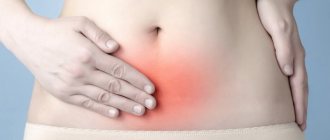
- lower abdominal pain;
- discomfort and pain during sexual intercourse;
- cramping pain when the polyp enters the cervical canal;
- the appearance of watery whitish discharge in large quantities.
Menstrual irregularities in young women are manifested not only by bleeding between periods, but also by metrorrhagia, too heavy menstruation. Blood may also be released during sexual intercourse. With the onset of menopause, bleeding from endometrial polyps can be episodic. Also, the consequence of polyposis can be infertility in women, recurrent miscarriage. Since the endometrial polyp has scant symptoms, you need to pay attention to the slightest disturbances and immediately refer the patient for additional examinations.
Diagnostics
At the first appointment, the doctor is unlikely to be able to make a diagnosis, since it is impossible to determine the location of the polyp in the internal cavity of the uterus without a thorough examination. There are objective and subjective reasons that may raise suspicions of uterine polyposis.
Subjective signs:
- pain in the lower abdomen;
- general malaise.
Objective signs:
- results of general and biochemical blood tests;
- analysis of vaginal smears;
- gynecological ultrasound results;
- hysteroscopy.
If the diagnosis is confirmed by the gynecologist, the doctor gives a referral for a biopsy. Further treatment will depend entirely on the results of histological examination. In most cases, the treatment method for adenomatous and glandular polyps is surgery.
Diagnosis of endometrial polyps
In order to clarify the presence and type of polyp, it is necessary to undergo an external examination:
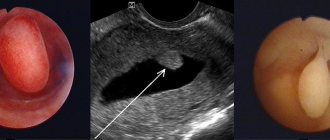
- ultrasound examination of the pelvic organs - makes it possible to detect fibrous and glandular-fibrous polyps;
- hysteroscopy is the best diagnostic method, which also allows removal of the tumor;
- histological examination of scrapings (histology of endometrial polyps) - allows you to determine the structure of the polyp.
Ultrasound signs of an endometrial polyp are quite typical, but a final diagnosis cannot be made. During examination, varying degrees of expansion of the uterine cavity can be detected. Inside, neoplasms of a homogeneous structure with clear contours are found. To clarify the diagnosis of endometrial polyp, hysteroscopy is necessary. It is carried out using a system with special optics, which is inserted into the uterine cavity. Neoplasms are located mainly in the area of the fundus and corners of the uterine cavity. Hysteroscopy can also be used to remove endometrial polyps. After this, it is imperative to conduct a histological examination, since approximately 0.7% of polyps have signs of malignant degeneration.
Vulvar polyp
The vulva is the collection of external genitalia of a woman. Polyps often inhabit the areas of the labia, clitoris, and vestibule of the vagina.
Pathological growth of the mucous membranes is accompanied by the appearance of a growth or tubercle, which can increase in size or form lobular structures that look like cauliflower. Due to the open localization of tumors, they, unlike polyps of the uterine body or cervical canal, are much easier to diagnose and, as a rule, are removed at an early stage, which avoids the degeneration of the tumor into a malignant form.
Causes of vulvar polyps
Polyps are often called not classic polyps, but genital warts, the primary reason for the formation of which is the penetration of the human papillomavirus into the patient’s blood. Against the background of a reduced immune response and hormonal imbalance, the papillomavirus causes pathological division of epithelial cells, which ultimately leads to the growth of condylomas. At the initial stage, such structures may take the form of multiple tubercles, which, as they grow, merge into one voluminous tumor-like papilloma.
The main factor in the formation of vulvar polyps is mechanical trauma to the external female genitalia. The healing of the wound in this case is accompanied by the formation of a scar, the basis of which is rough connective tissue. It is this area that becomes the localization zone for single polyps.
Also, vulvar polyposis can occur against the background of hormonal changes, such as pregnancy, puberty or menopause, and inflammatory and infectious processes, usually sexually transmitted infections.
Diagnosis and treatment of vulvar polyps
Diagnosing vulvar polyps is not difficult. A gynecologist can make a diagnosis after a simple physical examination, which is often combined with a gynecological examination, in order to exclude the possibility of proliferation of vaginal and cervical polyps.
Removal of vulvar polyps can be done in the following ways:
- Laser technique -
the pathological growth is destroyed under the action of a directed laser beam. The technique is considered the least traumatic and, accordingly, the safest among all currently known methods of excision of epidermal tumors. - Radioknife
- radio wave therapy is preferable for any gynecologist. It has many advantages, including over laser removal. In particular, a radioknife can be used to take polyp tissue for analysis, while a laser evaporates the entire tumor. - Cryodestruction is
the freezing of pathological tissue areas by exposure to extra-low temperatures through applications of liquid nitrogen. - Electrocoagulation -
the technique involves the use of electric current as an aggressive destructive agent. - Surgical excision is
used relatively rarely due to the high level of trauma and the long period of tissue healing.
Surgery to remove endometrial polyp
The operation to remove an endometrial polyp is technically simple and does not take much time. The intervention is carried out using general anesthesia. The patient's uterine cavity is scraped out, polyps and the endometrial layer are removed. The polyps are sent for histology, and the patient is prescribed care and rehabilitation therapy. Pregnancy after surgery is possible – both after IVF and during natural conception.
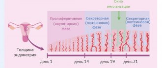
Uterine polyps and pregnancy
Small polyps do not interfere with conception, the implantation of the egg on the uterine wall, and the development of the fetus. However, sometimes it is the polyp that causes infertility. Therefore, when planning a pregnancy, it is advisable to make sure that there are no endometrial and cervical polyps. But if pregnancy has already occurred, cervical polyps are usually not removed.
Removal of the polyp, in turn, also does not prevent subsequent pregnancy.
Removal of endometrial polyp by hysteroscopy
Hysteroscopy for uterine endometrial polyp is not only a diagnostic, but also a therapeutic procedure - a minimally invasive surgical intervention. It is performed during an examination, when a thin optical instrument is inserted into the uterine cavity and the condition of the endometrium is determined. Hysteroscopy also involves taking bioptic material from altered areas of the inner lining of the uterus or curettage of an endometrial polyp. As a rule, a simple diagnostic hysteroscopy of an endometrial polyp is performed without anesthesia and lasts from 5 to 20 minutes.
If surgical hysteroscopy is performed to remove an endometrial polyp, then a hysteroscope with a micro-instrument in a tube is inserted into the uterine cavity. This allows for minor removal operations. Using special microscissors, you can remove a tumor or separate small adhesions in the uterine cavity. This therapeutic hysteroscopy, in contrast to a simple diagnostic one with curettage of the endometrial polyp, allows you to specifically get rid of the pathology inside the uterus. This cannot be done using a conventional curette, as there is a high risk of injury to the inner lining and cervix.
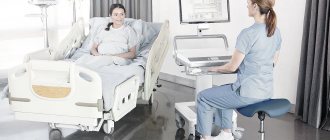
Surgical hysteroscopy or surgery to remove an endometrial polyp can only be performed in large clinics or centers that are authorized to use narcotic analgesics and equipped with resuscitation equipment. After removing the polyp using this method, conservative treatment and observation of the woman’s condition in the hospital lasts for 24 hours.
Hysteroresectoscopy of an endometrial polyp is a full-fledged surgical operation, which is indicated for patients with gross pathology in the uterine cavity, as well as for abnormalities in the development of this organ. During the operation, a special instrument is used, which, under video control, will allow for optimally precise manipulations in the uterine cavity. Hysteroresectoscopy of an endometrial polyp lasts from 30 to 90 minutes. It can only be performed under general anesthesia. After removal of the endometrial polyp in this way, treatment is conservative and the patient is monitored in the hospital for 2-3 days.
Simple and therapeutic hysteroscopy, as well as hysteroresectoscopy for endometrial polyps, is indicated for:
- treatment of fibrous polyp;
- treatment of endometrial glandular polyp;
- treatment of glandular fibrous polyp;
- treatment of adenomatous polyp.
Vaginal polyp
Vaginal polyps are benign neoplasms that grow on the mucous membranes and epithelial (integumentary) tissues. One of the places where polypous structures are localized is the vagina. The formations in this case most often represent multiple small tubercles with a wide base.
There are several reasons that provoke the development of pathological proliferation of vaginal tissue, however, regardless of the factor that caused the process, polyps in the vagina must be removed.
Other treatments for endometrial polyp
When an endometrial polyp should be removed, hysteroscopy is not the only method. In urgent situations or when the size of the tumor is too large, curettage is resorted to. The operation is more traumatic and more likely to cause relapses, which is why it is now rarely used or combined with hysteroscopy.
A more modern technique is laser removal of endometrial polyps. It is used only in some clinics. The operation is indicated for women of childbearing age, since after it the chance of secondary infertility decreases. If an adenomatous endometrial polyp is identified, removal of the uterus, sometimes with appendages, is indicated. This type of neoplasm is mainly typical for older women, so the operation does not affect fertility.
Polyps in the uterus: surgical treatment methods
In modern gynecological practice, there are several ways to remove polyps of the cervix and uterine cavity.
Hysteroresectoscopy
Hysteroresectoscopy is a modern technique that involves inserting an endoscope equipped with a video camera into the uterine cavity.
When performing this procedure, the doctor has the opportunity to assess the condition of the uterine cavity, visualize the polyp itself, and also remove it using mechanical, electrosurgical or laser instruments. Then a histological examination of the formation is carried out.
Advantages of hysteroresectoscopy:
- removal occurs under visual control and not blindly;
- the polyp is removed along with its stem, which prevents its re-formation in this particular place;
- there is no negative or harsh effect on the uterine mucosa, which is extremely important for women planning a pregnancy;
- the risks of trauma to the uterus and the addition of an infectious process are minimized;
- after the operation, the woman recovers quickly and can be discharged from the hospital in the near future;
- there is no pain;
- This minimally invasive intervention allows us to minimize risks for patients with somatic pathologies.
Fractional diagnostic curettage
This surgical intervention involves blind removal of a uterine polyp by curettage of the uterine cavity and cervical canal.
While conducting this study, the doctor is not able to exercise visual control over what is happening in the uterine cavity.
After separate curettage of the canal and the cervical cavity, the resulting material is also sent for histological examination.
According to statistics, after the procedure of curettage of the uterine cavity, recurrence of polyps occurs in more than 50% of cases. That is why in modern gynecology, if there is a diagnosis of “uterine cavity polyp” or “cervical polyp,” treatment involves hysteroresectoscopy of the polyp.
Treatment after endometrial polyp removal
If surgery to remove an endometrial polyp was performed by hysteroscopy or laser, the risk of complications is minimal. But in the first three days, patients are advised to take no-shpa three times a day. The fact is that after the intervention, spasm of the muscles and neck may occur, which leads to the accumulation of blood in the cavity of the organ. No-spa allows you to relax the muscles and prevents this phenomenon. Short-term antibiotics and anti-inflammatory drugs are also prescribed in the postoperative period.
If an endometrial polyp, its symptoms arose due to hormonal imbalances, combined oral contraceptives may be prescribed as regulators. Women who do not plan to become pregnant in the coming years are recommended to have an intrauterine device installed, which releases gestagen. Hormonal therapy is mandatory for adenomatous polyps in young women if the uterus has not been removed.
Pregnancy with endometrial polyposis
Patients with endometrial polyposis may develop infertility. This is due to changes in hormonal levels and difficulties that arise during the implantation of a fertilized egg into the wall of the uterus. According to statistics, removal of polyps significantly increases the chances of both a natural pregnancy and IVF success.
If you have any questions related to polyposis, you can ask them to the doctors at Nova Clinic. You can make an appointment with a doctor by calling the number listed on the website or using the booking button.
Treatment with folk remedies
Traditional treatment can sometimes be a fairly rational solution, because alternative methods sometimes turn out to be very effective. Among the folk remedies for the treatment of endometrial polyps, garlic pulp is used, which is placed in gauze, a tampon is made with a long string and inserted into the vagina overnight. The course of folk treatment with garlic is 4 weeks.
Egg yolks and pumpkin seeds are also used. This folk method of treating endometrial polyps has been effectively used for decades. You need to take 6 chicken eggs, boil them, separate the yolks. Add 5 tablespoons of crushed pumpkin seeds and pour in 0.5 liters of sunflower oil. Heat this mass in a water bath and cool. You need to take one teaspoon orally in the morning and evening for 5 days. Then take a break for 1 week and repeat the 5-day course again.
These traditional methods of treatment, as a rule, do not produce any side effects, but may not always be effective.
Prevention of polyps
The appearance of polyps is associated with disruption of the ovaries and excess estrogen.
Prevention of this disease includes many factors. Find out what you need to do to avoid polyps
- Do not eat foods contaminated with dioxide and meat containing hormones.
- Live in an area with good ecology.
- Avoid hypothermia, dress appropriately for the weather and do not sit on cold surfaces.
- Do not be promiscuous.
- To live an active lifestyle. Physical exercise does not allow blood to stagnate in the genitals.
- When choosing hormonal birth control pills, be sure to consult your doctor.
- Visit your gynecologist regularly.
Women's health is an essential component of a happy life. Take care of yourself and be healthy!
Do cervical polyps need to be removed?
If the polyps are asymptomatic, they are removed anyway (polypectomy).
Small polyps can be removed in primary care right at your gynecologist's appointment. The polyp is grabbed with special forceps and twisted several times, and then cauterized. Large and difficult-to-reach polyps are removed by excision with a radioknife or laser during colposcopy. Removed polyps should be sent for histological examination to exclude malignancy. The recurrence rate of pathology is 6-12%.
Operation stages:
- To examine the walls of the uterus, it is stretched.
For better visibility, a contrast medium is used - saline solution or 5% glucose. Most serious complications during hysteroscopy arise from the selection of an inappropriate environment. For example, the use of carbon dioxide as a medium is more suitable for performing hysteroscopy in the absence of bleeding from the genital tract. The presence of even slight bloody discharge will cause clouding of the environment, which will make examination of the uterine cavity impossible. In addition, carbon dioxide when stretched can cause embolism due to the thermal energy effect on tissue. Therefore, I prefer liquid media, particularly non-electrolyte solutions, as they are not susceptible to hemolysis and will not cause electrolyte disturbances. The saline solution I use does not irritate tissues and is quickly removed from the vascular bed, increasing the volume of circulating blood only temporarily. In a modern hysteroscope, thanks to its flow-through design, it is possible to remove blood and tissue particles without impairing visibility even during bleeding. - During the examination of the uterine cavity, in addition to assessing its size and shape, the relief of the uterine wall, the condition of the endometrium, and the accessibility of the mouths of the fallopian tubes are determined. The fundus of the uterus, angles, isthmic region, and cervical canal are also examined. If there are suspicious areas, tissue samples are taken using special forceps for subsequent histological examination.
- Removal of endometrial polyp using a bipolar hysteroresectoscopy loop
Using a bipolar resectoscope, the body and base of the polyp are removed; this approach prevents the development of bleeding from the polyp bed, and scar formation is excluded in the future. Multiple polyps can be removed by curettage. For better results, a combination of different methods is possible. For large polyps with a wide stalk, removal is carried out in parts - fragmentation. When removing polyps located in the area of the mouth of the fallopian tubes, technical difficulties arise, since the loop of the hysteroresectoscope may be too large for manipulation in the corners of the uterus. To destroy polyps localized in this area with underlying basement membrane, I use a loop electrode or coagulation with a ball electrode. This approach also reduces the risk of relapse in the future.
- Deviations discovered during the examination: synechiae, intrauterine septa, submucosal myomatous nodes are also removed.
Endometrial polyps are often combined with other diseases of the reproductive system: fibroids, ovarian cysts, endometriosis, etc. In this case, in our clinic it is possible to perform hysteroscopy with laparoscopy; During one anesthesia, correction of all disorders is carried out using transvaginal and laparoscopic access - through punctures of the anterior abdominal wall.
Tactics for managing patients in the postoperative period
If doctors have removed a polyp in the cervix under anesthesia, the woman is left under observation until the next day. Further management tactics depend on the result of histological examination. In the presence of fibrous and glandular-cystic endometrial polyps, an ultrasound examination of the pelvic organs is performed once a year. These formations do not require treatment or additional examination. If morphologists have identified glandular and adenomatous polyps or a combination with endometrial hyperplasia, hormonal therapy is carried out taking into account the patient’s age and the severity of proliferation. In the presence of mixed ones, they do not prescribe hormonal drugs, provided that the patient’s hormonal background is normal, there is no obesity and diabetes mellitus.
A polyp on the cervix during pregnancy negatively affects the gestation of the fetus. Although polyps are benign formations, they require mandatory medical observation and treatment. The danger of the pathology is that when polyps grow, a miscarriage is possible. For this reason, treatment of polyps should be carried out at the stage of pregnancy planning.
After hysteroscopy, the patient may experience spasmodic pain for some time. Minor discharge may be observed for two to three weeks. A week after the operation, the patient must undergo a gynecological examination. The doctor, guided by the patient’s age, the cause of the formation of the polyp and its nature, prescribes restorative therapy.
If your temperature rises, there is heavy bleeding, dark mucus with an unpleasant color, or severe and acute pain, you should consult a doctor. In this case, we can assume the presence of a complication - inflammation of the uterus, its perforation or hematometer (accumulation of blood in the uterus). Inflammation after removal of a cervical polyp occurs very rarely. It is usually provoked by the fact that the operation was carried out against the background of a latent infection, an inflammatory process, or the rules of antiseptics and antiseptics were violated during the surgical intervention. Perforation is formed as a result of a puncture of the wall of the organ due to the looseness of its structure or poor expansion of the neck. Small perforations heal on their own, and large ones are sutured by doctors.
In order for the recovery period to proceed faster and without complications, the patient after the operation is advised to strictly observe the rules of personal hygiene. At this time, she is not recommended to take baths, visit saunas and steam baths, or swim in open water. It is recommended to take a warm shower. For a month, taking aspirin, excessive physical activity, and lifting heavy objects is contraindicated. Douching and sexual intercourse should be avoided. In order to undergo examination and treatment of a uterine polyp using modern methods, call the contact center of the Yusupov Hospital.
Symptoms
The severity of external manifestations of polyposis depends on the following factors:
- woman's age;
- the presence of regular ovulation;
- normal functioning of the ovaries;
- gynecological pathology (uterine leiomyoma, endometrioid disease, adnexitis).
The main complaint with endometrial polyposis will be pathological changes in the menstrual cycle:
- a significant increase in the abundance of critical days;
- intermenstrual bleeding;
- premenstrual bleeding;
- uterine bleeding;
- the appearance of bleeding during menopause.
Rarely occurring pain in the lower abdomen is caused by the following reasons:
- cessation of blood flow in the endometrial polyp;
- prolapse of a large polyp through the cervix (“birth”).