- home
- Gynecology
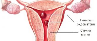
- Endometrial polyps
- Endometrial polyp removal
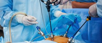
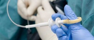
Surgical removal of the endometrial polyp is the only way to get rid of the formation, which is a focal thickening of the mucous layer of the uterine wall. Among the various methods for removing polyps in the endometrium, hysteroscopy is considered the most gentle method. The indication for surgical treatment is the presence of an endometrial polyp according to ultrasound data for 2 consecutive cycles or more.
What is a polyp in the uterus
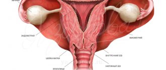
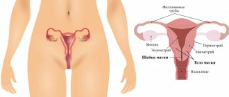
A uterine polyp is a benign space-occupying formation that forms in the uterine cavity against the background of local hyperplasia of cells in the growth layer of the endometrium. The pathology can affect patients of different age groups. Detectability is approximately 6-20%.
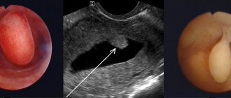
The sizes of endometrial polyps range from 1-2 millimeters to several centimeters.
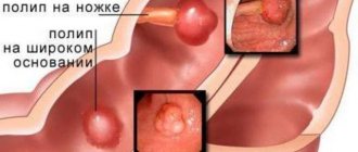
The formation is attached to the uterine wall through a thin stalk or a wide base. Nutrition is provided by a vessel in the center of the polyp. Pathological structures can be either single or multiple. When several formations are detected, they speak of polyposis. Polyps can cause intermenstrual bleeding, abdominal pain, and problems with conception. Usually pathology is diagnosed during ultrasound. In gynecology, polyps are considered potentially malignant formations that need to be removed surgically.
Polyps by type of attachment
Preparation
The procedure is usually carried out on days 6-10 of the cycle. This is the most favorable period, since it is at this time that the endometrium is thin. This approach greatly simplifies the process of removing polyps. If surgery is needed urgently, the doctor can prescribe it on any day of the cycle. The main condition is the absence of severe bleeding.
In preparation for the procedure, the woman is examined by a gynecologist, anesthesiologist, and therapist. Additional studies may be required as directed by your doctor to identify concomitant diseases. The vagina is carefully examined. A repeat ultrasound is performed. Laboratory tests are also being carried out and test results are awaited.
A day before the operation, the woman should begin preparation. Do not eat heavy foods that can lead to gas formation. In the evening, you should refuse to eat, but continue to drink still water as needed. An enema is given at night. On the morning of the operation, smoking is prohibited and you should refuse food and liquid. The enema is also repeated. After or before the procedure, in order to avoid the development of inflammatory processes, the patient may be prescribed a course of antibacterial drugs.
Causes
The reasons for the appearance of polypous growths are still being studied, but an important role in etiopathogenesis is played by:
- hormonal imbalance;
- injuries, including medical procedures (curettage);
- infectious, inflammatory diseases of the genital organs;
- uncontrolled use of oral contraceptives, etc.
Diagnosis of polyps is difficult; often they do not manifest themselves at all, and the disease is asymptomatic. Reasons for examination include excessive discharge (leucorrhoea) and intermenstrual bleeding. The diagnosis is made by ultrasound examination of the uterus. Hysteroscopy is used to identify small polypous growths. If a polyp is detected, treatment is required. Sometimes reverse development of the tumor is observed after conservative therapy. Removal is resorted to if there is no result.
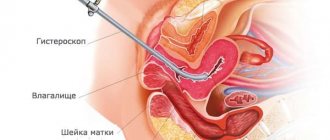
Hysteroscopy is usually used for diagnostic examination and resection of a mass in the uterus. It can also be removed with a laser, but the prices for such a service are higher due to the high cost of the equipment.
There are several theories for the development of cervical and uterine polyps. Most often, a polyp in the uterus appears as a result of hormonal imbalance - this is the most common version of the causes of the pathology. Under the influence of high levels of estrogen, the endometrium of the uterus (inner layer) grows. The endometrium can grow evenly, then hyperplasia appears. In case of uneven growth of the endometrial layer, polyps form in the uterus and cervix. Progesterone deficiency ensures the active growth of benign formations.
The following factors contribute to the appearance and growth of polyps in the cervix and uterus:
- Fibroids and myomas.
- Ovarian dysfunction.
- Polycystic ovary syndrome.
- Endometrial hyperplasia.
- Chronic infection in the genitourinary organs, including sexually transmitted infections and human papillomavirus. During inflammation, there is a sharp increase in the number of leukocytes (immune cells), which, along with the destruction of pathogenic microorganisms, promotes the growth of cells of the inner uterine layer.
- Cervical polyps most often appear after unsuccessful curettage.
- Chronic stress leads to an increase or decrease in hormone levels.
- Vascular obstruction - epithelium grows around a blocked vessel.
- Hereditary factor - polyps of the uterus and cervix are more often formed in those patients whose relatives suffered from polyposis. This category of women should be especially attentive to their health and undergo regular preventive examinations.
- An inactive lifestyle contributes to the appearance of congestion in the pelvic organs, as a result of which the uterus and appendages experience a lack of oxygen, which leads to hormonal imbalance and cell proliferation.
- The drug Tamoxifen is used in the treatment of cancer; it causes hormonal disorders. In some patients, a polyp may form in the cervical canal of the cervix or in the uterus.
Diseases of the endocrine system (diabetes mellitus, thyroid disease, obesity) contribute to the development of polyposis. The endocrine glands function in close connection, so disturbances in one gland lead to a malfunction of the other, in particular in the ovaries, which begin to produce sex hormones at an accelerated pace. In diabetes mellitus, microcirculation is disrupted, cell hypoxia occurs, which contributes to their proliferation and atypical changes. With excess weight, adipose tissue produces estrogens, which provoke an increase in cervical polyps. When ovarian function is disrupted, a large amount of estrogen is synthesized, which is released into the blood constantly (normally, the hormone is produced in the first 14 days of the cycle).
As a result, during menstruation, individual fragments of the endometrium remain in the uterine cavity, rather than exfoliating and coming out. Gradually, connective fibers and vessels grow into these areas. This is how a uterine polyp appears.
Appearance
The appearance of polyps is varied: most grow on a thin stalk, hanging into the intestinal lumen. Less common are neoplasms with a wide base, and even less common are tumor-like forms that slightly rise above the surface of the rectal mucosa. The size of a typical polyp is from 0.5 to 2 cm, but there are also giant forms that can block the intestinal lumen. As the size of the formation increases, the risk of transformation into a malignant tumor increases. Small formations (up to 0.5 cm) are represented predominantly by hyperplastic polyps , the cells of which differ little from normal epithelial cells and are characterized by a minimal risk of cancer transformation.
Large pathological elements (more than 2 cm) often have a villous surface, which is easily injured and bleeds. Such neoplasms are especially dangerous in terms of transformation into rectal cancer .
Symptoms of polyps in the uterus
The main signs of polypous growths are:
- spotting at the end of sexual intercourse of varying intensity;
- bleeding;
- nagging pain in the lower abdomen, above the pubis;
- intense menstruation, and, as a result, signs of anemia - pale skin, general weakness, shortness of breath, dizziness.
Often, polyps occur without any external signs and are an accidental “find” during a routine examination.
Signs of polyp formation
Preoperative diagnosis
The proctologist diagnoses formations located near the anus during a digital examination. However, the main method for diagnosing rectal polyps is a hardware examination using sigmoidoscopy, which will provide a volume of information sufficient to select a treatment method: • localization, size and morphological appearance of growths; • presence of complications (bleeding, suppuration); • histological type of tumor (tissue sampling is carried out according to indications); • condition of the rectum.
If the doctor decides to undergo surgery, it will be necessary to undergo a standard preoperative examination, which includes: • a general blood and urine test; • blood chemistry; • determination of blood group and Rh factor; • coagulogram + determination of blood sugar levels; • tests to detect blood-borne infections (hepatitis B and C, HIV, syphilis); • ECG; • fluorography; • consultation with a therapist, and for women – consultation with a gynecologist.
Additionally, you may need: • gastroscopy (EGD); • colonoscopy; • CT scan of the abdominal cavity; • Doppler ultrasound of the veins of the lower extremities; • ECHO-KG.
Treatment of uterine and cervical polyps with conservative methods
There are conservative and surgical methods of therapy. Symptoms and treatment of a polyp in the uterus are two interrelated factors. The brighter the clinical picture, the more important the treatment measures. If there is a small formation caused by excess estrogen, hormonal drugs are prescribed, with the help of which it is possible to reduce their size or completely disappear. Treatment of uterine polyp without surgery (removal of the polyp) with the help of medications is recommended for adolescent patients who have not given birth.
If the disease is of infectious-inflammatory origin, a course of anti-inflammatory and antibacterial therapy is necessary. Treatment of uterine polyps with drugs is carried out for women of any age category. When conservative therapy is ineffective, the polyp is removed.
Complications
• Large polyps can block the intestinal lumen and cause a fatal condition - acute intestinal obstruction. • Bleeding from villous growths contributes to the development of anemia. • Secretion of mucus by adenomatous tumors leads to dehydration and symptoms of hypokalemia : ___• increased fatigue; ___• muscle weakness up to the development of paralysis; ___• heart rhythm disturbances. • The inflammatory process associated with fibrous formations depletes the body and can be complicated by purulent inflammation of the perirectal tissue and sepsis. • Adenomatous and villous polyps inevitably develop into rectal cancer 5-15 years after their occurrence. Treatment for rectal cancer often involves “life with a colostomy bag” and painful courses of chemotherapy. High mortality is associated with early liver metastases, in such cases the prognosis becomes hopeless.
How much does it cost to treat polyps in the uterus?
The cost of treatment differs in different clinics. It primarily depends on the type of formation and the choice of treatment method. Thus, with conservative therapy, it may include control examinations to monitor the effectiveness of therapy. If the patient’s condition does not improve during therapy, surgical removal of the polyp may be required. Different surgical treatment methods also have an impact on cost. The use of expensive equipment increases the price. In fact, it is possible to find out how much it will cost to remove a polyp only after a complete examination of the patient. In addition, prices are influenced by the clinic’s pricing policy and the level of qualifications of doctors.
Types of surgical interventions
All types of surgical interventions can be divided into two large groups: traditional operations and minimally invasive techniques. Intervention using a minimally invasive method involves the use of endoscopic techniques and the latest technologies: • radio wave radiation, • laser, • electrocoagulation loop.
Such techniques are called “treatment without surgery” because they: • do not involve opening the abdominal cavity and intestinal cavity; • carried out using a bloodless method; • have a short recovery period with minimal severity of unpleasant side effects (pain, weakness, etc.).
traditional interventions to for impressively sized polyps, as well as for malignant neoplasms. If the growth is located in close proximity to the anus (up to 10-12 cm), then the operation is performed through the rectum. Otherwise, surgeons obtain surgical access by opening the abdominal cavity.