Classifications of precocious puberty
An attempt to systematize individual varieties of this pathological condition made it possible to identify two main forms of premature puberty
:
1. True,
or
premature puberty of central origin
, the development of which is associated with premature activity of the pituitary gland and hypothalamus. In this case, an increase in the production of sex hormones by the gonads occurs due to the stimulation of the internal sex glands by gonadotropic hormones produced by the pituitary gland and hypothalamus.
2. False,
or
peripheral precocious puberty
, which is caused by enzymatic defects in the synthesis of steroid hormones by the adrenal cortex or tumors of the gonads, which leads to an increase in the production of sex hormones, and is not associated with the level of gonadotropins.
3. Precocious puberty associated with gene code disorders
, leading to autonomous activation of the activity of the gonads, independent of the level of gonadotropins.
There is no clear boundary between true and false forms of precocious puberty; they can transform into one another or occur in combination, however, for the convenience of diagnosis and choice of treatment tactics, a working version of the classification of precocious puberty has been developed, according to which the following forms are distinguished:
1. True precocious puberty
, which includes:
- idiopathic premature puberty;
- cerebral precocious puberty, which developed due to tumors, as well as non-tumor lesions of the central nervous system (hamartomas, gliomas, encephalitis, hydrocephalus, meningitis, toxoplasmosis, radiation, surgery, arachnoiditis). Cerebral precocious puberty can also be caused by congenital syndromes such as neurofibromatosis, tuberous sclerosis and a number of others;
- true premature puberty, developing as a result of prolonged exposure to sex hormones due to untimely correction of congenital dysfunction of the adrenal cortex, or late removal of tumors that produce hormones.
2. False precocious puberty
which occurs:
- in male patients due to testicular tumors, neoplasms located in and outside the skull that produce chorionic gonadotropic hormone, adrenal tumors and congenital dysfunction of the adrenal cortex;
- in female patients - as a result of malignant neoplasms of the ovaries, adrenal tumors, or the presence of ovarian follicular cysts.
3. Gonadotropin-independent forms of precocious puberty
:
- McCune-Albright syndrome;
- testotoxicosis.
4. Incomplete forms of premature puberty
:
— accelerated pubarche (hair growth);
- early thelarche (enlargement of the mammary glands).
All of the listed forms of premature puberty are characterized by the main signs of progression of puberty, namely, the appearance of secondary sexual characteristics, an increase in the volume of the external genitalia, accelerated growth and maturation of bone tissue.
Depending on the completeness of clinical manifestations, the full form of precocious puberty
, in which all of the above clinical signs can be identified, and
an incomplete form of precocious puberty
, which is characterized by the development of only premature pubarche (secondary hair growth) or thelarche (increase in the size of the mammary glands).
In addition, there are forms of precocious puberty that do not fit into any of the above, for example, precocious puberty that developed against the background of uncompensated primary hypothyroidism.
Pubarhe
The onset of pubic hair growth usually occurs after thelarche, around 11–12 years of age, and is often accompanied by axillary hair growth. Pubarche usually occurs after thelarche, but simultaneous development of thelarche and pubarche is also normal. In representatives of the black race, pubarche may precede thelarche, which is not a pathology.
The development of pubic and axillary hair occurs secondary to an increase in the concentration of circulating androgens. Sometimes the concepts of pubarche and adrenarche are used as synonyms, but this is not entirely true. Pubarche is the development of pubic hair, which occurs under the influence of adrenal androgens after the age of 10 years in the stage of pubertal development, and adrenarche is the activation of androgen synthesis in the adrenal glands, which occurs between the 6th and 8th years of life and precedes the onset of pubertal development.
Causes of premature puberty
True, or gonadotropin-dependent precocious puberty is due to the fact that the activation of the pulsed secretion of gonadotropin-releasing hormone produced by the hypothalamus occurs earlier than expected. An increase in the secretion of this GnRH leads to an increase in the production of luteinizing and follicle-stimulating hormones of the pituitary gland, which are gonadotropic. These, in turn, activate the production of sex hormones in the gonads, thereby leading to the development of secondary sexual characteristics that change the appearance of the child. The most common cause of true precocious puberty is hypothalamic hamartoma.
The cause of false precocious puberty is most often a disruption in the enzymatic processes of production of steroid hormones in the adrenal cortex, less often - tumors of the gonads and adrenal glands that produce sex hormones.
The cause of the development of forms of precocious puberty, independent of the level of gonadotropin, is considered to be genetic mutations that lead to the constant activation of the production of sex hormones by the cells of the male and female gonads without the participation of gonadotropic hormones.
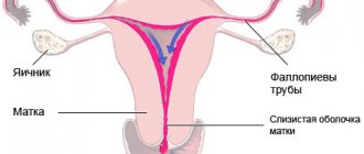
Menstrual cycle
The hypothalamus, pituitary gland, ovaries and uterus are components of the female reproductive system that take part in the establishment and regulation of the menstrual cycle and function through positive and negative forward and feedback connections. In a genetic female fetus, at the 20th week of development, the ovaries contain 6-7 million germ cells, the number of which decreases to 2 million at the time of birth and to 300,000 before the onset of puberty.
The menstrual cycle normally lasts 28 days (variations from 21 to 35 days). Only 15% of women have a 28-day menstrual cycle. The menstrual cycle is most irregular during the 2 years after menarche and 3 years before menopause (the last menstruation in a woman’s life). Anovulatory cycles during these periods are 6-35%.
The menstrual cycle is divided into two 14-day phases: I - follicular and II - luteal, characterized by changes in the ovaries during the cycle. These phases are also called proliferative and secretory, reflecting synchronous changes in the endometrium during the same period of time. These cyclic changes occur in the superficial, functional layer of the endometrium (both its compact and spongy layers), whereas the basal layer of the endometrium is insensitive to hormonal stimuli and remains intact throughout the menstrual cycle (regenerative zone).
During the follicular phase of the cycle, the release of FSH by the pituitary gland causes the development of primary ovarian follicles. Ovarian follicles produce estrogens, which, in turn, stimulate endometrial proliferation. Under the influence of FSH, usually only one of these follicles (dominant) reaches maximum development - the stage of a mature (tertiary, Graafian) follicle; others stop developing at different stages (primary, secondary follicles).
In the middle of the menstrual cycle, around the 14th day, in response to the achievement of the maximum concentration of estrogens synthesized by granulosa cells of the follicles, a peak in LH secretion by the pituitary gland occurs. This LH peak stimulates ovulation - the rupture of the wall of the mature follicle and the release of the egg, which almost immediately enters the lumen of the ampullary part of the fallopian tube.
After ovulation, the second, luteal phase of the menstrual cycle begins. The dominant follicle in which ovulation took place accumulates luteal pigment and develops into the corpus luteum. The corpus luteum, next to the secretion of estrogen, begins to produce progesterone, which promotes secretory changes in the endometrium (decidualization, decidual reactions - accumulation of glycogen, as well as increased vascularization) to ensure implantation of a fertilized egg.
If fertilization does not occur, the corpus luteum degenerates and progesterone (and estrogen) levels decrease. With a sharp decrease in the level of progesterone and estrogen in the endometrium, ischemia and desquamation of the epithelium of the functional layer develops - menstruation (menstrual phase).
Follicular phase
Canceling the influence of estrogen and progesterone at the end of the luteal phase of the previous menstrual cycle leads to a gradual increase in the release of FSH by the pituitary gland. In turn, FSH stimulates the growth of 5 to 15 primordial follicles (eggs, the development of which is stopped at the diplotene stage in the prophase of the first meiotic division, surrounded by a single layer of granulosa cells), which means the beginning of the first, follicular, phase of the new menstrual cycle. The development of primordial (primary) follicles to the Diploten stage of prophase of the first meiotic division does not depend on the action of gonadotropins.
Of these primordial follicles, usually only 1 becomes dominant and matures into a preantral, secondary follicle (an oocyte surrounded by the zona pellucida with several layers of granulosa and theca cells). The development of preantral follicles is gonadotropin dependent.
Selection of the dominant follicle occurs on days 5-7 of the menstrual cycle. The preantral follicle secretes estrogens, accumulates follicular fluid and matures to the stage of ovulatory (tertiary, Graafian) follicle. FSH is responsible for inducing the synthesis of LH receptors and the aromatase enzyme, which is responsible for the conversion of androgens to estrogens in the growing follicle. Estrogens act synergistically with FSH and increase the number of FSH receptors in granulosa cells of the follicle, as well as their mitotic activity.
The follicle develops and reaches preovulatory maturity through the production of estrogens, which enhance its maturation and also stimulate the formation of FSH and LH receptors in an autocrine manner. Estrogens are produced by the “two-cell”, “dagonadotropin pathway”. The cells of the inner theca membrane of the follicle (papa cells) produce androstenedione in response to LH stimulation, and the granulosa cells of the follicle, under the influence of FSH and with the help of the aromatase enzyme, convert this androstenedione into estradiol.
The synthesis of androgens, which are also converted into estrogens, stimulates LH. Androgens promote atresia of non-dominant follicles. Androgens in high concentrations are subject to 5a-reduction into more active androgens. A premature increase in LH secretion also reduces the mitotic activity of granulosa cells and contributes to degenerative changes in the follicles.
An increase in the level of circulating estrogens, according to the law of central negative feedback, affects the secretion of FSH by the pituitary gland. This leads to the withdrawal of gonadotropins supporting other developing follicles. The dominant follicle is protected from a decrease in FSH levels due to the increased concentration of FSH receptors in it. In addition, increased vascularization of the inner layer of theca cells also contributes to greater sensitivity of the dominant follicle to the action of FSH. By suppressing the secretion of gonadotropins by increasing its own estrogen production, the dominant follicle optimizes its own development by stopping the growth of other follicles.
Ovulation
At the end of the follicular phase, estrogen levels increase significantly and reach a critical (peak) value (> 200 pg/ml), which is observed for about 50 hours, which is a trigger for the anterior pituitary gland to release the maximum amount of LH. This positive feedback between rising estrogen levels and rising LH concentrations is enhanced by low progesterone levels.
Ovulation occurs due to LH-induced rupture of the follicle and release of a mature egg approximately 34-36 hours after the onset of the LH rise or 10-12 hours after the LH peak. The LH surge initiates the resumption of meiosis in the oocyte, causes luteinization of the granulosa cells of the follicle and stimulates the synthesis of prostaglandins and progesterone necessary for follicle rupture. Degenerative changes in the follicle wall can develop due to the destruction of collagen, which leads to both passive reduction and rupture of the follicle. Prostaglandins and lysosomal enzymes (proteases) cause degenerative changes in the follicle wall.
Ovulation is accompanied by the completion of the first meiotic division with the release of the first polar body. The egg usually enters the fallopian tube and, thanks to the movement of the cilia of its epithelium, moves into the uterus. This process usually lasts 3 to 4 days. If fertilization of the egg does not occur within 24 hours after ovulation, it degenerates.
Luteal phase
After ovulation, the luteal phase of the cycle begins. Granulosa cells and the inner thecal membrane of the follicle line its wall and, under the influence of LH, form the corpus luteum. Life expectancy and steroidogenic activity of the corpus luteum depend on prolonged tonic LH secretion, which leads to an increase in the secretion of progesterone by the corpus luteum. Normal function of the corpus luteum requires optimal preovulatory development of the follicle (i.e., adequate FSH stimulation) and constant tonic LH support.
The corpus luteum synthesizes estrogens and a significant amount of progesterone, causes further development of glands and secretory changes in the endometrium (vascularization, accumulation of glycogen), which are preparatory processes for implantation of a fertilized egg. The peak secretion of progesterone by the corpus luteum occurs around the 8th day after the LH peak, which affects the maturation of the secretory endometrium and suppresses follicular growth.
Each day of the luteal phase is accompanied by certain microscopic changes in the glands and stroma of the endometrium. Implantation usually occurs on days 22-23 of the menstrual cycle, coinciding with the maximum intracellular apocrine secretory activity of endometrial cells.
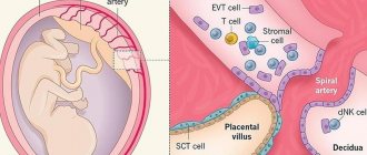
If fertilization occurs, the developing trophoblast begins to synthesize human chorionic gonadotropin (hCG), a glycoprotein similar to LH, which provides support for the function of the corpus luteum (development of the corpus luteum of pregnancy), i.e. stimulates the secretion of estrogen and progesterone, which is necessary to maintain the endometrium until the development of the placenta and the beginning of its hormone-producing function (8-10 weeks of gestation). Insufficient function of the corpus luteum causes the so-called luteal phase defect of the cycle (luteal phase insufficiency), which is considered one of the possible causes of abortion at an early stage.
If fertilization, and therefore the synthesis of hCG caused by it, does not occur, the corpus luteum degenerates (corpus luteum of menstruation), the level of progesterone and estrogen decreases, hormonal support of the endometrium does not occur, ischemic changes develop in it and menstruation occurs - rejection of the functional layer of the endometrium.
Clinical signs of precocious puberty
In true forms of precocious puberty, girls experience enlargement of the mammary glands and external genitalia. In most cases, pubertal hair develops, but its intensity is significantly less than in girls during normal puberty. Other androgen-dependent symptoms of puberty, such as acne, oily seborrhea, and increased functioning of the sweat glands, are also uncharacteristic. These symptoms in girls with early puberty develop after 6–7 years of age because physiological activation of androgen function of the adrenal glands occurs during this period.
With high activity of the pathological process, menstruation begins, which is regular. For example, in girls with hypothalamic hamartoma, menstruation begins extremely early - several months after the noticeable enlargement of the mammary glands. However, half of girls with true precocious puberty may not menstruate for several years after the appearance of secondary sexual characteristics.
In patients with false forms of premature puberty, menstruation can be observed along with breast enlargement. In this case, the discharge is erratic, there is no cyclicity, it can be copious or spotting.
With isolated thelarch in children under two years of age, the only secondary sexual characteristic is enlargement of the mammary glands; after three years, a milder version of true early puberty is observed.
With isolated adrenarche, girls develop hair growth in the pubic and sometimes axillary areas. In boys, there is an increase in the size of the testicles and penis, secondary sexual characteristics quickly develop, such as sexual hair growth, acne, deepening of the voice, increased muscle mass, increased sweating with a specific odor.
False forms of precocious puberty in boys are characterized by unchanged testicular volume against the background of rapidly developing secondary sexual characteristics. With testotoxicosis, the gonads can significantly increase in volume, but there are options for the course of the pathological process in which the enlargement of the testicles is insignificant compared to the development of secondary sexual characteristics.
In girls and boys with all forms of early puberty, growth acceleration of up to 10-15 cm per year is observed, which is characteristic of a growth spurt during physiological puberty, and it may precede the development of secondary sexual characteristics. Skeletal bones also undergo early differentiation, leading to premature closure of growth plates, which can lead to a significant reduction in final height.
Telarche
The first stage of thelarche is the development of the mammary gland buds, usually occurring around 10-11 years of age. Thelarche is usually the first phenotypic change in a series of events during puberty and occurs due to an increase in circulating estrogen levels. Simultaneously with thelarche, estrogenization of the vaginal mucosa and development of the vagina and uterus occur. Further development of the mammary glands occurs during puberty and adolescence. Corresponding changes in the mammary glands are divided into 5 stages, according to the classification of Marshall and Tanner.
Methods for diagnosing premature puberty
At the first stage of treatment of patients with precocious puberty, the form of the disease should be determined and the nature of activation of gonadotropic function should be identified. The next step is to determine the source of the increased production of gonadotropic and sex hormones. To go through all these stages, a series of diagnostic studies should be carried out.
When collecting anamnesis, attention should be paid to the nature of sexual development in relatives. Early puberty among males on the paternal or maternal side often becomes evidence of testotoxicosis. If, when collecting anamnesis, a specialist receives information about the presence of brothers and sisters in the family with signs of premature puberty, this suggests that the patient has congenital dysfunction of the adrenal cortex. Hypothalamic hamartoma usually manifests itself with an early onset of the disease and the rapid development of secondary sexual characteristics.
The physical examination involves a clinical assessment of sexual development based on the Tanner-Marshall classification, which assesses the degree of development of the mammary glands, external genitalia, menstruation, and body type in girls. In boys, attention is paid to the volume and consistency of the testicles, the size of the penis, the presence and frequency of erections, the degree of development of the muscular system, changes in voice, the presence of acne on the skin, as well as the degree of pubic and axillary hair. In addition, growth dynamics are determined in patients of both sexes.
During an external examination, you should also pay attention to the presence of symptoms of diseases accompanied by clinical signs of premature puberty. For example, the presence of large spots with uneven borders on the skin may indicate the presence of McCune-Albright-Braitsev syndrome, and a large number of small pigment spots and subcutaneous nodules are characteristic of neurofibromatosis.
Laboratory diagnosis of precocious puberty consists primarily of determining the level of sex hormones, although this type of study does not reveal the form of early puberty. Another laboratory method for diagnosing the disease is to determine the content of dehydroepiandrosterone sulfate, an increased level of which is observed in androgen-producing adrenal tumors. Measurement of 17-hydroxyprogesterone concentration provides evidence of the presence of congenital adrenal dysfunction in patients. If patients are suspected of having tumors that produce chorionic gonadotropic hormone, it is recommended to determine its content in the blood, which, in the presence of a tumor, exceeds normal levels tens of times.
Experts consider the test with luliberin to be an informative method for laboratory diagnosis of premature puberty, which allows one to obtain objective information about the state of gonadotropins. To carry it out, a natural luliberin preparation can be used, which is administered intravenously at a dose of 50-100 mcg or its artificial analogues of daily action - diferelin or buserelin. When administering luliberin intravenously, before its administration, as well as half an hour, an hour, one and a half and two hours after, blood samples are taken, in which the maximum rise of luteinizing hormone is recorded at 30 minutes, and follicle-stimulating hormone - an hour and a half after administration of the drug.
Since buserelin is administered through the nose, test results may be falsely negative if the patient has swelling or atrophy of the nasal mucosa, so its use is limited. More reliable results can be obtained with subcutaneous administration of daily action of diferelin, while measurements of the level of luteinizing and follicle-stimulating hormones are carried out in blood samples taken one hour and four hours after administration of the drug.
In the case of a true form of precocious puberty, the level of maximum concentration of luteotropic hormone is characteristic. In false gonadotropin-independent forms of early puberty, the level of luteotropic hormone is reduced to minimally detectable values. Incomplete forms of precocious puberty syndrome are characterized by levels of the above hormone that correspond to the norm. In the case of isolated thelarche, an increase in the level of follicle-stimulating hormone is observed against the background of a slight increase in the level of luteinizing hormone.
Instrumental methods for studying patients with precocious puberty include radiographic examination of the bones of the hands to determine bone age. In patients with incomplete forms of precocious puberty, bone age coincides with chronological age. Tumors of the hypothalamus, testotoxicosis, congenital dysfunction of the adrenal cortex due to high concentrations of sex hormones lead to a sharp increase in bone age, which is clearly observed on x-rays.
To exclude neoplasms of the central nervous system, computer and magnetic resonance imaging methods are used. Ultrasound examination in girls makes it possible to assess the degree of enlargement of the ovaries and uterus, to identify the presence of follicular cysts and space-occupying formations of the ovaries. In boys, ultrasound can be used to diagnose testicular tumors and adenomas characteristic of testotoxicosis, as well as to identify tumors in the adrenal glands.
The influence of various factors on the onset of puberty
| : Incorrect or missing image | This section is missing references to information sources. Information must be verifiable, otherwise it may be questioned and deleted. You may edit this article to include links to authoritative sources. This mark is set May 14, 2011 . |
K:Wikipedia:Articles without sources (type: not specified)
- Historical shift: The average age of onset of puberty has dropped significantly since the 1840s, decreasing by about 4 months per decade.[1]
- Genetics: 46% of girls start puberty at the same age as their mothers, suggesting genetic influences. The specific gene responsible for the transmission of this information by inheritance, however, remains unknown. It is believed that this is the androgenic hormone gene.
- Environment: there is a hypothesisK:Wikipedia:Articles without sources (type: unspecified)[ source unspecified 3764 days
] that earlier onset of puberty can be caused by cosmetics containing placenta extract or estrogens, as well as chemicals phthalates, which are used in production of cosmetics, toys and plastic containers.
Waste from pharmaceutical industries that produce sex steroids may also be incompletely processed and found in the environment in significant concentrations K: Wikipedia: Articles without sources (type: not specified) [ source not specified 3764 days
].
Sex hormones are sometimes used on livestock farms and can accidentally pollute the environment. Concerns expressed by [ who?
] regarding bisphenol A, which is used in the manufacture of baby bottles, sporting and medical products, and in the production of some food products. - Diet: Dietary patterns have the greatest influence on the onset of puberty, especially in girls. Additional calories, in addition to those required for growth and activity, are stored in the form of subcutaneous fat, and fat is necessary for bearing and feeding offspring. Therefore, a certain amount of fat signals that the body has sufficient energy reserves for puberty and fertility. In vegetarian families, for example, there is a later onset of puberty in girls than in families that eat animal proteins.
- Society: The timing of the onset of puberty has racial, national and social differences. The average age of menarche varies among different peoples from 12 to 18 years: the earliest is observed in representatives of the Negroid race, and the latest in representatives of the Asian race living in high mountain areas.