Cytological examination (cytology) is the main method of screening for assessing the condition of the cervical epithelium. The main task of cytological screening is to search for altered epithelial cells (atypical, having a structure different from normal epithelial cells).
The term “atypical cells” includes both cells with signs of dysplasia - mild, moderate or severe (precancerous cells), and cancer cells themselves. The difference between them is the degree of severity of changes in the structure of cells.
Cytological screening must be performed by all women (excluding virgins and patients who have undergone hysterectomy), starting at 21 years of age and ending at 69 years of age (if there are no changes in the studies), the frequency of testing is once a year, according to order 572n ( November 1, 2012), however, it is permissible to take an analysis once every three years (order of the Ministry of Health of the Russian Federation No. 36 an, dated February 3, 2015).
Currently, there are two alternative methods for fixing and studying biological material, the key difference between which for patients is their effectiveness.
Why do a cervical smear?
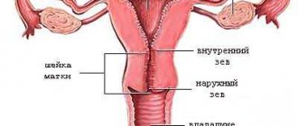
The cervical canal is a tubular section of the cervix that connects the uterine cavity to the vaginal cavity. It is easily accessible to the gynecologist during an examination with an obstetric speculum. The question of examining a smear from the cervical canal arises when it is necessary to detect:
- infectious pathogens,
- human papilloma virus,
- inflammatory processes of the genital area;
- benign formations.
The human papillomavirus can cause cervical erosions and cause malignant processes. A smear from the cervix (PAP test, cervical smear) helps to identify dangerous cancer processes of degeneration long before the onset of oncology. This is a highly effective way to prevent cervical cancer.
What else is prescribed with this study?
Antigen for squamous cell carcinoma (SCC) (carcinoma of the cervix, nasopharynx, esophagus, ear and other localizations)
8.14. Ven. blood 1 day
2,200 ₽ Add to cart
HPV 16/18/31/33/35/39/45/51/52/56/58/59 type, DNA (HPV, PCR, genotyping) scraping, count.
19.57. Scraping 1 day
1,700 ₽ Add to cart
Gynecological smear for flora
16.1. Scraping 2 days
620 ₽ Add to cart
When to take a cervical smear: indications
It is important that a woman does not forget to visit a gynecologist annually and have cervical smears. Indications for the study are:
- vaginal discharge;
- pulling pain in the lower abdomen;
- disruptions of the menstrual cycle;
- absence of such studies in the anamnesis;
- heredity burdened with cancer;
- presence of HPV (human papillomavirus);
- taking combined oral contraceptives;
- first trimester of pregnancy;
- rash, redness of the external genitalia.
Interpretation of cytological examination results
The most common at present is the Bethesda classification (The Bethesda System), developed in the USA in 1988, to which several changes have been made. The classification was created to more effectively transfer information from the laboratory to clinical doctors and ensure standardization of treatment of diagnosed disorders, as well as follow-up of patients.
The Bethesda classification distinguishes squamous intraepithelial lesions of low grade and high grade (LSIL and HSIL) and invasive cancer. Low-grade squamous intraepithelial lesions include changes associated with human papillomavirus infection and mild dysplasia (CIN I), high-grade - moderate dysplasia (CIN II), severe dysplasia (CIN III) and intraepithelial carcinoma (cr in situ). This classification also contains indications of specific infectious agents that cause sexually transmitted diseases.
To designate cellular changes that are difficult to differentiate between reactive states and dysplasia, the term ASCUS - atypical squamous cells of undetermined significance (squamous epithelial cells with atypia of unclear significance) has been proposed. For a clinician, this term is not very informative, but it directs the doctor to the fact that this patient needs examination and/or dynamic monitoring. The Bethesda classification has now also introduced the term NILM – no intraepithelial lesion or malignancy, which combines normal, benign changes, and reactive changes.
Since these classifications are used in the practice of a cytologist, below are parallels between the Bethesda classification and the classification common in Russia (Table 22). Cytological standardized report on material from the cervix (form No. 446/u), approved by order of the Ministry of Health of Russia dated April 24, 2003 No. 174.
The reasons for receiving defective material are different, so the cytologist lists the types of cells found in the smears and, if possible, indicates the reason why the material was considered defective.
Cytological changes in the glandular epithelium
| Bethesda Terminology developed in Bethesda (USA, 2001) | Terminology adopted in Russia |
| ASSESSMENT OF SWIM QUALITY | |
| Full material | The material is adequate (a description of the cellular composition of the smear is given) |
| The material is not complete enough | The material is not adequate (a description of the cellular composition of the smear is given) |
| Unsatisfactory for evaluation | Cellular composition is not enough to confidently judge the nature of the process |
| Satisfactory to evaluate, but limited by something (identify reason) | |
| Within normal limits Metaplasia (normal) | Cytogram without features (within normal limits) - for reproductive age Cytogram with age-related changes in the mucous membrane: - atrophic type of smear - atrophic type of smear with leukocyte reaction Estrogenic type of smear in a postmenopausal woman Atrophic type of smear in a woman of reproductive age |
| BENIGN CELL CHANGES | |
| Infections | |
| Trichomonas vaginalis | Trichomonas colpitis |
| Fungi morphologically similar to the genus Candida | Elements of Candida fungus detected |
| Cocci, gonococci | Diplococci located intracellularly were found |
| Predominance of coccobacillary flora | Flora coccobacillary, possibly bacterial vaginosis |
| Bacteria morphologically similar to Actinomyces | Flora of the Actinomycetes type |
| Other | Flora of the type Leptotrichia |
| Flora – small sticks | |
| Flora – mixed | |
| Cellular changes associated with Herpes simplex virus | Epithelium with changes associated with Herpes simplex |
| Possibly chlamydial infection | |
| Reactive Changes | |
| Inflammatory (including reparative) | The changes found correspond to inflammation with reactive changes in the epithelium: degenerative, reparative changes, inflammatory atypia, squamous metaplasia, hyperkeratosis, parakeratosis, and/or others. |
| Atrophy with inflammation (atrophic | Atrophic colpitis Atrophic type of smear, leukocyte reaction Mucosal epithelium with hyperkeratosis Mucosal epithelium with parakeratosis Mucosal epithelium with dyskeratosis Reserve cell hyperplasia Squamous metaplasia Squamous metaplasia with atypia |
| Radiation changes | Epithelium of the mucous membrane with radiation changes |
| Changes associated with the use of intrauterine contraceptives | |
| PATHOLOGICAL CHANGES IN THE FLAT EPITHELIUM | |
| Squamous epithelial cells with atypia of undetermined significance (ASC-US*) Squamous epithelial cells with atypia of undetermined significance not excluding HSIL (ASC-H) | The changes found are difficult to differentiate between reactive changes in the epithelium and dysplasia. Cells were found that were difficult to interpret (with dyskaryosis, enlarged nuclei, hyperchromic nuclei, etc.) |
| Changes in squamous epithelium (non-tumor, but worthy of dynamic observation) | |
| Low grade squamous intraepithelial lesion (LSIL): human papillomavirus infection, mild dysplasia (CIN I) | Epithelium of the mucous membrane with signs of papillomavirus infection. The changes found may correspond to mild dysplasia. |
| High-grade squamous intraepithelial lesion (HSIL): moderate, severe dysplasia and intraepithelial carcinoma (CINII, CIN III) | The changes found correspond to moderate dysplasia. The changes found correspond to severe dysplasia. The changes found are suspicious for the presence of intraepithelial cancer. |
| Invasive cancer | |
| Squamous cell carcinoma | Squamous cell carcinoma Squamous cell carcinoma with keratinization Small cell squamous cell carcinoma |
| Glandular hyperplasia The changes found correspond to endocervicosis | |
| Atypical glandular epithelial cells (possible assumptions): – Unclear significance (AGUS); – suspicious for neoplasia; – endocervical adenocarcinoma in situ (AIS); – adenocarcinoma | Glandular hyperplasia with atypia of the dysplasia type (I, II, III) Adenocarcinoma |
| Endometrial cells are cytologically benign (in a woman in menopause, etc.) | |
| Endometrial adenocarcinoma Adenocarcinoma of another location Adenocarcinoma without additional characteristics | Adenocarcinoma, possibly endometrial adenocarcinoma Adenocarcinoma NOS (not otherwise specified) |
| Other malignant tumors (if possible, determine the nosological form) | |
| Assessment of hormonal status | |
* whenever possible, ASCUS should be defined as similar to reactive, reparative or precancerous processes;
** changes associated with exposure to human papillomavirus, previously designated as koilocytosis, koilocytic atypia, condylomatous atypia, are included in the category of mild changes in squamous epithelial cells;
*** If possible, it should be noted whether the changes relate to CIN II, CIN III, whether there are signs of cr in situ;
****hormonal assessment (carried out only on vaginal smears): – the hormonal type of smear corresponds to age and clinical data; – the hormonal type of smear does not correspond to age and clinical data: (decipher); – hormonal assessment is impossible due to: (specify the reason).
Preparing for a cervical smear test
The optimal period for visiting a gynecologist is 5 days before the start of your period or 5 days after its end (the beginning of the cycle). Taking a smear in the middle of the cycle prevents blood from entering the test material. 2-3 days before the procedure you must:
- abstain from sexual intercourse;
- carefully observe intimate hygiene;
- stop using vaginal medications (suppositories, etc.);
- do not use vaginal contraception.
After meeting all the above conditions, you can make an appointment with a gynecologist to take a smear from the cervix.
When and who needs such screenings? According to modern clinical guidelines, cervical cancer screening with cytological examination is recommended for all women over 21 years of age. In general, it is recommended to start it 3 years after the first sexual intercourse. So early entry into intimate life is the basis for the early start of preventive gynecological examinations. In the first 2 years, screening for cervical cancer is carried out annually. Subsequently, if the results of repeated cytological studies are negative, preventive examinations become more rare and are carried out once every 2-3 years. After 65 years, the frequency of screening studies is determined individually. The reason for conducting liquid cytology is the presence of background and precancerous diseases of the cervix in a woman (cervical erosion, changes in the cervix identified during examination, an atypical colposcopic picture - examination of the cervix under magnification, the presence of human papillomavirus of high carcinogenic risk - (12 types) , since there has been a proven connection between the presence of the virus and the development of cervical cancer, precancerous changes according to the traditional oncocytogram) In this case, the patient is classified as a high-risk group for the development of cancer, and a cytological examination of gynecological smears is carried out on her annually. Additional screening activities are carried out during the preparation of a woman for conception. How to prepare for research. Preparation for liquid cytology of the cervix begins 2-3 days before the test and includes: sexual rest for 2-3 days, refusal to douche, stopping the use of any means for vaginal insertion. When is the best time to do screening? Cytological examination is not carried out during menstruation, 5 days before it and 5 days after it. Preference is given to the first half of the menstrual cycle, although this recommendation is not strict. If the patient has had a colposcopy, a cytological examination is permissible no earlier than 24 hours after it. And in the case of a cervical biopsy - only after 3 weeks. Methodology and advantages: automated preparation of cytospecimen. Includes vacuum filtration of a portion of the suspension from a test tube, centrifugation, application of the resulting cell sediment in a uniform layer on a glass slide, staining using the Papanicolaou method using wet fixation. Microscopy of a cytospecimen. The PAP test based on liquid cytology is carried out according to the same principles as in the case of the traditional technique. But at the same time, the peculiarities of coloring, position and size of cells after wet fixation of the cytopreparation are taken into account. In a traditional oncocytological study, the information content depends on many factors - human (the doctor’s eye may simply be “blurred”), the quality and adequacy of the collection of material and the preparation of the drug in the laboratory. The results of the analysis are deciphered only by a gynecologist or oncologist. An answer from the laboratory can be received in 5-10 days after taking the material. But often this period is extended to 2-3 weeks. If necessary, an express study is carried out, in this case the doctor will know the result within the first 24 hours. And what after the study? The recovery period after liquid cytology is not fundamentally different from such when taking a regular smear for oncocytology or a biopsy of the cervix. It is recommended to maintain sexual rest for 1.5 weeks, refuse to use vaginal tampons and douching. In the first days after the test, light bleeding from the vagina is acceptable, so it is advisable for a woman to use sanitary pads. An increase in body temperature, prolonged or heavy bleeding, pain in the lower abdomen is an alarming sign. The appearance of such symptoms requires immediate consultation with a doctor. How does liquid-based cytology differ from conventional cytology? The key differences between these screening methods include: during a conventional cytological examination, tissue samples are taken in a targeted manner, and the areas for examination are selected by the doctor based on visual changes in the mucous membrane. In the case of the liquid technique, material from any woman is obtained from the entire circumference of the cervix. This significantly reduces the likelihood that any modified section will be missed. When performing conventional cytology, the biomaterial is dried on glass at room temperature before sending. And in liquid cytology, it is placed in a special test tube (bottle) with a special stabilizing medium, which extends the permissible period of transportation and storage of the resulting sample. The biomaterial placed in a test tube is suitable for research for several months and does not require special conditions. With the traditional method, no filtration is performed. Therefore, if there are inflammatory elements, a large amount of mucus and other impurities in the smear, the result of a cytological study is not reliable enough and usually requires a repeat PAP test after treatment. The liquid method does not have this drawback. With the traditional method, not the entire volume of the resulting tissue ends up on glass and is subjected to subsequent examination. Liquid cytology is performed using an automated method. With traditional oncocytology, up to 35-40% of cells remain on the instrument and the doctor’s gloves. This creates the possibility that existing malignant tissue will remain undiagnosed. With the liquid method, such loss of biomaterial does not occur! This is ensured by placing the cytobrush in a stabilizing and suspending medium, followed by automated centrifugation of the sample and the formation of a special cytopreparation with a standardized, even layer of cells on a glass slide. When taking a traditional smear for oncocytology, cells on a glass slide are usually located in several layers, overlapping each other and thereby impairing visualization. Liquid cytology does not have this drawback; the resulting cytopreparation is monolayer. Possibility of re-analysis of the same biomaterial or other studies using liquid cytology. After all, the suspension in a test tube does not lose its properties for several months, and its volume is sufficient to obtain several cytopreparations. With the traditional method, the tissues being examined are not protected in any way, and there is a high risk of damage during storage. In general, liquid-based cytology using automatic screening is a significantly more informative technique compared to traditional collection of smears from the cervix for oncocytology. And its main advantage is the low percentage of false-negative results of gynecological cancer screening, which is ensured by the progressive technological features of the test with strict adherence to the rules for collecting biomaterial. Diagnostic capabilities: liquid cytological screening is aimed at identifying a variety of cellular atypia in the early stages, which indicates that a woman has a precancerous condition or cervical cancer. The single-layer and uniformity of the cytospecimen ensures a high degree of visualization, allowing the laboratory technician to reliably determine the nature of the changes. This minimizes the likelihood of diagnostic errors and false negative results. The presence of the suspension and its sufficient volume allow additional studies to be carried out according to indications: analysis for tumor markers; any PCR studies; HPV testing; immunocytochemical studies with determination of proliferation markers. Liquid cytology is recommended by WHO, FDA and global anti-cancer communities as the “gold standard” for the early diagnosis of cervical cancer due to the high efficiency of the method.
Taking a cervical smear
The procedure is carried out quickly and almost painlessly. The gynecologist takes a cytological smear from the cervical canal with a special spatula. He obtains several epithelial samples from different parts of the cervical canal. The biological material is then applied to a glass slide and sent to the laboratory for detailed cytological examination. Compliance with the rules for taking a smear is very important, since neglecting them can lead to false results of the smear analysis or it will be uninformative and the study will have to be repeated.
What does cytology analysis show?
Cytological analysis may have different tasks, depending on which cells are taken for microscopy. First of all, laboratory staff evaluate how well the material being tested corresponds to the standard. This is indicated by the shape and structure of the cells, the presence or absence of certain inclusions in them[2].
A cause for concern will be the presence of leukocytes (blood cells that perform a protective function) or microorganisms in the sample, which will indicate the occurrence of an infectious process. The most ominous sign of pathology is the identification of atypical cells with signs of malignant degeneration. In this case, the results of the cytological analysis will become the basis for oncological search: diagnostics aimed at identifying cancer, which may still be at an early stage and does not manifest itself as changes in the patient’s well-being.
A close “relative” of cytological research is histology analysis. The difference between these two types of diagnostics is that during histological examination, doctors study not individual accumulations of cells, but tissues of different organs or formations. Such an analysis requires preliminary removal of material - a biopsy (“pinching off” a piece of tissue) or even surgical intervention. Preparation of a histological specimen requires more effort and time than sample preparation for cytological analysis. Therefore, such a study is carried out less frequently and only if there are sufficient grounds, while “cytology” is often prescribed for preventive purposes - to make sure that the patient is healthy.
Indications for cervical cytology
In addition to detecting modified cellular structures and determining a precancerous condition, this type of analysis can be used as a method for additional diagnostics of intracellular changes.
He is appointed:
- To identify abnormalities in cell development in body tissues.
- To determine the nature of pathogenic microflora.
- To determine the cause of changes in the cyclicity of menstruation.
- To identify pathologies caused by papillomavirus.
- In order to identify a woman’s inability to conceive a child.
- To determine the pathology of the epithelial layer of the cervix.
- To determine the cause of bloody vaginal discharge.
This type of analysis is also prescribed:
- Before a planned pregnancy.
- With frequent labor processes.
- If the birth occurred at an early age (the woman giving birth was under 18 years old).
- Before the onset of menopause.
- Before inserting a contraceptive device.
- If a woman has not contacted an antenatal clinic for more than 3 years.
- If a visual examination of the cervix using a vaginal speculum raises doubts about the health of this organ.
- With a positive test for HIV infection.
- With genetic burden (illness of close relatives with cancer).
If a cytological examination suspects the presence of a tumor, the patient must undergo this type of examination at least twice a year.
How does the procedure work?
A smear for oncocytology is taken during a standard examination or colposcopy. The gynecologist scrapes cells from the cervix with a disposable medical brush. The procedure for taking a smear for oncocytology lasts 30 seconds and is absolutely painless for the patient. The brush has stiff bristles to effectively collect material, so after a smear, scanty spotting may appear. They usually go away within 24 hours and are not a cause for concern. Our doctors will definitely warn you about this minor discomfort and offer the appropriate hygiene product.
Main indicators of cervical cytology
The cytological smear is subjected to microscopic examination.
This determines:
- The presence of pathogenic microflora.
- The number of red blood cells and leukocytes.
- Condition of the columnar epithelium.
If the number and shape of cells does not cause abnormalities, the study is considered negative, which is the norm.
When the analysis shows that morphological changes have occurred in the cells, with a violation of their structure, this result is considered positive. In this case, a repeat type of analysis is prescribed in combination with additional types of research.
Doctors' recommendations
All experts in the field of medicine (gynecology and oncology) argue that this type of analysis should be done once a year.
This will make it possible to identify cancer pathology at an early stage of development. Only timely detection of this disease will allow for a complete recovery.
Cervical cytology allows you to identify women who are at risk. Register them and carry out monitoring to monitor the progression of the oncological process.
Cervical cytology and pregnancy
This test is taken three times during pregnancy:
- An initial smear is taken during a visit to the antenatal clinic for registration.
- At week 30, testing is performed again.
- To avoid infection of the child during childbirth, cytology is performed at 37 weeks of pregnancy.
This frequency of this test is due to the fact that during pregnancy women may experience a hormonal imbalance, and as a result, this leads to changes in the vaginal microflora. A weakened immune system may be a favorable factor for the development of vaginal candidiasis and other undesirable consequences.
Pregnant women should be aware of the special importance of conducting this type of analysis, that this is a safe type of diagnosis, it is performed with a sterile instrument and cannot be a source of infection for the woman.
It is very important to take a PAP test before pregnancy. If during its passage an increased content of leukocytes, erythrocytes, and morphologically changed cells is detected, pregnancy should be postponed. Its planning is allowed after complex therapy, in the event that a repeat analysis is negative.