April 5, 2020
Neoplasms (neoplasia) is the medical name for tumors, i.e., excessive growth of any tissue in the body. Tumors are the result of uncontrolled proliferation of cells that have not yet reached maturity and therefore have lost their ability to fully perform their functions.
Tumors can occur in internal organs and on the surface of the skin. Many people, not knowing what types of skin tumors there are, when any skin tumor appears, mistakenly believe that it is cancer. In fact, this is not always the case.
According to the main classification, skin tumors are divided into benign and malignant. There are also precancerous formations - borderline between the two main types. Each type has its own subtypes and characteristics, and correct diagnosis is needed to make an accurate diagnosis.
What is keratosis?
Let's start with the fact that cutaneous keratosis is a collective term that refers to a whole group of non-inflammatory diseases associated with impaired keratinization of the skin. Clinically, the process can manifest itself as slight peeling or the formation of thickened horny layers. There are follicular, seborrheic, and actinic keratoses.
Keratosis follicularis is characterized by the formation of horny plugs at the openings of hair follicles.
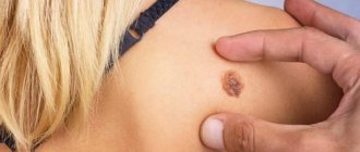
Seborrheic keratoses are plaque-like or nodular formations with a warty surface, covered with dry, horny masses that are flesh-colored, brown, or black in people over 50 years of age.
Actinic keratosis appears on exposed areas of the skin, initially appearing as bumpy, rough skin that eventually develops into rough, scaly patches ranging in color from normal skin tone to reddish brown. They are often limited in size and vary in size.
Why is keratoma dangerous and why is it necessary to remove it?
Keratoma itself does not pose a threat to human life and health. However, due to the fact that over time it is increasingly exposed to negative factors, such as mechanical damage and exposure to ultraviolet radiation, this benign neoplasm can degenerate into a malignant tumor.
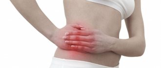
Sometimes a keratoma can cause significant discomfort to a person. Itching, burning sensation, inflammation, bleeding, pain - all these are alarming signs. The appearance of these symptoms may indicate that the process of degeneration of the keratoma into a malignant neoplasm has begun.
If you notice new moles, spots on your skin, or strange growths, be sure to visit a dermatologist-oncologist who can conduct research and determine whether the formations on the skin are malignant. Next, it is important to undergo regular medical examinations to prevent the occurrence of cancerous tumors.
Also, in a medical institution, you may be offered to remove keratomas on the face and other parts of the body and get rid of the need to regularly see a doctor.
For what reasons do acquired keratoses occur?
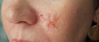
The main cause of almost all keratoses is said to be chronic exposure to ultraviolet rays. In the literature, all changes associated with sun exposure are often grouped under the term dermatoheliosis. Thus, the damaging effect can affect the epidermis (senile, actinic keratosis), dermis (solar elastosis), blood vessels (telangiectasia), sebaceous glands (porokeratosis) and melanocytes (dyschromia).
The effects of sun damage to the skin gradually accumulate as the total amount of time spent exposed to UV rays increases year after year. This leads to the fact that the peak incidence of this nosology occurs at the age of 50 years and older.
However, nowadays, actinic keratosis has become much more common in young people. As a rule, these are people of the first and second phototypes (with fair skin, blond or red hair, and blue, green or gray eyes). There is a high likelihood of developing keratoses in young people exposed to sunlight for a long time.
The incidence is slightly higher in men because they tend to use little or no sun protection. Clinical studies estimate that about 60 percent of predisposed individuals by the age of forty have at least one element of actinic keratosis. Some experts believe that almost everyone over the age of 80 suffers from some form of keratoses.
In addition, persons whose immune defenses are weakened by chemotherapy, extensive exposure to x-rays or a number of industrial chemicals, patients with AIDS, patients who have undergone organ transplants, patients with disorders of the nervous and endocrine systems, etc. less able to combat the effects of radiation and, therefore, more prone to developing keratoses.
Treatment of keratosis: what methods exist
The most relevant type of treatment for keratomas offered by modern medicine is their removal. However, not every tumor of this type must be removed - most people who have this tumor live a long life and die for completely different reasons.
Thus, it is possible to cure a keratoma only by removing it, but such a need does not always exist. The only type of formation that must be removed in any case is the cutaneous horn.
Only those tumors that threaten to become malignant are subject to removal procedures. In order to identify them in time, it is recommended to visit a dermatologist once or twice a year for the purpose of a preventive examination and monitoring of neoplasms over time, and to remove only dangerous keratomas.
At the request of the patient, the doctor can also remove those keratomas that create a significant cosmetic effect, or are located in places where they are often injured due to friction or mechanical impact.
The most commonly used methods for getting rid of keratomas are:
- laser removal;
- electrocoagulation;
- cryodestruction;
- radio wave removal;
- destruction by acids;
- surgical intervention.
In each specific case, the doctor selects the most suitable method for the patient to get rid of the tumor. Firstly, the existing contraindications of each method and their presence in the affected person are taken into account. Secondly, removal methods may be suitable for one type of keratoma, but not for another. For example, if a malignant lesion is suspected, the doctor will prescribe the patient a radio wave, laser or surgical method, since other techniques are ineffective in this case. Laser and radio wave methods are less traumatic, and if the patient does not want the appearance of a noticeable scar after the procedure, one of them is chosen.
Keratomas that cause discomfort as a cosmetic defect can be removed with acids, cryodestruction or electrical cauterization.
Laser removal of keratomas: how it happens
This method is considered one of the most effective - during exposure to a directed light beam, a sharp increase in temperature occurs in the tumor cells, as a result of which they are completely destroyed. The percentage of relapses after laser removal is no more than 5-8% of all cases. Usually one session is enough to completely get rid of the tumor. Skin restoration after the procedure lasts 1-2 weeks, after which only a small, inconspicuous scar remains at the site of the keratoma.
Laser destruction is recommended for patients who have been diagnosed with the beginning of the process of malignant degeneration of keratoma.
The procedure can be carried out in an outpatient setting, and for this it is not at all necessary to place the patient in a hospital hospital. Today, the offices of dermatologists and oncologists in clinics, hospitals, cancer centers and private clinics are equipped with laser installations.
Electrocoagulation as a method of treating keratomas
Electrocoagulation is the process of cauterizing a tumor with an electric current. During the process, the keratoma is exposed to an electric current discharge. The doctor uses a precise and powerful electric knife to cut out the affected tissue.
As a result, tumor cells are burned. After the procedure, a crust remains at the site of formation, under which the wound heals. The crust disappears on its own after about 1.5-2 weeks, and in its place a pink or red patch of skin becomes visible. After about a month, the affected area acquires a normal flesh color. When treating small keratomas, this removal method leaves virtually no traces.
When performing operations on the face and hands, that is, in places where the formation of significant cosmetic defects is unacceptable, the electrocoagulation technique is not used due to the risk of extensive scars.
Cryodestruction: treatment of formations with liquid nitrogen
In the process, the doctor uses a special can with a spray and a nozzle that jets liquid nitrogen onto the treated area at a certain temperature. The procedure takes place mainly without the use of anesthesia, and the patient feels cold and tingling at the treatment site. The formation tissues are destroyed due to exposure to very low temperatures, and the keratoma dies. After this method of treatment, a crust also remains, which after about a month and a half is replaced by skin of normal color.
The disadvantages of the method include the inability of the doctor to control the depth of freezing, as a result of which a wound that is too painful can form, which after healing will leave a noticeable large scar. In addition, cryodestruction has a high probability of relapse, since it is difficult for the doctor to control whether he managed to freeze all the affected cells.
However, to remove small tumor foci that are localized on the torso or limbs, it is quite advisable to use the cryodestruction method.
What is the radio wave method
In this case, removal of the keratoma occurs through the use of a radioknife. The instrument cuts out the affected area from the skin, acting like a scalpel during surgery. In this case, smaller incisions are formed on the skin, which practically do not bleed. After the procedure, there are no noticeable scars left, and the procedure itself is almost painless.
The radio wave knife, in addition to destroying pathologically altered tissues, simultaneously coagulates damaged blood vessels and affects nearby tissues. Considering that affected cancer cells tend to spread into the plane of adjacent tissues, removal of basal cell carcinoma using the radio wave method eliminates the possibility of cancer recurrence.
Treatment of keratoma with acids and special substances
This method eliminates surgical intervention in the skin - the effect on the tumor is produced by aggressive components: acids and cytostatics. For these purposes, creams, ointments, lotions and emulsions are used externally.
Most often, the active ingredients in these drugs are trichloroacetic or glycolic acid, Podophyllin or 5-fluorouracil. Treatment of the affected area should be carried out by a doctor, since only a doctor can correctly calculate the dosage in order to prevent the occurrence of a chemical burn.
The application of the drugs lasts for some time, and gradually an ulcer forms at the site of the keratoma, which must be lubricated with special medications to speed up healing.
Surgical removal of keratomas
This method is carried out as an operation, during which the doctor uses a scalpel to cut out the tumor itself and a small area of skin around it, and then stitches the edges of the wound. Surgical removal by skin excision is certainly effective, but the wound after it takes much longer to heal than, for example, after a radio wave procedure. In addition, a postoperative scar remains at the site of the keratoma.
A surgical intervention of this nature is usually performed using local anesthesia, but in some cases, if there are no contraindications, the doctor may offer the patient general anesthesia, for example, if the area affected by the group of keratomas is very large.
What are the symptoms of this disease? In what areas do they appear most often?
The patient, as a rule, may miss the onset of the disease and not pay attention to small irregularities, roughness, sometimes invisible to the eye, on the skin of the cheeks, bridge of the nose, ears, forearms, upper arms and forearms, back of the hands, back of the neck, upper chest, even on the scalp. Moreover, actinic keratosis can also develop on closed areas of the body that have been repeatedly exposed to the sun.
Developed actinic keratosis is represented by neoplasms from 0.1 cm to 2 cm or more. Over time, the spots become red or brownish in color and flake, and may rise above the skin in the form of growths. We most often see such patients at clinic appointments. Actinic keratosis usually develops very slowly and usually does not cause any discomfort other than aesthetic ones. Itching or burning in the affected area usually occurs in areas of long-term and severe keratinization. Most often, elements of skin keratosis develop slowly, but can disappear and reappear with repeated exposures that damage the skin. They can become inflamed and, in rare cases, even bleed.
Treatment of keratoma with folk remedies
To remove keratomas on the head or body at home, you can use not only pharmaceutical drugs, but also popular folk remedies. You can prepare an effective ointment yourself using simple and affordable ingredients.
Homemade ointments for removing skin lesions are prepared based on various acids and chemicals with a “burning” effect. The most commonly used are celandine, table vinegar, onions or garlic.
Ointment with celandine - to prepare it, 50 g of dried celandine herb must be thoroughly ground to a powder and mixed with 50 g of pre-melted pork fat. The prepared product must be applied to the area of neoplasms three times a day.
Walnut ointment - to prepare a medicinal ointment you will need 10 green walnuts and 10 tablespoons of olive oil. The nuts should be finely chopped along with the green peel, the oil should be heated, then the ingredients should be mixed and left to infuse for 24 hours in a dark place. The product should be applied to the area of neoplasms twice a day for 4-5 weeks.
Proper skin care plays an important role in the treatment of neoplasms. For this purpose, it is best to use natural oil - castor, sea buckthorn, fir or sunflower. This product effectively softens the skin, eliminates dryness and discomfort, and prevents further development of tumors.
Are there any new developments in this direction on the world market?
Well, for example, from 1998 to the present, the Australian biopharmaceutical company Peplin has been studying the topical treatment of actinic keratosis with the drug Ingenol Mebutate, which is the first in a new class of formulations and is derived from milkweed juice. This ingredient has a long history of traditional use for a variety of skin conditions, including the topical treatment of skin cancer and precancerous skin lesions. The company plans to enter the third phase of the trial soon.
What are destructive (ablative) methods?
All skin tumors must be examined by a dermatologist before removal due to the risk of skin cancer. Treatment performed in a timely manner will help prevent the development of basal cell carcinoma (BCC) or squamous cell carcinoma of the skin (SCC).
- Cryosurgery using liquid nitrogen is the most common method for treating keratoses, but is not suitable for hyperkeratotic lesions. Liquid nitrogen is sprayed directly onto the affected areas of the skin using a cryodestructor or applied using the “reed” method (application with a cotton swab on a wooden stick). Cryosurgery is easily performed on an outpatient basis, shows excellent cosmetic results and is well tolerated.
- Radio wave, electro- and diathermo-laser destruction.
- Photodynamic therapy involves the application of a photosensitizing agent, methyl aminolevulinate, and then exposure to light of a specific wavelength, which leads to tissue necrosis. Photodynamic therapy is well tolerated and has excellent cosmetic results.
- Surgical removal . The surface of the skin is cleaned with a special instrument (curette).
- Chemical peeling:
- Jessner's solution (resorcinol, lactic and salicylic acids in ethanol);
- trichloroacetic acid solution 35%.
- Dermabrasion . The affected areas of skin are removed using a fast-moving abrasive brush.
- Phototherapy (IPL) and fractional photothermolysis - coagulation of keratosis elements using light energy. Suitable for the initial manifestations of actinic keratosis.
I monitor one mole, remove another: how not to miss dangerous neoplasms on your skin
Where do they come from
On the Internet you can find a version that our moles initially grow... due to tanning. They say that a person is born with clear skin, and over time it is because of the sun that moles appear on it.
“In fact, the formation of moles and their number is determined primarily by heredity,” says dermato-oncologist at the Clinic of Skin Diseases, assistant at the Department of Skin Diseases at Sechenov University, candidate of medical sciences Ekaterina Vertieva.
- That is, those who have a lot of new growths on the skin in their family - mom, dad, grandmother or grandfather - will have more moles.
The second factor is the sun. Under the influence of its rays, the number of moles increases.
At the same time, there are several peaks in a person’s life when most moles appear. Such surges are associated with hormonal changes: at a younger age (3 - 6 years), adolescence and from 25 to 35 years. Women may experience additional peaks during pregnancy.
— Is it true that there is a “safe limit” of moles, and if there are more than normal, then a person has an increased risk of skin cancer?
— If there are more than a hundred moles on the skin, then the risk of malignant neoplasms is really higher. But not so much that those with a rich scattering of moles should immediately sound the alarm. The logic is simple: the more tumors there are, in principle, the higher the likelihood that among them there will be nevi with malignant potential (how to protect yourself from danger - see below).
STAY IN TOUCH
What is what
Among the names of growths on the skin you can find the classic, familiar word “mole,” as well as nevus and keratoma. What is the difference?
“ Nevus is the medical name for the same mole,” explains Ekaterina Vertieva. - Essentially, this is a benign skin tumor. A mole consists of pigment cells of melanocytes, which produce the coloring pigment melanin (because of it, our skin darkens when tanning. - Ed.) A mole is a cluster of such melanocyte cells. That is why the appearance of nevi is associated, among other things, with exposure to the sun.
— Keratoma is a benign superficial formation on the skin that can never degenerate into something malignant. Keratomas occur more often in old age. They consist of keratinocyte cells (these are the surface cells of the upper layer of the skin of the epidermis). Externally, a keratome usually looks like a brown formation rising above the surface of the skin. Keratomas often become crusty and flaky. Pieces can fall off, which scares people. In fact, this is a natural desquamation process. There is no need to be scared, the doctor reassures.
On a stem and with hairs - what can you expect from them?
- There are moles that seem to move away from the skin and rise above it - is this dangerous?
- Look: we have superficial layers of skin, the epidermis, and deep layers, the dermis. Common, classic dark moles are superficial, so-called border nevi. They are found in the epidermis. Over time, some superficial nevi absolutely physiologically, that is, safely, naturally grow into the deep layers of the skin, the dermis. And then they begin to rise above the surface of the skin. Nothing wrong with that.
Moreover, over time, by about 50-60 years, dermal nevi completely lose their pigment. They cease to be brown and acquire the color of healthy skin. This is the so-called mole involution. All this is absolutely safe.
- When do hairs grow from a mole? They say this is a good sign, such nevi are definitely safe and will not degenerate into melanoma.
- This is a misconception. Melanoma can easily grow hair. Therefore, if a nevus has suspicious signs (see below), then the hairs are not a reason to calm down and not check the mole.
By the way, doctors categorically do not advise pulling hair out of a nevus; this is unnecessary trauma. You can only cut it carefully.
Meyerson phenomenon
This is the name of the condition when redness appears around the mole, like an inflamed pink corolla, our expert explains. And it reassures: as a rule, it is not dangerous. Most often, the Meyerson phenomenon occurs in patients with a tendency to skin diseases: eczema, atopic dermatitis. If you experience any discomfort or itching, you should consult a doctor. Usually, dermatologists prescribe atopic corticosteroids to relieve such symptoms; with their help, the symptoms are easily removed.
— If you damage a mole and it bleeds, do you need to see a doctor urgently?
— It is necessary, but not necessarily right on the same day or the next day, up to 7 days is quite normal. There is no need to be afraid that after injury, even with bleeding, your mole will definitely degenerate into something bad. Do not believe the stories about a grandmother who picked off a mole, it immediately metastasized into the blood, and death quickly occurred. It is a myth. If a person injures a benign mole, then nothing bad will happen to it. Adverse consequences occur if it was initially melanoma.
— What to do immediately after a mole is damaged, before going to the doctor?
- Treat with a clear antiseptic. Hydrogen peroxide, for example. Just don’t smear it with fucorcin, iodine or brilliant green. Because it will then be difficult for the doctor to examine and assess the condition of your mole due to staining with these substances.
How often to check with a doctor
There are tips on the Internet to show your moles to a doctor every 6 to 12 months.
“If a person has 100 or more nevi and is advised to visit a doctor every six months or a year, then this is still too much,” says Ekaterina Vertieva. - In most cases, moles will not turn into anything bad. But, to be sure of this, it is important to undergo a full examination once by a dermatologist, or even better, an oncodermatologist. Have a specialist carefully check all moles and confirm that none of them are cause for concern. Or he indicated those that could potentially be dangerous, and then told how often they need to be monitored.
If the doctor, after examination, says that all moles are calm, then, as a rule, it is recommended to see the next time in 3 - 4 years. At the same time, no one canceled the self-examination.
THIS WILL BE USEFUL
The golden rule for self-examination
This is the international standard for assessing moles. It can be used as a test at home. The mole is tested according to the ABCDE criteria.
A (asymmetry, asymmetry) - has the mole become asymmetrical?
B (border) - have the borders of the mole become uneven?
C (colour) - has the color of the mole changed: several shades of brown, black, blue, pink have appeared?
D (diameter, diameter) - has the mole grown more than 6 mm in diameter?
E (evolution, evolution) - does the mole change intensively over 4 - 6 months (in shape, color, size)?
Important : if the answers to all questions are “yes,” this is reason to suspect the mole is malignant and immediately consult a doctor. If you have several of the symptoms, then the risk is lower, but you also need to see a doctor.
Delete cannot be saved
“There are three reasons for removing moles,” says Dr. Vertieva. — Cosmetic removal — if a person simply doesn’t like the way a mole looks. We also recommend removing benign moles if they are located in places where they are often injured. For example, for men in the beard area, for women in the bra clasp area. The third option is if we see suspicious symptoms. In these cases, the doctor conducts an examination using a dermatoscope (a device similar to a microscope. - Ed.). If a mole is causing concern, removal is done for medical reasons.
— What are the current methods for removing moles?
- Laser, radio wave method, electrocoagulation (cauterization of the nevus with high-frequency current) are used - these methods destroy the mole. As well as surgical excision.
If the patient has a suspicious nevus, then surgical removal is necessary. Only in this case, the tumor tissue is fully preserved, and histologists will be able to give a reliable conclusion as to whether there are malignant cells in the mole.
In other cases, when the mole is definitely calm, the least traumatic method is the radio wave method or laser (coagulation is a rougher method). After removing the nevus, a neat wound remains; it must be treated with a solution of potassium permanganate or fucorcin. A crust forms, which falls off after an average of two weeks. You can wet the wound site with water from 3 to 4 days.
I strongly do not recommend self-medicating at home and using celandine to remove tumors. At best, this technique leads to a chemical burn, and sometimes to more serious consequences in the case of malignant potential of the formation.
QUESTION ON THE TOPIC
Is it possible to sunbathe if you have a lot of moles?
“Yes, this is not a contraindication for tanning,” our expert rejoices. — The main thing is to follow safety rules: stay in the sun before 11 a.m. and after 4 p.m. And use sunscreen.
BREAKTHROUGH AND PROSPECTS
“Now there is a real revolution in the treatment of melanoma, the most aggressive skin cancer,” says Ekaterina Vertieva. — Previously, chemotherapy was used, which was very toxic and at the same time extended the life of patients by only a couple of months. Since the beginning of the 2010s, targeted drugs have been used. They do not hit all cells of the body, like chemotherapy, but selectively hit specific targets. These drugs are available in Russia under compulsory medical insurance and prolong the life of our patients very significantly. The main trend now is the study of new targets for the fight against melanoma. And also immunotherapy (the method for which the Nobel Prize in Medicine was awarded this year. - Ed.).
CONGRATULATIONS!
The oldest department of Sechenov University is 150 years old
On May 27, the Department of Skin and Venereal Diseases named after. V.A. Rakhmanov Sechenov University - 150 years. Already in the first years of its work, the department was recognized as the best in Europe. Over a century and a half history, vast experience in teaching the science of dermatovenereology has been accumulated here. Today, about 2,500 skin and venereal diseases are known, say department employees. The most common are psoriasis, atopic dermatitis, eczema and fungal infections; In recent decades, there has been a steady increase in the incidence of allergic dermatoses.
The head of the department, Professor Olga Olisova, lists modern promising scientific developments of the department's employees: improving the early diagnosis of lymphoproliferative skin diseases (T-cell lymphomas) using genetic markers; development of hardware methods for diagnosing melanocytic skin neoplasia; studying the pathogenetic connections of HPV with the development of skin tumors using molecular genetic detection methods; development of diagnostic markers for severe bullous dermatoses and orphan skin diseases (using the example of mastocytosis).
BY THE WAY
At the Department of Skin and Venereal Diseases of Sechenovka, a unique plaster museum has been created, containing more than 3,000 wax plaster casts with images of various skin and venereal diseases. In terms of the quality and quantity of exhibits, the dummy museum is a national treasure that has no equal not only in Russia, but throughout the world.