Skin neoplasms are usually understood as benign or malignant skin lesions of a tumor nature against the background of abnormal proliferation of dermal cells. It must be said that, as a rule, it is recommended to remove the resulting benign neoplasm, since the slightest injury or exposure to sunlight significantly increases the risk of its malignancy. Malignant neoplasms of the skin include skin cancer.
Skin cancer is one of the few that can be almost completely cured by consulting a doctor in time. Yusupov Hospital provides a wide range of diagnostic and treatment services for all forms of skin cancer. Experience in managing patients with this pathology allows us to demonstrate excellent results. Doctors develop a comprehensive individual program for the management of each patient. It includes both diagnosis and treatment, as well as rehabilitation. At the Yusupov Hospital, a diagnosis is made as quickly as possible and, preventing the progression of the disease, an effective treatment method is selected. The living conditions are comfortable, and all the medical staff are specialists in their field.
Reasons for the development of the disease
Currently, there is no clear answer to the question of what causes skin cancer. Like many other oncological diseases, skin tumors are considered a multi-etiological pathology. There are several predisposing factors, the presence of which increases the risk of developing a tumor focus. These include:
- Excessive exposure to ultraviolet rays on the skin. A similar situation occurs with prolonged and frequent exposure to sunlight, visiting a solarium, or working outside. Residents of the southern regions are at risk of developing skin cancer.
- Having light skin. Lack of melanin production increases the likelihood of skin tumors.
- Skin burn. A high degree of burn is accompanied by scarring of the skin. This process contributes to the emergence of latent carcinogenesis.
- Irradiation. Exposure to radioactive, ionizing rays has a detrimental effect on the skin. The risk of radiation dermatitis increases.
- Presence of skin contact with toxic substances. This group of carcinogens includes arsenic, aluminum, titanium, nickel and other heavy metals.
- Immunodeficiency. Conditions in which the body's protective functions decrease predispose to the formation of a tumor focus.
- Age. Most often, skin tumors affect people over 50 years of age.
- Concomitant systemic diseases. Doctors identify a group of pathologies in which the risk of developing skin cancer increases significantly. These include systemic lupus erythematosus, leukemia, and chronic skin diseases.
- Heredity. The presence of a skin tumor in previous generations of relatives is not a major risk factor. However, family history in combination with other predisposing conditions increases the possibility of developing skin cancer.
- Tattooing. In this case, there are two risk factors. This is a violation of the integrity of the skin and the introduction of paint with carcinogenic substances. Cheap tattoo ink may contain impurities of aluminum, titanium, and arsenic.
- A large number of nevi. Doctors urge you to monitor the condition of moles and contact a specialist if there is the slightest change. Traumatization of nevi increases the possibility of developing skin cancer.
- Excessive alcohol consumption, smoking. Chronic intoxication has a detrimental effect on the body as a whole. Against this background, the risk of tumor formation increases several times.
- Eating foods high in nitrates.
Expert opinion
Author:
Alexey Andreevich Moiseev
Head of the Oncology Department, oncologist, chemotherapist, Ph.D.
Every year, 9,000 cases of newly diagnosed melanoma are registered in Russia. This aggressive malignant tumor is responsible for the death of 40% of patients. Such statistics indicate that the country's population is not sufficiently aware of the symptoms of melanoma. As a result, a visit to a doctor occurs in the later stages, when treatment is considered ineffective.
Doctors at the Yusupov Hospital determine the location of melanoma and prescribe treatment appropriate to the stage of tumor development. An individual approach to the problem of each patient allows us to reduce the number of deaths. A tumor detected in time is characterized by a favorable prognosis for recovery. The later melanoma is detected, the longer the treatment process will be. Its success depends on many factors. The main treatments for melanoma are surgery, radiation and chemotherapy. Depending on the condition, symptomatic therapy is carried out.
You can diagnose the presence of skin cancer yourself at home. Doctors recommend regularly examining the skin, especially nevi, for the appearance of pathological formations.
Reviews about the treatment of basal cell carcinoma
Maria Alexandrovna, 57 years old “ Around the end of the summer of 2021, a reddish spot the size of a pea, with jagged edges, appeared on my right cheek. Over time, the spot began to increase in size and peel off. This alarmed me and I went to the clinic to see a surgeon, who examined me and did not give me a clear answer that it was he who did not give me. He prescribed me ointments, but they had no effect. As time passed, the spot became inflamed and turned into a weeping wound, which began to get worse. I was tired of all this and on the Internet I came across the LazerVita clinic and made an appointment with a surgeon. On the day of the appointment, the surgeon Gafarov Sarkhan Vidadievich saw me, examined the wound, he immediately did not like it and took cytology. After explaining everything to me and reassuring me, he made an appointment with oncologist Violetta Aleksandrovna Purtskhvanidze in 3 days, by which time the cytology will be ready. He prescribed me to treat with antiseptic solutions and antibiotics for now. Three days later, I came to see Violetta Alexandrovna. After examining the wound, she diagnosed Basal cell carcinoma, which was confirmed cytologically. She explained my diagnosis to me in detail, reassured me, and explained to me all the treatment options. Then, on the same day, Dr. Sarkhan Vidadievich performed an operation on me: laser removal. The operation went without complications, everything healed perfectly. I want to express my gratitude to the entire LazerVit team, and especially to doctor oncologist V.A. Purtskhvanidze. and doctor surgeon S.V. Gafarov Thank you very much!
» All reviews about the treatment of basal cell carcinoma and other diseases.
Kinds
Skin tumors are divided into three types: benign, borderline or precancerous tumors, and malignant. All of them differ in their ability to metastasize to other organs, complications, and the ability to lead to death. It should be added that a large number of moles on the skin or other neoplasms of a benign nature (papillomas, warts) is evidence of a given person’s predisposition to cancer. Benign neoplasms are considered:
- moles or nevi;
- atheromas;
- adenomas;
- lymphangiomas;
- hemangiomas;
- fibroids;
- neurofibromas;
- lipomas;
- papillomas.
Borderline tumors include:
- keratoacanthoma;
- senile keratoma;
- cutaneous horn;
- xeroderma pigmentosum and other not very common neoplasms.
Malignant neoplasms are represented by melanoma, sarcoma, epithelioma, basal cell carcinoma.
During a general examination of the patient, an oncodermatologist
EVERYONE is watching! Skin on the torso, face, neck, limbs, mucous membranes, scalp.
- Dermatoscope.
- SIAscopically.
- Cytologically.
- Histologically!!
Biopsy of a skin tumor
A mandatory diagnostic step for skin tumors is morphological verification of the process; for this, a biopsy
. The oncologist takes cells or tissues from the site of the tumor and submits them for examination to pathologists, who form their conclusion.
The morphological report contains information about the tumor:
- localization of the process (where the tumor is located);
- diameter (what size is the tumor);
- growth type;
- characteristics of the resection margin (the tumor was completely removed or not).
The oncologist makes a final diagnosis and forms a treatment plan only after receiving the results of the biopsy.
Benign skin tumors
Benign neoplasms have a slow growth rate; during development, they put pressure on nearby tissues, but do not penetrate them. Benign neoplasms are as follows:
- Lipoma is a neoplasm from the fatty layer;
- papillomas and warts. Externally they look like growths on a leg (if injured they often turn into cancer) or bulges, the origin is viral;
- Dermatofibroma develops from connective tissue. Most often it is detected in representatives of the fair sex at a young and mature age. Distinctive features are small size (0.3-3 cm), slow growth, and insignificant subjective sensations. Mainly affects the lower extremities. Dermatofibroma should be distinguished from nevus, basal cell carcinoma and dermatofibrosarcoma;
- Nevi are various sharply limited hyperpigmented areas of skin that have different shapes and colors. The surface is both striped and smooth. Warty nevus growths up to two centimeters in diameter may be observed. They can be distinguished from soft fibromas by the hyperkeratotic layers present on the surface (dense crusts resembling peeling). The most dangerous representative is considered to be a pigmented borderline nevus, in which melanin is present and which can degenerate into melanoma;
- Lentigo usually occurs in adolescence on any part of the body. Outwardly it looks like a smooth oval spot, the diameter of which can reach up to one and a half centimeters. If this neoplasm occurs in old age, it is called senile lentigo;
- Atheromas develop from the sebaceous gland. Atheroma or epithelial cyst has a high potential for malignancy into liposarcoma. Most often it occurs in areas of the skin where many sebaceous glands are concentrated (scalp, face, forehead). This is a single, painless formation that rises above the surface. In case of inflammation and the beginning of the process of suppuration, the skin becomes red, and pain occurs;
- Hemangioma. There are capillary and cavernous hemangioma. The capillary can reach significant sizes, but the cavernous, despite its deeper location, does not reach large sizes. The color of the tumor depends on the structure and can vary from red to bluish-black. Surgical treatment with excision of the tumor and underlying layers is indicated.
At the moment, there are two most common forms: superficial and nodular
Superficial (newly identified) BCC:
- most often develops on the skin of the trunk and limbs;
- less common: head and neck;
- in younger patients, with a predominance of women.
Important!
The main development factor is genetic - this is a hereditary mutation that is transmitted to descendants or arises in connection with environmental or other adverse effects of the external environment. Nodular form (first identified) of BCC:
- most often develops on the scalp
- in elderly and senile patients
Removal of benign skin tumors
Removal of benign skin tumors must be carried out to eliminate a cosmetic defect, prevent compression of adjacent structures and permanent damage.
Surgical removal is carried out in different ways:
- Classic method (scalpel);
- Electrocoagulation;
- Radio wave method;
- Laser method, etc.
As a rule, preference is given to laser and radio wave methods, since they are the least traumatic and have the lowest relapse rate.
The choice of treatment method should be made only after a thorough examination and taking into account all the individual characteristics of the patient.
The Yusupov Hospital provides diagnostic and treatment services for all benign skin tumors. Our specialists will help you eliminate both a cosmetic defect and prevent the consequences of incorrect treatment tactics in a short time.
4.Prevention of basal cell carcinoma
Methods for preventing basal cell skin cancer are well known to everyone. They are often talked about simply as ways to avoid skin cancer during the hot season:
- Try not to be in the sun during the hottest hours - from 10 am to 2 pm;
- Wear hats (hats with brims) and thick clothing when in the open sun;
- Use sunscreen;
- Be regularly examined by an oncodermatologist and tell your doctor about any suspicious or changing appearance of skin lesions (moles, spots, etc.).
Precancerous skin growths
Precancerous skin conditions are divided into two main groups: facultative, the risk of malignancy of which is minimal, and obligate - precancerous conditions, which will ultimately become cancerous.
Precancerous skin conditions include:
- Xeroderma pigmentosum. This tumor develops due to excessive sensitivity of the skin to solar energy, as a result of which the skin loses its ability to regenerate. The disease is congenital in nature, it is easy to diagnose it in children of the first year of life by the abundance of freckles on the surface of the skin, which is most often exposed to solar radiation. In almost every case of this disease, cellular and squamous cell carcinoma occurs. There is a very high mortality rate in people under twenty years of age with this disease;
- Buschke-Levenshtein condyloma, the causative agent of which is considered to be the human papillomavirus. This neoplasm has rapid growth, enormous size, and also secretes a cloudy liquid with an unpleasant odor. This disease is characterized by a progressive course, tends to grow into nearby tissues and can recur even after complete surgical removal. Additionally, the condition quickly progresses to squamous cell skin cancer;
- Actinic keratoma or actinic keratosis (or solar). Typically occurs in older people. Externally, this condition appears as orange or yellow rashes on the skin no more than one centimeter in diameter. Subsequently, scales and dry crusts form at the site of the rash, and when mechanically peeled off, slight bleeding is observed. If a compaction appears at the base of the tumor, it is considered that this is the beginning of malignancy of the tumor. But this phenomenon is observed quite rarely;
- Paget's disease. After 42 years, women may experience areas of redness around and on the nipple with accumulation of biological fluid, signs of peeling, and weeping. Then crusts form in this area, and nipple retraction is observed. The development of this disease can take years. According to some oncologists, this condition is a development of early stage cancer;
- Cutaneous (senile) horn. This disease is usually observed in very old age. It also occurs on open areas of the skin that are constantly squeezed or subject to friction. Primary cutaneous horn occurs on healthy skin, while secondary horn occurs after certain diseases (for example, lupus erythematosus, solar keratosis). At the end of its formation, the tumor has the appearance of a cone-shaped horny formation, the length of which is significantly greater than the diameter of its base. This disease is characterized by a long course and has a tendency to become malignant.
Risk factors for basal cell carcinoma
- Light skin type.
- Red or blond hair.
- Prolonged exposure to ultraviolet radiation.
- Exposure of skin to radiation.
- Chronic trauma to the skin, for example, rubbing with uncomfortable clothing or shoes.
- Exposure of the skin to chemical carcinogens, including household chemicals.
The development of basal cell carcinoma may be due to genetic predisposition. This pathology is called Gorlin syndrome. It is characterized by the following symptoms: multiple foci of basal cell carcinoma, endocrine pathology, skeletal and mental disorders. Pathology begins to manifest itself at a young age.
Malignant neoplasms of the skin
Tissue cells of such tumors are difficult to differentiate at the initial stage of development. They have lost the ability to perform their own functions, can penetrate into nearby tissues and organs, and often metastasize into the blood and lymph vessels, forming tumors throughout the body. The main signs that may indicate the degeneration of benign neoplasms (nevus, pigment spots, etc.) into malignant ones are changing pigmentation of the mole, spontaneous and rapid increase in size of the neoplasm, its spread to other areas, bleeding, ulceration, that is, those manifestations that were not there before.
Malignant skin neoplasms are divided into several groups, according to their cellular structure and clinical manifestations:
- Melanoma is the most common malignant tumor. Localized in the skin. In most cases, melanoma is a consequence of the degeneration of a nevus against the background of a severe burn or injury. Therefore, trauma to the nevus is the main risk factor for malignancy of the neoplasm. Formations are especially dangerous in areas that are constantly exposed to friction. Treatment is surgical, sometimes with the use of radiation and chemotherapy. The prognosis of the disease is directly dependent on the time of detection of the tumor and its treatment;
- Basalioma. Occurs in areas of the skin that are often exposed to excessive sun exposure. Heredity contributes to the development of the disease. Over the course of several years, it degenerates into squamous cell skin cancer. At the initial stage, the formation has the appearance of a whitish nodule, on the surface of which a dry crust forms;
- Squamous cell carcinoma or epithelioma is observed less frequently and has a severe course. The focus of localization is most often the perianal region, the external genitalia. It is almost impossible to distinguish it from another type of cancer visually; it metastasizes quickly. At the initial stage, the tumor looks like a ball in the thickness of the skin with a diameter of no more than a centimeter. As they grow, warts and ulcerations form, after which the edges become dense and uneven, and severe pain appears. As soon as the formation metastasizes to the lymph nodes, the patient's condition quickly deteriorates. Death can occur due to bleeding due to tumor disintegration and vascular damage, as well as as a result of rapid exhaustion of the body. For treatment purposes, surgical removal of the tumor and lymph nodes is indicated, often in combination with radiation and chemotherapy;
- Kaposi's sarcoma or angioreticulosis. The disease in most cases develops in patients with AIDS, but the usual form of the disease clinically has an identical clinical and histological manifestation. The risk group includes men. The site of the disease is the lower extremities. First, violet, sometimes lilac-colored spots are formed that do not have clear boundaries. Gradually, dense, rounded nodules of a bluish-brown color appear, reaching a diameter of up to two centimeters. These nodules often aggregate and ulcerate. In patients with AIDS, the disease has an aggressive course, often with severe damage to the lymph nodes and metastases throughout the body.
Treatment methods
The main methods of treating basal cell carcinoma are surgery and radiation therapy. The choice of method will be determined by the location of the tumor. In addition, other removal methods can be used: local chemotherapy, photodynamic therapy, etc.
Surgical removal of basal cell carcinoma
Surgery is the preferred method of treating basal cell carcinoma, as it allows you to control the radicality of the intervention. The following methods are used:
- Classical removal - the tumor is excised with a scalpel at a distance of 0.5-1 cm from its edges. The wound is sutured with cosmetic sutures, and the removed material is sent for histological examination. If pathologists issue a conclusion that there are no malignant cells in the cut-off edges, the treatment is considered radical and no other measures are required. However, this method is problematic to use if the basal cell carcinoma is located in the face, and even more so on the eyelids, nose or ear.
- Curettage with electrocoagulation. In this case, the basalioma is scraped out with a curette, and the bottom of the wound is cauterized with an electrocoagulator. After healing, a whitish scar remains.
Regional metastases in the lymph nodes are also surgically removed. Typically, during such an operation, the fatty tissue around the affected nodes is excised.
Radiation therapy for basal cell carcinoma
If surgical treatment is not possible, radiation therapy is prescribed. Considering the good sensitivity of basal cell carcinoma to the effects of ionizing radiation, the effectiveness of such treatment reaches 90%. Radiation therapy is carried out in long courses over several weeks. In this case, irradiation sessions are carried out daily for 5 days, and then a 2-day break is taken in order for healthy tissue to be restored. Indications for radiation therapy are the following conditions:
- Elderly age.
- An inconvenient location for basal cell carcinoma from the point of view of surgical removal is the face, especially the nose, lips, eyelids and ears.
- High risks of recurrence after surgical removal.
Chemotherapy
Local and systemic chemotherapy can be used to treat basal cell carcinoma. Local chemotherapy is used to treat superficial forms of the disease. It involves the use of ointments containing antitumor drugs, for example, fluorouracil.
Systemic therapy is used for persistent recurrent basal cell carcinomas and in the presence of distant metastases. As part of the treatment, drugs are prescribed that act on the processes that ensure the uncontrolled proliferation of malignant cells. For basal cell carcinoma, the effectiveness of drugs from the group of inhibitors of the Hedgehog signaling pathway has been proven, these are Vismodegib and Sinidegib.
Photodynamic therapy
Photodynamic therapy is a relatively new, but very promising method for the treatment of malignant neoplasms of superficial localizations. Its effectiveness is based on the selective destruction of pathologically altered cells, which occurs as a result of a photochemical reaction in which the photosensitizer enters under the influence of light with a certain wavelength.
When treating basal cell carcinoma, the photosensitizer is applied superficially in the form of an ointment or application. Then the tumor is exposed to a lamp emitting light of specified parameters. As a result, the cell activates the production of free radicals, which damage membranes and organelles, leading to the death of the neoplasm as necrosis. After a few days, a scab forms in its place. When it falls off, young skin will form in its place. Photodynamic therapy gives a very good therapeutic effect and aesthetic result. It is recommended for use in the treatment of superficial basal cell carcinoma in the face and hands.
Symptoms and signs
The clinical picture of skin cancer development depends on its type. Often the first signs of a tumor are mistaken for other skin diseases. This results in untimely consultation with a doctor and the spread of the malignant process. Common signs for all types of skin cancer are:
- The appearance of a small spot or lump on the skin. The color of the formation can be pinkish, gray-yellow. It is noteworthy that the color of the neoplasm differs from moles and freckles.
- Uneven shape and asymmetric contours of the pathological focus.
- The appearance of itching and slight discomfort in the area of the tumor process. This symptom appears some time after the formation of cancer.
- Progressive growth of the tumor.
- Fatigue, weakness, sudden loss of strength.
- Decreased appetite, resulting in sudden weight loss.
- As cancer develops, regional lymph nodes are affected.
If any of the above symptoms appear, it is recommended to seek medical help. Depending on the type of skin cancer, the following symptoms are distinguished:
- Basalioma. It is the most common form of skin tumor. It is characterized by a favorable prognosis when detected at the initial stage. Appears in the form of a nodule, painless on palpation. It has a grayish-pink color. The surface of the nodule is usually smooth. Scale formation is possible. As the tumor process spreads, the affected area increases. The neoplasm is covered with a bloody film. The predominant localization of basal cell carcinoma is the face, neck, and arm area. Basalioma does not cause a significant change in condition. This is the reason for late seeking medical help.
- Squamous cell carcinoma. At an early stage, it looks like a dense red or brown nodule. A tumor of this type is prone to rapid decay, and therefore, as the process progresses, an ulcer forms. It often bleeds, its edges are heterogeneous. Squamous cell carcinoma quickly grows into nearby tissues. If detected at a late stage, metastasis to regional lymph nodes or organs is possible.
- Melanoma. Refers to an aggressive type of tumor. Most often, melanoma occurs at the site of an injured nevus, which increases in size, changes color and acquires uneven contours. A characteristic feature of this type of cancer is the asymmetric form of formation, as well as a tendency to bleed. The malignant neoplasm itches and increases in size. As the process progresses, the tumor turns into an ulcer. Melanoma progresses rapidly and often metastasizes.
- Adenocarcinoma. At the initial stage of development, it looks like a dense nodule. It is most often localized in the armpit area, under the breast. With the development of the tumor process, adenocarcinoma grows into nearby tissues. This type of tumor is detected extremely rarely and is characterized by slow growth.
Clinical signs of basal cell skin cancer
Surface form
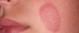
- the least aggressive of all “carcinoma” forces the patient to consult a doctor when the tumor is already a fairly significant cosmetic defect or begins to cause inconvenience due to the addition of ulceration. The superficial form of BCC is also called basal cell carcinoma of the trunk and extremities. On the body, most often, it has a multiple character. Radial, extremely slow tumor growth predominates, occupying an area of several centimeters before the onset of invasion and confirmation of the diagnosis. This form is often considered by a specialist as a superficial keratoma and is ignored in senile and elderly people. Visually, it is defined as a flat, pinkish-brownish, slightly scaly formation, with a barely detectable ridge-like edge on palpation, thread-like nodules like a “pearl necklace” along the perimeter of the tumor, pearlescent color and, sometimes, visualized telangiectasia.
With nodular form
basal cell skin cancer, the patient consults a specialist if the tumor size is quite small, since the exophytic component is often subject to constant trauma and, as a consequence, contact bleeding. The process begins with a pearly-light or pink papule, on a wide base, a glossy surface with the absence of a skin pattern and the presence of telangiectasia.
The center of the node often ulcerates, followed by the formation of an eschar and hyperkeratosis. The perimeter roller is also often defined by the classic “pearl necklace”.
Scleroderma-like/sclerosing form of BCC
. The least common but most aggressive form. The skin formation resembles a scar, pale yellow in color, waxy to palpation, glossy. At the same time, the “string of pearls” characteristic of basaliomas in general and telangiectasia are most often absent. It recurs extremely often, due to the spread of the tumor both in area and in depth (the “iceberg” phenomenon). Very often the disease is not diagnosed for a long time due to its atypical picture.
For ulcerated basal cell carcinoma
patients complain of weeping and bleeding in the earliest stages of the tumor, lack of pain, which increases in proportion to the enlargement of the lesion. Subsequently, a secondary infection occurs, which can visually increase the area of the lesion. When this form of tumor occurs on the extremities, it is most often considered by primary care physicians for a long time as trophic ulcers in elderly people, even in the absence of other clinical signs of chronic venous insufficiency.
Complaints of pain appear with locally widespread tumors and are associated with involvement of nerve trunks or bones in the process. Arrosive bleeding is also characteristic of advanced forms of basal cell carcinoma.
Diagnostics
Diagnosis of malignant skin tumors is in most cases simple. The examination of the patient should begin with a history and medical examination, with special attention paid to a complete examination of the skin and regional lymph nodes. The Yusupov Hospital diagnoses and treats benign, precancerous and malignant skin tumors. The Yusupov Hospital has all the necessary equipment for an accurate diagnosis and the most effective therapy.
The main diagnostic measures to confirm skin cancer include:
- Dermatoscopy is a visual assessment of the tumor, zoomed in using special magnifying glasses;
- Thermography - measuring the temperature of a tumor;
- A smear is an imprint. A method in which a glass slide is applied to a crusted ulcer with gentle pressure. Several pieces of glass and different areas of the suspected tumor are used. The collected prints are then examined using microscopy;
- Scraping Using a special wooden spatula, scrape off a certain amount of content from the bottom of the ulcerated surface and transfer the material to a glass slide for subsequent study;
- Biopsy. In the puncture form, using a needle and syringe, cellular material is taken from the depth of the formation. In the excision option, which is possible when the size of the formation is tiny, it is excised within tissues not affected by the process, followed by examination of the removed area. With the incisional option, a large tumor is removed in a wedge shape, capturing healthy tissue structures;
- Imaging methods necessary to clarify and verify the spread of the oncological process. These include: ultrasound screening, computed tomography examination.
Complications and prognosis of basal cell carcinoma
We have already said that basal cell carcinoma is a semi-malignant tumor. It grows slowly and rarely metastasizes. With timely treatment, the prognosis is favorable. Relapses develop in less than 10% of patients. The situation is worse in patients with large basal cell carcinomas or with an infiltrative form of the disease. They have a high risk of relapse and distant metastases. Moreover, such forms of basal cell carcinoma can destroy the underlying tissues, leading to disfigurement of appearance, the development of bleeding, chronic inflammation and dysfunction of the affected segment.
Book a consultation 24 hours a day
+7+7+78
Treatment at the Yusupov Hospital
The Yusupov Hospital provides 24-hour diagnostic and treatment services for all benign skin tumors. Specialists of the highest level will help in a short time to eliminate both a cosmetic defect and prevent the consequences of incorrect treatment tactics. Most often, a surgical technique is used to treat skin tumors, in which the affected tissue is completely removed with a small inclusion of healthy tissue. The choice of surgical procedures is quite wide, their use depends on the stage and type of tumor:
- Cryotherapy is the removal of neoplasia using liquid nitrogen. An effective and safe method, used in the initial phases.
- Electroexcision - excision of a tumor using an electric knife;
- Laser destruction - removal of pathology using a special laser, without affecting surrounding, undamaged tissue;
- Excision is a method in which neoplasia is excised with a scalpel to the extent necessary for a given case, possibly including healthy tissue and even subcutaneous tissue, as well as fascia with regional lymph nodes. A radical method can help in late stages of the disease.
The prognosis can be very varied, it all depends on the type and stage of the disease. In the absence of metastases and adequate treatment, the chances of a favorable outcome reach 80-90%. The more widespread the cancer process, the lower the chances of a good outcome.
At the Yusupov Hospital, patients are diagnosed and treated in the most productive and comfortable conditions. Specialists work on modern technology, follow innovations in the medical industries, and conduct their own research and dynamic observations. The wards are equipped for maximum comfort, and attention is paid to the leisure of patients. All staff are friendly, responsive and competent.
2.What does basal cell carcinoma look like?
Basal cell carcinoma usually begins as a small, dome-shaped lump, often with small superficial blood vessels. The skin in this area may look shiny and translucent, as if pearlescent. Some basal carcinomas contain the pigment melanin, and because of this they appear dark rather than shiny. Because of these variations, it can be difficult to distinguish basal cell carcinoma from benign skin lesions without a biopsy.
Basal cell skin cancer grows slowly. It may take months or even years before it reaches a serious stage. Although basal cell carcinoma metastasizes to other organs and tissues very rarely, skin changes can damage or disfigure the eye, ear, or nose if the tumor grows nearby.
Visit our Oncology page
Mohs micrographic surgery – how the operation works
The technique is one of the most expensive treatment options for basal cell carcinoma, with tumor recurrence occurring in only 1-2% of operated patients. Initially, the procedure begins as an elliptical excision or surgery, followed by closing the wound with skin flaps. Tissues cut off along with the tumor are immediately sent frozen for histological analysis. If basal cell carcinoma cells are found at the edges of the excised skin, excision of the skin occurs at the site of detection. Thus, in one operation, as many incisions and tissue removal can be made as necessary to completely remove all cancerous structures. In many cases, the final incision turns out to be much larger in size than the tumor was initially visible to the eye.
Curettage and electrodissection
This removal method is usually used for superficial and nodular basal cell carcinomas. The operation is performed with local anesthesia. After the anesthesia begins to take effect, the surgeon uses a special curette in the form of a miniature spoon with a hole to scoop out the tumor tissue. Due to its loose and soft structure, tumor tissue is easily amenable to the process of mechanical removal in this way. After the entire area of basal cell carcinoma visible to the eye is removed, the remaining tissue and bleeding vessels are cauterized with an electric knife. Due to cauterization, the skin around the tumor also becomes loose and soft - this way, the required indentation in depth and area is achieved to eliminate the possibility of the presence of cancer cells in the surrounding structures.
The edges of the wound are not sutured; it heals under a crust that forms from electrical cauterization. If the surface of the basal cell carcinoma is very small, only electrodissection can be used.
The disadvantage of this method is the high probability of relapse if the doctor did not perform the curettage with sufficient qualifications. The tumor recurrence rate ranges from 7 to 40%.


