- home
- Treatment of cancer (oncology)
- Mediastinal tumors
—
—
Mediastinal tumors are a concept that combines benign and malignant neoplasms localized in the middle (mediastinal) part of the chest between the pleural cavities. Here are the lungs and trachea, heart, aorta, many arteries, esophagus, thymus (thymus gland), and lymph nodes. Accordingly, the group includes formations from different tissues, different in etiology, degree of malignancy, preferred treatment tactics, etc.
Hospitalization of cancer patients. Daily. Around the clock
9,500 patients trust us annually.
Call me back!
Malignant tumors of the mediastinum account for about 20–40% of all cases of neoplasms in this localization. In total, up to 100 types of neoplasia are found in this area, including cysts and pseudotumors - aneurysms of large vessels and conglomerates of lymph nodes. They are most often diagnosed in people aged 20–40 years; no gender dependence has been identified.
Classification and varieties
Primary tumors initially arise in this area, secondary tumors arise as a result of metastasis of tumors located in other organs. They can develop from different types of tissues: epithelial, nervous, connective, muscle, lymphatic, etc. Based on their location, they are divided into localized in the upper, middle or lower, as well as the anterior or posterior mediastinum.
Most often in connection with the mediastinal cavity they talk about thymomas - the most common type of neoplasm of the thymus gland. Also common formations of this localization include neuromas. While in adults the bulk of such neoplasias are benign, mediastinal tumors in children are often malignant. To determine their development, the traditional 4 stages for oncology are used. Some lung lesions are also classified as mediastinal cancer.
Our expert in this field:
Moiseev Alexey Andreevich
Oncologist, Ph.D.
Candidate of Medical Sciences
Experience: More than 19 years
Call the doctor
Call the doctor
Publications in the media
Benign mediastinal tumors are detected more often than malignant ones (4:1). Among benign neoplasms, the most common are teratomas and neuromas, and among malignant neoplasms, tumors of lymphoid tissue are most common. In the anterior mediastinum, tumors are registered 2 times more often than in the posterior one.
Classification • Benign tumors •• Thymomas •• Teratomas •• Neurogenic tumors (neurinomas) •• Connective tissue tumors (lipomas, chondromas) •• Vascular tumors (hemangiomas) •• Bronchogenic cysts •• Pericardial cysts • Malignant tumors •• Lymphomas •• Germinomas •• Neuroblastomas.
Tumors of the anterior mediastinum • Dermoid cysts (teratomas) •• Frequency. Teratomas are more often detected in adolescents. In 80% of cases, these tumors are benign •• Etiology. Teratomas arise from the same embryonic pharyngeal pouch as the thymus gland (III–IV pairs) •• Pathological anatomy. The teratoma includes derivatives of all germ layers, including elements of ectodermal, endodermal and mesodermal origin •• Diagnosis. Teratomas are diagnosed by radiography. On photographs they appear as smooth-walled cysts or dense homogeneous shadows, often with calcified walls. A variety of structures (for example, teeth) can be found inside the tumor. These cysts are usually asymptomatic until infectious complications or malignant changes develop ••
Treatment: thoracotomy, complete removal of the tumor. • Thymomas (tumors of the thymus gland) •• Frequency. Among neoplasms of the anterosuperior mediastinum in adults, thymomas are most often detected. Thymomas can occur at any age, most often at 50–60 years of age. In 40–50% of patients, thymoma causes concomitant pseudoparalytic myasthenia gravis •• Morphology ••• Thymus tumors, by cellular nature, can be lymphoid, epithelial, spindle cell or mixed ••• 2/3 of thymomas are regarded as benign; of these, 10% are represented by simple cysts ••• With epithelial thymomas the prognosis is unfavorable, with spindle cell thymomas it is much better ••• It is easiest to distinguish a benign tumor from a malignant one by its tendency to damage adjacent tissues. Benign tumors are encapsulated. Malignant tumors are invasive, they grow into nearby organs or pleural cavities. Distant metastasis is relatively rare •• Diagnosis ••• Most patients with thymomas are asymptomatic; The tumor is discovered accidentally during a chest x-ray. The appearance of symptoms is due to invasion of a malignant tumor. Chest pain, shortness of breath, and superior vena cava syndrome occur ••• Abnormalities detected during X-ray, CT, or MRI of the chest organs help in the diagnosis; concomitant pseudoparalytic myasthenia gravis. In such circumstances, it is necessary to examine the mediastinum for the presence of a thymic tumor. The greatest help is provided by lateral radiography of the chest organs, since in the direct projection, small tumors can be hidden in the shadow of large vessels •• Surgical treatment. Most thymic tumors are removed through a median sternotomy approach (thoracotomy is possible) ••• Thymomas not accompanied by myasthenia gravis: mediastinal examination and tumor removal are necessary ••• Benign tumors are removed ••• Malignant tumors. If possible, all areas of tumor spread should be removed. When an infiltrating thymic tumor cannot be completely or partially removed, postoperative radiation therapy is resorted to. Chemotherapy and immunotherapy have no clinical benefit ••• Thymomas associated with myasthenia gravis should be removed en bloc along with the rest of the thymus.
• Connective tissue tumors •• Frequency. They occupy third place among mediastinal tumors •• Morphology. Tumors are represented by fibromas, lipomas, chondromas, myxomas •• Diagnosis. Usually patients have no complaints. Typically, such tumors are detected by x-ray. Tumors are localized in both the upper and lower parts of the anterior mediastinum. They often do not have clear boundaries, their capsule is not expressed in all parts •• Treatment is surgical •• The prognosis is favorable.
Tumors of the posterior mediastinum • Neurogenic tumors (neurinomas) •• Frequency. They occupy the second place among mediastinal tumors. Neuromas can be a manifestation of neurofibromatosis (von Recklinghausen's disease) •• Etiology. Tumors develop from the nerve elements of the sympathetic trunk, branches of the vagus nerve, meninges and intercostal nerves. Most often located in the upper mediastinum •• Morphology. Schwannomas, neurolemmomas, ganglioneuromas, neurofibromas •• Diagnosis. Most patients with neuromas have no complaints. When a tumor grows in the lumen of the spinal canal (in the form of an hourglass), neurological symptoms are possible. The diagnosis is made radiographically, using a CT scan of the chest. On photographs, tumors appear as round, clearly defined shadows located in the area of the costovertebral angle •• Treatment - thoracotomy, tumor removal ••
The prognosis is favorable. • Vascular tumors (hemangiomas, lymphangiomas) are quite rare •• Etiology. Tumors develop from elements of the wall of blood or lymphatic vessels •• Morphology. Tumors are represented by vascular formations. Quite often their malignant transformation occurs •• Diagnosis is made by x-ray and CT scanning •• Treatment is surgical. • Bronchogenic cysts are detected quite rarely, more often in women •• They develop from mixed germinal buds of the foregut or tracheal kidney •• Morphology. Thin-walled formations filled with transparent (sometimes mucous) contents. The inside of the cyst wall is lined with ciliated epithelium •• X-ray diagnosis •• Surgical treatment.
• Pericardial cysts are thin-walled formations with transparent contents that easily rupture when released. The inside of the cysts is lined with single-layer squamous or cubic epithelium and is located in the lower parts of the mediastinum. Malignant tumors of the mediastinum
• Lymphoma. In 50% of patients with lymphomas (including Hodgken's disease), the mediastinal lymph nodes are involved in the process. Of these, only 5% are affected exclusively by the mediastinum •• Symptoms: cough, chest pain, fever and weight loss •• Diagnosis is confirmed by radiography and lymph node biopsy. The latter is performed with mediastinoscopy, anterior mediastinotomy or thoracoscopy •• Surgical treatment is not indicated. Patients are subject to combined chemotherapy and radiation therapy.
• Germinomas are tumors arising from embryonic germs that normally differentiate into sperm and eggs. These neoplasms account for less than 1% of all mediastinal tumors. Their metastases are recorded in the lymph nodes, pleura, liver, bones and retroperitoneum •• Histological types ••• Seminoma ••• Embryonic cell sarcoma ••• Teratocarcinoma ••• Choriocarcinoma ••• Endodermal sinus tumor •• Symptoms: chest pain, cough and hoarseness due to damage to the recurrent laryngeal nerve •• Diagnosis. Various methods of radiation diagnostics are used (radiography, MRI, CT of the chest organs) •• Surgical treatment . If possible, complete removal of the tumor •• Adjuvant therapy. Seminomas are very sensitive to radiation. For other cell types, chemotherapy gives good results.
ICD-10 • C38 Malignant neoplasm of the heart, mediastinum and pleura • C78.1 Secondary malignant neoplasm of the mediastinum • D15.2 Benign neoplasm of the mediastinum
Mediastinal tumors: symptoms and diagnosis
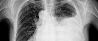
At the beginning of the development of a neoplasm, symptoms may be absent; in such cases, the disease is sometimes detected by fluorography. As they grow, they appear:
- pain in the chest, which can radiate to the shoulder girdle, neck, and area between the shoulder blades;
- painful sensations in the joints;
- arrhythmia, heart rhythm disturbances, including bradycardia or tachycardia;
- cough, shortness of breath;
- asthenia, fatigue;
- redness and sagging of the face;
- headaches and dizziness;
- specific signs that appear with specific types of tumors - night sweats, itching, low blood sugar, hypertension, thyrotoxicosis, muscle weakness, etc.
To establish a diagnosis, radiography, CT, MRI, and endoscopy are performed. The most accurate way to determine the nature of formations is a biopsy - taking biomaterial for histological and cytological studies.
Operative assistance in oncology
- If you are denied chemotherapy and surgery
- If treatment does not work
- If you are sent under the supervision of an oncologist at your place of residence
Call me back!
Diagnostics
The following methods are used in the diagnosis of mediastinal tumors:
• Detailed blood test.
• Bone marrow puncture (collection of a small amount of cerebrospinal fluid using a thick needle) followed by a myelogram (study of the cellular composition of the resulting fluid).
• Reaction with tuberculin antigen (a laboratory method that allows you to determine whether tuberculosis has become one of the causes of the disease).
• Wasserman reaction (a laboratory method that allows you to determine whether syphilis is one of the causes of the disease),
• Laboratory determination of tumor markers (unique proteins that appear in the blood only if there is a certain type of tumor in the body).
• Chest X-ray and CT scan allow us to determine the location of the tumor and make assumptions about its origin and the stage at which the tumor process is located.
• CT of the mediastinal vessels allows you to clarify the stage of the disease, identify the presence of metastases and get an idea of the potential outcome of the disease.
• MRI is especially important in making a diagnosis if the tumor is vascular in nature, as it allows you to examine the entire area of the mediastinum and see the location of each organ and vessel in a three-dimensional perspective.
• PET-CT is perhaps the most complex method for diagnosing tumors, but it is also the most effective, as it allows you to see even the smallest tumor and metastases, consisting of only a few cells.
• Biopsy - removal of a small area of diseased tissue to determine the type of tumor, treatment options and prognosis for the patient.
• Videothoracoscopy – when the diagnosis cannot be established in any other way, doctors perform a small operation: they place a camera inside the patient’s chest under anesthesia. The image from the camera is displayed on the screen, and the surgeon sees exactly which organ is affected by the tumor.
Our prices
- Primary appointment with an oncologist* RUB 3,250.
- Repeated appointment with an oncologist* RUB 3,000.
- Primary appointment with a leading specialist/candidate of medical sciences (oncology)* RUB 4,250.
- Appointment with an oncologist-mammologist KMN* RUB 5,300.
- Repeated appointment with a leading specialist/candidate of medical sciences (oncology)* RUB 4,000.
- Preventive appointment with an oncologist* RUB 1,490.
- Primary appointment with an oncologist-chemotherapist* RUB 4,600.
- Repeated appointment with an oncologist-chemotherapist* RUB 4,000.
- Consultation with an oncologist on palliative care* RUB 2,000.
How does the da Vinci surgical robot work?
The operation of the da Vinci surgical robot is fully controlled by an experienced surgeon through small incisions no larger than 2 cm in size. An endoscope video camera inserted through one of the incisions transmits to the doctor a detailed three-dimensional image of the organ. As a result, the doctor can carefully plan the operation. The surgeon controls the instruments, which have 7 degrees of freedom of movement, thanks to EndoWrist technology and controls their movements inside the patient’s body remotely using special joysticks. Performing an operation using the da Vinci robot requires a highly qualified surgeon and special skills.
Book your room today
6 seats
1 local ward
- 4 meals a day
- Bathroom in the room
- Anti-decubitus mattresses
8200 rub.
Book
13 places
2 local ward
- 4 meals a day
- Bathroom in the room
- Anti-decubitus mattresses
5700 rub.
Book
2 seats
VIP chamber
- a guest room
- 4 meals a day
- Bathroom in the room
- Anti-decubitus mattresses
17700 rub.
Book
Reviews
16.11.2021
Oncology clinic NACFF. The real story about “not giving a damn”
In 2021, on August 16, my older sister flew to Lobnya to undergo examination and, if necessary, treatment at the NACFF oncology clinic.
She lives in the capital Kyrgyzstan...
Read full review
27.10.2021
At the clinic, like at home
30.09.2021
Feedback on the treatment of ovarian cancer at the NACFF clinic
07.09.2021
NACFF patient about treatment, quick examinations and attentive attitude
View all reviews
Hospital, hospitalization, emergency medical care - 24/7, 7 days a week
+7 (495) 259-44-44
Treatment methods
The vast majority of mediastinal tumors require surgery:
• Resectable tumors, that is, those tumors that can be completely removed, are often removed by surgeons along with the organ in which it formed, or even part of neighboring organs and tissues.
• Unresectable tumors, that is, those tumors that for some reason cannot be removed, are treated with chemotherapy and radiation therapy. If the tumor shrinks and there are indications for its removal, the surgeon performs an operation.
Surgeries to remove mediastinal tumors are complex surgical interventions associated with technical difficulties due to the fact that all mediastinal organs are compactly located and often fuse together in the event of pathology. Because of this, there is a high risk of affecting neighboring tissues, which will lead to serious complications. Therefore, recently, minimally invasive approaches, such as laparoscopy and operations using the da Vinci surgical robot, have been recommended for the removal of mediastinal tumors.