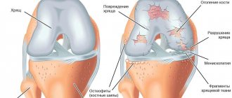
Arthritis of the knee joint is an inflammatory disease that affects the joint and periarticular tissues. If left untreated, inflammation invades neighboring tissues and can spread.
Arthritis of the knee joint on x-ray.
In this regard, it is extremely important to carry out differential diagnosis and make the correct diagnosis.
There are several basic forms of arthritis:
- Infectious . This form occurs when infection is introduced through open wounds (external route) or through blood (internal route) during an infectious disease.
- Post-traumatic . It develops as a result of chronic overload of the knee joints (hereinafter referred to as KS), due to falls, blows, and bruises.
- Rheumatoid . It stands out as a separate item as the most dangerous form due to its widespread manifestations. Caused by autoimmune aggression of the body as a result of a violation of defense mechanisms. In this case, antibodies (hereinafter referred to as AT) themselves destroy the structures of the joint.
- Septic . For this form, the characteristic symptom is an acute onset and severe swelling of the periarticular tissues.
- Gouty . Caused by a violation of metabolic processes in the body. As a result of these disturbances, salt crystals with sharp edges (osteophytes) are formed in the CS bursa. These growths systematically injure tissue and lead to regular inflammatory reactions and pain.
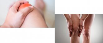
Arthritis of the knee - external manifestations.
Separately, it is worth carrying out a differential. diagnostics with knee joint arthrosis:
- Both conditions are characterized by stiffness, pain and limited joint function, but arthritis also causes local hyperemia and swelling.
- in the case of arthritis, inflammation is the main damaging factor in the development of pathology; in the case of arthrosis, inflammation is a consequence that contributes to the development of destructive-degenerative changes (hereinafter referred to as DDI) of bone structures.
- arthritis is a reversible condition, the treatment of which achieves full restoration of joint function; with arthrosis, the development of existing changes can only be slowed down, but the pathology is prone to progression.
Causes
Now that we know that one of the main reasons for the development of arthritis is infectious diseases, let’s talk more specifically about what organisms have a destructive effect:
- fungi;
- viruses;
- gram-negative bacteria;
- streptococci;
- staphylococci;
- gonococci;
- brucellosis;
- Reiter's disease (most often affects the hip joints, but in a proportion of cases the knee joint is also involved in the process);
- helminthic and protozoal infestations;
- chlamydia;
- syphilis;
- hepatitis.
As for non-infectious arthritis, their development is due to many factors:
- DDI of cartilage structures (age factor).
- Injuries of varying severity and their complications.
- Inflammatory processes in tissues close to the joint capsule 4. Genetic
- predisposition
- Disorders of calcium metabolism (for example, rickets).
- Behçet's disease.
- Ankylosing spondylitis.
- Cappilatotoxicosis.
- Osteomyelitis.
Arthritis statistics
Despite the fact that arthritis is a very common disease, collecting statistics on it is not easy. Many people ignore the symptoms of the disease, citing the fact that such manifestations are “natural” at a certain age. There are approximately 314 million people diagnosed with arthritis in the United States, and approximately 39-42 million cases are reported each year.
In Russia this figure is close to 143 million people. The number of registered patients per year is approaching 17 million. At the same time, a much smaller segment of the population is susceptible to arthrosis of the knee joint (for the USA - 21 million, for Russia - 1.5 million).
Interpreting these data, we can say that 1% of Russians are diagnosed with arthritis every year and the number of healthy Americans is decreasing by approximately the same amount. In developed countries, up to 90% of injuries are due to falls, and the remaining 10% are non-traumatic injuries.
The issue of rheumatoid arthritis (hereinafter referred to as RA) is particularly acute. According to statistics, about 1.3 million Americans are susceptible to this disease, which is equivalent to 41 cases per 100 thousand population. Women suffer from pathology 2-3 times more often than men, and the life risk is: 3.6% for women and 1.7% for men. The worldwide prevalence of the disease is 0.5-1% (up to 5% among the elderly population).
Symptoms
The symptoms of arthritis largely depend on the cause that caused it. Acute ones begin suddenly and, unlike chronic ones, are characterized by severe intoxication. A clear symptom expressed in arthritis of any etiology is pain. At the beginning of the disease, it may be small and appear during exercise.
However, there are a number of nonspecific signs characteristic of arthritis of any origin. The symptoms can be seen more clearly at a certain stage:
- Initial. The articular cartilage is slightly damaged. It manifests itself as slight pain during active movements, with slight lameness. There is no restriction of mobility, and the pain goes away with rest. There is moderate swelling with unchanged skin. The temperature can reach 37.3-37.5 degrees.
- Pathological changes are more noticeable, swelling and pain increase with limited mobility. X-rays show erosion, drying of the hyaline cartilage with thickening of the capsule, and narrowing of its lumen. The temperature can rise to 38.5.
- Terminal. DDI of articular and bone tissues is typical. Inflammation leads to persistent muscle tension and subsequent atrophy of the muscle layer. X-rays show narrowing of the joint space and proliferation of osteophytes. The joint reacts to weather changes (meteodependence). The temperature rises to 39.0 and above.
Stages of knee arthritis on x-ray.
Stages of bilateral damage
As we have already found out, pathological resorption of tissues of the knee joint can occur either exclusively in one lower limb or simultaneously in both legs. Simultaneous damage to the right and left joints is usually called bilateral deforming arthrosis. It is not difficult to understand that when both joints are affected by the disease, the pathology is more complicated and more difficult to tolerate, and it threatens the loss of locomotor-support functions of two limbs at once
Both knee joints are destroyed almost symmetrically.
Such pathogenesis sometimes belongs to the idiopathic or primary form of gonarthrosis. In other words, the disease appeared for no apparent reason, which does not happen very often. Often it has a post-traumatic nature of origin. This suggests that the degenerative-dystrophic process is caused by previously suffered knee injuries, which at one time were treated incorrectly, incompletely or not treated at all. No less often, it is facilitated by congenital pathologies of the musculoskeletal system (curvature of skeletal components, genetic defects of muscle-ligamentous elements, congenital inferiority of the joint, etc.). Another common consequence of disorders are certain diseases, especially rheumatoid arthritis and diabetes.
Good to know! Knee arthrosis requires competent differential diagnosis. To prescribe therapeutic measures that will help improve the quality of life, it is necessary to correctly diagnose and accurately determine the root cause of the pathology. And this is strictly within the competence of the orthopedist-traumatologist. It is impossible to find out on your own whether the problem really lies in gonarthrosis. In addition, there are a lot of movement disorders, for example, Parkinson's disease, which can easily be mistaken for arthrosis. Thus, you can make a mistake and trigger a very dangerous neurodegenerative brain disease. Therefore, if you have not done so already, undergo a high-quality diagnosis, and never prescribe treatment on your own!
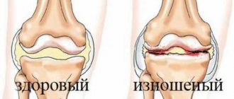
Stages of gonarthrosis of the knee joint, from complete absence to complete destruction.
Like left- or right-sided osteoarthritis, bilateral disease occurs in several stages, and the clinical picture is not always equally expressed on the right and left limbs. Simply put, one of the joints suffers from irreversible changes to a greater extent than the other. Let's look at what happens to a large and complex joint at each stage, and how it affects a person. Note that the characteristics presented below are universal, therefore they are also applicable for left- and right-sided types, and not just bilateral arthrosis. The article will highlight the European classification of stages according to Kelgren.
Initial stage (1st stage)
The painful syndrome is not very pronounced or absent. I may experience mild signs of impaired mobility, mostly in the morning and after prolonged physical activity. At the edges of the articular surfaces, X-rays reveal small osteophytes (bone growths), in the peripheral part of the articular bones - cysts of insignificant size and mild osteosclerosis. The gap between the articular surfaces has a slight narrowing or is not changed. At this stage, conservative methods promise a favorable prognosis for preventing the progression of the disease.
Second degree
At the second stage, the radiographic signs listed above become more distinct, namely: rough marginal spines and cystic formations are well visualized, osteosclerosis is sharply expressed, the radiograph clearly shows that the gap between the bone units has been significantly reduced, and the congruence of the joint is distorted. Painful sensations in the 2nd phase can no longer be confused with simple fatigue; they occur much more often and are more intense. A person notices a tendency for their appearance: pain is associated in particular with walking, going up and down stairs, and forced long-term standing. The flexion function is most impaired, the person begins to limp. To help the patient, specialists already at this stage often resort to surgical intervention methods, since non-surgical tactics sometimes refuse to “work”, and the disease progresses.
Third stage
The pain becomes persistent and almost constant, lameness progresses, and the muscles are seriously atrophied. The patient is no longer able to cope without the use of supporting devices. The third degree of severity provokes curvature of the legs, more often varus deformity, when the limbs acquire an O-shape, as they say simply, “legs like a wheel.” At the same time, the interarticular space is critically narrowed, there are practically no “living” zones of cartilage, and osteophytes are of massive size. The exposed articular surfaces of the bones are in contact with each other, each movement is accompanied by strong friction. Often non-viable tissues and pathological bone formations break off and fall into the joint cavity, which causes terrible pain and joint blockages. At this stage, knee replacement surgery is inevitably needed. Otherwise - lifelong disability with excruciating physical suffering.
Fourth stage
This is the end point, which is characterized by the final loss of the joint - the complete death of all its functional structures. That is, the hyaline cartilage is erased, there is no joint space, and the bony epiphyses are terribly deformed. This stage can be characterized in two words - hellish pain, which even powerful painkillers cannot extinguish. Regarding the motor potential of the problematic limb, we can say the following: there is a persistent flexion-extension contracture, the maximum possible loss of support, coordination and the ability to move. The patient is mostly confined to a wheelchair; at best, he moves with the help of a walker or crutches.
4th degree of gonarthrosis is the last one on the right. A pronounced deformation of the joint and the axis of the tibia is noticeable.
I would like to note that advanced pathology also negatively affects the spinal system and the musculoskeletal framework as a whole. Therefore, it is not surprising when a patient complains that, in addition to unbearable pain in his legs, he has pain in his lower back or another part of the spine. At the final stage of degenerative processes, since the pronounced pain syndrome “does not go away” either day or night, total bilateral endoprosthetics is indicated as an emergency.
The diagnosis is based on an assessment of the patient’s complaints, his individual data (professional activity, concomitant and past diseases, level of daily physical activity, etc.), as well as on the information that the orthopedic doctor receives during examination, after instrumental and laboratory diagnostics.
Diagnostics
Taking into account the multiplicity of causes that provoke the development of arthritis, some forms were especially actively studied by doctors in order to develop an optimal diagnostic algorithm.
Juvenile arthritis (hereinafter referred to as JA) is one of the most disabling rheumatic diseases of childhood.
Clinical blood test
- JA with systemic onset – pronounced leukocytosis (30-50 thousand) with a neutrophilic shift to the left (up to 30% of band leukocytes). ESR increases to 50-80 mm/h, hypochromic anemia, thrombocytosis.
- Juvenile polyarthritis, JRA – hypochromic anemia, neutrophilic leukocytosis (up to 15*109/l), ESR > 40 mm/h.
- Pauciarticular juvenile arthritis - usually laboratory parameters remain normal, but sometimes typical changes characteristic of JA occur.
Immunological and immunogenetic analysis
- JA with systemic onset – the content of CRP, IgM and IgG increases.
- Juvenile polyarthritis - sometimes ANF (antinuclear factor) is positive, RF is negative. Increased levels of CRP, IgM and IgG.
- Pauciarticular juvenile arthritis - 80% of cases are positive for ANF, RF is negative, a high titer of HLA A2 is detected.
X-ray examination of joints
Changes in bone structures are assessed according to Stein-Broker.
- stage – epiphyseal osteoporosis is observed.
- stage – osteoporosis is associated with the breakdown of cartilage, narrowing of the joint space, and isolated erosions.
- stage – DDI of cartilage tissue and bone, osteochondral erosions and subluxations in the joints are formed.
- stage – similar to III with the inclusion of fibrous or bone ankylosis. Reactive arthritis (hereinafter referred to as ReA)
Patient examination scheme
- clinical blood test;
- proteinogram (total protein and protein fractions);
- CEC titer;
- immunological markers of RA;
- immunological markers of SLE – antinuclear factor, antibodies to DNA, LE cells;
- HLA typing (HLA B-27);
- diagnosis of intestinal infections and latent genitourinary infections (PCR, RNGA, RIF);
- X-ray of the affected joints, sacroiliac joints, and spine.
With a long course of ReA, laboratory parameters similar to JA are always found: increased ESR, dysproteinemia, hyperimmunoglobulinemia, high titer of CEC. One of the most important diagnostic signs of ReA is seronegativity for immunological markers of RA and SLE.
Treatment of knee arthritis
To achieve maximum therapeutic effect, you should follow an integrated approach to the treatment of arthritis.
Medication link:
- NSAIDs. With their help, it is possible to relieve pain, especially at night. Among the many drugs, none showed specific benefits, which means that any NSAID drug is suitable for use.
- Glucocorticosteroids (GCS). Usually not prescribed, only in short courses, if the course of arthritis affects the activity of the cardiovascular system.
- Antibiotics. Short courses (1-2 weeks) against a specific pathogen.
- Chondroprotectors. Prescribed to restore the integrity of articular cartilage. However, numerous studies (for example, data from 10 large studies in the British Medical Journal) show no effect even compared to placebo!
- Hyaluronic acid. An important element of cartilage tissue. It is injected into the joint cavity, creating a protective effect and preventing the joint from further damage. However, the therapy does not involve stimulation of one’s own acid, so it seems very expensive (from 30 to 250 thousand rubles annually).
How do diseases differ?
A disease such as arthrosis is age-related. Over time, the integrity is compromised and degenerative disorders of cartilage tissue appear. It is impossible to cure arthrosis, just as it is impossible to stop the aging process. Arthrosis is a chronic disease that occurs in waves: an acute attack is followed by a period of remission.
If you do not seek medical help, arthritis can greatly ruin your life: joint mobility will decrease, and you will be haunted by constant pain. In particularly severe cases, the disease can lead to disability. Therefore, do not delay going to the doctor, make an appointment on time. Our clinic’s specialists will help slow down irreversible changes and maximize the youth and health of your joints.
As for arthritis , it is an inflammatory disease that can affect one or more joints, adjacent tissues, ligaments and muscles. It is often a concomitant symptom of other diseases or joint injuries. The disease is treatable, especially if it is started on time. The peculiarity of arthritis is that it can spread to other joints and tissues, and also become chronic. In some cases, the disease develops gradually, in others it develops quickly and abruptly.
To cure arthritis, you first need to determine its root cause. To do this, our doctors conduct a full examination. Based on the results obtained, you will be prescribed complex therapy that will help you cope with the disease.
Physiotherapy
Phonation
Vibroacoustic therapy involves the transmission of sound microvibration using a special medical device. It creates microvibrations that, with their physical characteristics, are identical to those created by muscle tissue under maximum static physical tension. In short, this therapy is a direct alternative to exercise.
Vitafon.
Phoning Effects:
- Improving lymph flow in the area of influence, which promotes accelerated tissue cleansing, has an anti-inflammatory effect.
- Improves blood flow and, accordingly, nutrition of the treated area.
- Has a beneficial effect on nerve pathways with prolonged exposure.
- Promotes the release of joint lubrication.
Galvanization and electrophoresis
The essence of the procedure is to activate the blood supply to joint tissues in chronic arthritis. Vasodilation occurs in the area of influence, increasing blood supply and improving recovery processes.
Electrophoresis.
UHF therapy
The affected joint is exposed to a continuous or pulsed electric field. For the knees, low-thermal doses are used at a current power of 20-30 W.
The procedure is aimed at reducing swelling, activating regenerative processes in the joint, improving nutrition and blood supply. The method allows you to achieve long-term remission.
Infrared laser therapy
Using a laser applicator, they act on biologically active points located along the lateral surfaces of the joint. The procedure activates blood flow, reduces pain sensitivity, and stimulates healing processes.
Ultrasound therapy
The method optimizes and accelerates the biochemical processes occurring in joint tissues, accelerates healing processes, and reduces swelling.
Hydrogen sulfide and radon baths, peloid therapy, massage, and manual therapy have also proven themselves well.
Knee structure
The knee is a complex structural element of the musculoskeletal system, which provides flexion and extension movements of the lower limb in this section. The articulation is based on the following bone components:
- Distal part of the femur (internal, external condyle);
- Proximal part of the tibia (also internal, external condyle, tibial plateau);
- The patella is a flat bone, which is surrounded on all sides by connective tissue fibers of the tendon coming from the femoral muscles (easily palpable through the skin on the anterior surface of the joint).
The articular bony surfaces of the femur and tibia are not compatible; for normal sliding, a cartilaginous layer is located between them. The cartilage in the knee takes the form of paired formations - menisci. The menisci have a horseshoe shape (the convexity is directed outward), and they are anatomically distinguished:
- Body (the most massive part);
- Front horns;
- Hind horns.
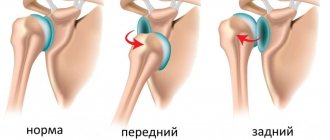
An important role in the knee is played by the tendon-ligamentous apparatus, which ensures the stability of the articulation in the sagittal and frontal planes. The posterior and anterior cruciate ligaments prevent pathological mobility in the anteroposterior direction. When they rupture, a “drawer” symptom appears, when, with a fixed tibia, the femur can be moved back. To combat the disease, you need to know exactly the symptoms and treatment of knee arthritis.
The medial and lateral collateral ligaments provide immobility of the knee in relation to the outer and inner sides. When the internal or external collateral ligament is torn, a valgus (towards the midline) or varus (away from the midline) deformity of the lower extremity can occur.
Endoprosthetics for arthritis
The main indications for knee replacement for arthritis are:
- Deforming arthrosis developed as a result of chronic inflammation.
- Rheumatoid arthritis.
Most often, total endoprosthetics is performed in these cases. At the initial stages of development of degenerative processes, the method of unicondylar endoprosthetics is used.
Knee replacement in the Czech Republic: guarantees, prices, rehabilitation, reviews and statistics.
Find out more
Noltrex injections:
- effective at any stage of knee arthrosis;
- give a long-lasting effect - from nine months to two years;
- do not cause allergies due to their synthetic origin;
- absolutely safe due to biocompatibility with body tissues;
- do not give adverse reactions;
- indicated, including for diabetes mellitus - under the supervision of an endocrinologist.
Even the last stage of gonarthrosis is not a death sentence. Medicine does not stand still, so it’s too early to despair! Consult a specialist you trust and choose the most suitable treatment for you. And most importantly, maintain a positive attitude and faith in recovery!
Prevention
- The most important factor in favorable disease control is control of BMI (body mass index), which reduces the axial load on the joints. You should avoid foods rich in starch and sugar (potatoes, sweets, sweet flour products). 2. It is worth adding more vegetables and fruits: apples, sea buckthorn, rowan, black currants, plums.
- Include fatty fish in your diet: tuna, salmon, cod, sardines, trout, herring.
- Rejection of bad habits.
- Taking vitamins.
- Hardening.
- Maintaining a healthy lifestyle.
- Strengthening the immune system.
- Maintain an active lifestyle (preferably including regular exercise).