What position of the baby in the stomach is considered normal?
The doctor may say that the baby is “poorly positioned” or “malpresented.” What is the difference? “Position” is the placement of the fetus relative to the long axis of the uterus: along, across, obliquely. “Previa” indicates that part of the baby’s body that is closest to the “exit”.
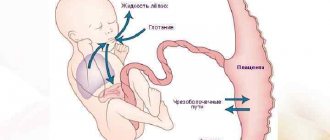
The ideal position of the baby in the uterus is longitudinal with an occipital presentation, that is, head down, with the chin pressed tightly to the chest. This is a physiological position, thought out by nature, when the risk of injury to the baby and mother during childbirth is minimal. And it occurs most often.
Incorrect position or presentation of the fetus is observed in approximately 3.5–6% of cases. The most common of the “non-standard” options is pelvic presentation, leg or breech. There is a facial presentation: the baby's head is thrown back, and it is not the back of the head that appears first, but the face. The most difficult case from the point of view of obstetricians is the transverse or oblique position of the fetus in the uterus.
Some women, whose baby “sat on its butt” or “lay across” during their first pregnancy, are afraid: what if the same thing happens next time? But it is important to understand that the incorrect positioning of the child is a feature of the course of a particular pregnancy and has nothing to do with subsequent ones.
Diagnostics
Head presentation of the fetus at the 20th or later week of pregnancy is determined during ultrasound. But at such an early stage, the location of the fetus does not have any serious significance.
The ultrasound report should indicate the presentation of the fetus, but this is just a description of the position in which the child was at the moment when the ultrasound examination was performed. In general terms, you can determine the type of presentation starting from the 28th week. In this case, an obstetrician-gynecologist talks with the pregnant woman.
A specialist can use external research methods. First of all, the height of the uterine fundus is measured. Also, an obstetrician-gynecologist can palpate the presenting part through the woman’s abdominal cavity.
If a breech presentation is diagnosed, then in this case the fetal buttocks will be clearly palpable in the lower part of the patient’s abdomen. The doctor cannot confuse them with the head, since they are less mobile and more soft.
If the location is transverse, then the child’s head will be located on the right or left side. Also, with this diagnosis, deviations from the norm in the height of the uterine fundus are often recorded.
The doctor can determine what position the baby is in by listening to the lower abdomen in the area below the belly button. If the heartbeat is observed exactly there, then the presentation is cephalic. With a pelvic or transverse position, the heartbeat will be strongest in the navel area or, conversely, above it.
To do this, at each examination, starting from the 28th week, the obstetrician-gynecologist must not only measure the diameter of the abdomen using a centimeter tape, but also palpate it to determine what position the fetus is currently in.
However, this diagnosis allows only to determine the general features of the situation. Even the best gynecologist cannot test the degree of extension of the head. That is why additional ultrasound is performed. It allows you to clarify the type of cephalic presentation, as well as approximately determine the weight of the unborn child and the structural features of his body.
Only an ultrasound can immediately reveal that the umbilical cord has become entangled or diagnose placenta previa. Therefore, ultrasound examinations are mandatory in later stages of pregnancy. Thanks to ultrasound, it is possible to determine whether a natural birth can be carried out.
Why me? Possible causes of presentation
This question worries every mother whose baby is settled in her stomach “not as it should be.” There are several possible reasons.
- Pathological hypertonicity of the lower segment of the uterus and decreased tone of its upper sections. The fetal head is pushed away from the entrance to the pelvis and takes a position in the upper part of the uterus. This happens after inflammatory processes, repeated curettage, multiple pregnancies, complicated childbirth, and a scar on the uterus after a cesarean section.
- Features of fetal behavior and development, for example, increased mobility due to polyhydramnios, small head size, prematurity.
- Structural features and anomalies of the uterus and pelvis: bicornuate, saddle-shaped uterus, the presence of septa or fibroids in the uterus, anatomical narrowing or abnormal shape of the pelvis.
- Limitation of fetal mobility: entanglement in the umbilical cord, oligohydramnios, etc.
Usually the position of the baby in the uterus is fixed by the 32nd–34th week of pregnancy. All these reasons only increase the risk that by this time the child will remain in the wrong position, but they cannot be considered a “final verdict.”
Features of preventing complications during childbirth
If a woman has any abnormal changes in the health of her gynecological organs, then there is a very high probability of a difficult birth that will require surgical intervention. For such women, a very careful diagnosis of the condition of the fetus, its location, and placement aspects is required. A woman in labor is most often placed on a hospital stay to avoid additional stress and irritation in the environment of everyday life. In addition, during this time, detailed rational tactics for childbirth are developed. With early detection of a complicated fetal presentation, the woman has time to prepare for a cesarean section, in which case the likelihood of subsequent psychological distress is reduced.
Waiting for hour "X"
During a routine examination, the doctor, even without the use of technology, is able to approximately determine the position of the baby in the tummy: head down or butt.
The diagnosis is clarified using ultrasound, simple and three-dimensional echography. Early diagnosis of the type of malpresentation will allow you to develop a corrective program or prepare for natural childbirth with an incorrect position or cesarean section according to indications, which will protect you from many injuries and complications. Until the 32nd-34th week, the baby can be in any position. He has plenty of room for a life-changing acrobatic flip and preparation for birth. Sometimes, causing mommy and doctors to worry, the baby turns over just before the contractions begin, and sometimes even with their onset.
Shoulder presentation of the fetus
If the fetus is in a transverse position, the fetal shoulder may be inserted into the pelvic inlet. The diagnosis of shoulder presentation must be confirmed by abdominal (Leopold's maneuvers), vaginal and ultrasound examination. If spontaneous conversion to occipital presentation does not occur, shoulder presentation is an indication for elective cesarean section due to the risk of umbilical cord prolapse and uterine rupture at the onset of labor.
"Coup" plan
If your due date is approaching and your baby is still in the wrong position, don't panic. You should never panic at all, especially if you are pregnant. There is an action plan!
Step 1. Corrective gymnastics...
... will help “persuade” the baby to take the correct position before childbirth. It is carried out after 24 weeks or at certain times in the third trimester. General contraindications to any set of exercises: threat of miscarriage, placenta previa. But there are other features of pregnancy in which doing gymnastics can be dangerous. Before performing any (!) exercises, be sure to consult your doctor!
With breech presentation
- Lie on your side, but not on a soft surface. Lie on one side for 10 minutes, turn to the other, lie down for another 10 minutes. Turn from side to side 3-4 times. Such simple exercises should be performed 2-3 times during the day.
- Lie on your back with your pelvis raised. To do this, place pillows under your legs and lower back. The legs should be 20–30 cm higher than the head. You can spend 10–15 minutes in this position 2–3 times a day.
- Take a knee-elbow position. Stay like this for 15–20 minutes. Repeat 2-3 times a day.
What happens: When performing such exercises, the motor activity of the fetus is stimulated, and it gets more opportunity to turn.
In transverse (oblique) position
- Lie on your side in accordance with the position of the fetus: the head on the left - on the right side, on the right - on the left. The legs are bent at the knee and hip joints. Lie down for 5 minutes.
- Take a deep breath, turn to the opposite side. Lie down for 5 minutes.
- Straighten the leg (in the 1st position - the right one, in the 2nd position - the left one), the other leg remains bent.
- Grab your knee with your hands and move it to the side opposite to the position of the fetus. Bend your torso forward. With your bent leg, describe a semicircle, touching the anterior abdominal wall, take a deep, extended exhalation and, relaxing, straighten and lower your leg.
What happens: A slight mechanical “pushing” of the baby by the muscles into the correct position.
Step 2: Additional steps
- In the transverse position, it is recommended to sleep on the side where the fetal head is located.
- With a breech presentation, turning the baby head down stimulates swimming (after consulting a doctor!).
Step 3. Visit to an osteopath
After the 35th week, a doctor in a hospital setting can rotate the fetus (in transverse and oblique cases, less often in breech presentation). During the entire “operation,” the condition of the mother and child is monitored. The procedure has contraindications and a high risk of complications and injuries, so it is performed in extreme cases.
Step 4. Consolidate the result
As soon as the efforts have been crowned with success and the little “striker” has decided to take the correct position, it is important to help him “get a foothold.” To do this, purchase a prenatal bandage, wear it throughout the day and do a special exercise (consult a doctor!).
Sit on the floor, spread your knees to the sides and press them as close to the floor as possible. Press your feet together. Stay in this position for 10–15 minutes. You can do this several times a day.
What happens: stretching of the ligaments and muscles of the pelvis, which promotes the insertion of the head into the pelvis.
Possible complications
Head presentation of the fetus at week 20 is not dangerous, but complications can arise if the fetus suddenly begins to change position. If the expectant mother is not diagnosed at a later stage of pregnancy, then it is very difficult to predict the outcome of the birth.
If a woman receives serious injuries during childbirth, she may suffer from severe bleeding and the appearance of fistulas. This negatively affects her health. You may develop fibroids or you may not be able to conceive again.
If a child suffers from hypoxia during a natural birth, this can lead to irreversible developmental changes and even a detailed outcome. When diagnosing an extensor cephalic presentation, birth at home or in water should be excluded.
This will further aggravate the situation and there will be less chance of a favorable outcome. But even if the child is positioned optimally comfortably, delivery should take place under the supervision of a doctor who can quickly respond to any unforeseen situation.
As a rule, at the 36th week, doctors recommend deciding on a maternity hospital. Today, women can choose any establishment, not necessarily in their region of residence or according to their registration.
If labor begins earlier than expected, and the patient is taken to another perinatal center by ambulance, then you must inform the doctor what presentation the fetus is in. This will save time during the examination. It is advisable to have the results of your latest ultrasound with you.
Thus, the cephalic presentation of the fetus can change both at the 20th week and later. It is the type of position of the child in the womb that is important. But even in the most difficult situations, if the doctor makes a timely decision to perform a caesarean section, the risk to the life of the woman and her child is much less.
https://youtu.be/JlqSAd3CSYY
Malposition of the fetus: truth and myths
...improper position of the fetus is a 100% indication for delivery through cesarean section
No! A caesarean section is recommended in 60–70% of cases of abnormal position of the fetus in the uterus. But most often, the indication for it is not only a non-standard location, but also a number of related reasons. Natural childbirth with breech presentation is classified as pathological: its course and outcome are significantly complicated, which forces the issue to be decided in favor of a cesarean section. And in case of transverse or oblique position, facial presentation, surgical intervention is absolutely necessary.
... natural birth with breech presentation is most dangerous for boys.
Yes! When a boy is born from this position, there is a risk of injury to the scrotum, especially if the buttocks and legs are raised high. This can lead to infertility and other problems in the future. Another danger is direct thermal and painful irritation of the baby’s scrotum during a vaginal examination of the mother, moving through the birth canal, which provokes premature breathing of the baby. Therefore, a caesarean section is indicated.
...if you put headphones “with music” on your stomach, the baby will become interested and roll over.
No! In most cases, if the baby turns over, it means that he has matured enough to prepare for childbirth and was able to physically perform this “trick”. And the music has nothing to do with it. If the baby is “unwilling” or unable to roll over, these methods will especially not work.
...breech presentation negatively affects the joints.
Yes! Possible underdevelopment of joints and congenital dislocation.
...pregnancy with breech presentation is more often accompanied by complications than with cephalic presentation.
Yes! About 3 times. Complications are often accompanied by hypoxia and delayed fetal development, an abnormal amount of amniotic fluid, and entanglement of the umbilical cord.
Types of cephalic presentation and their features
There are several types of placement of the fetus in the womb. These types differ, and their exact definition determines whether surgery will be required during childbirth.
Occipital
This is the best option, which occurs in more than 90% of cases. With an occipital presentation, the baby's head tilts slightly forward so that he will pass through the birth canal with a small fontanel, that is, with the back of his head forward.
With this position, the likelihood of receiving birth injuries is minimal. Also, with an occipital presentation, the risk of vaginal or perineal rupture in a future woman in labor is virtually eliminated.
Anterocephalic
This position is also called 1st degree of head extension. The child is most likely to move along the birth canal with a second or greater fontanel. This means that the birth process will take longer, since the area of this part of the head is slightly larger.
However, even in this situation, natural childbirth is allowed. But in this case, the likelihood that both the woman and the child will be injured increases. Another risk is that contractions may become weaker during labor.
This phenomenon is usually called the weakness of generic forces. If such a situation drags on for a fairly long period, then there is a risk of oxygen starvation in the child. Therefore, the doctor carefully monitors and evaluates the position of the fetus and only then makes the optimal decision about whether it is worth the risk and a natural birth.
Facial
This presentation is also commonly called the 3rd degree of head extension. She simply cannot straighten up further. In such a situation, the fetus will move along the birth canal with its chin forward. Theoretically, natural childbirth with a facial position is possible. However, this is only allowed in a situation where the child is light in weight and the woman’s pelvis is large enough.
Otherwise, there is a high risk of injury. With this diagnosis, doctors most often suggest not to take risks and perform a caesarean section procedure. This minimizes complications, both for the woman in labor and for her child.
Frontal
Head presentation of the fetus at 20 weeks of this type is also called the 2nd degree of head extension. This means that the largest part of the fetal head will enter the woman’s pelvic area. This can cause not only protracted labor, but also additional complications. The baby in this position will pass through the birth canal forehead first.
This very often causes injury to the spine, spinal cord and even brain. During labor, acute hypoxia may occur. If it continues for a long time, it can provoke not only irreversible consequences, but also the death of the child.
If a diagnosis of frontal presentation is made and natural childbirth is performed, then the woman is guaranteed to receive serious injuries, including very dangerous ruptures:
- perineum;
- uterus;
- cervix.
The pelvic ligaments may also be damaged. In some situations it even resulted in broken bones. Therefore, with this type of presentation, natural childbirth is extremely dangerous; the doctor insists on performing a cesarean section.
Low cephalic presentation
As a rule, it is diagnosed from the 20th to the 38th week. With a low cephalic presentation, there is a risk that birth will occur prematurely.
Therefore, in such a situation, the best solution would be increased monitoring of the woman by medical staff. Also during pregnancy, it is important to ensure that there are no harmful effects on the uterus and fetus. To do this, a woman will need to reduce physical activity and rest more often in a lying position.
Not allowed:
- run;
- jump;
- lift weights.
There is also such a thing as cephalic presentation with low placentation. This is a rather dangerous complication during pregnancy, which can appear with absolutely any type of fetal presentation, even if it is positioned absolutely correctly.
The deviation is characterized by the fact that the placenta is concentrated in the lower part of the uterus near the exit. The fetal head is located much higher. With this placement, it begins to block the internal os of the uterus, which can cause premature contractions and subsequent birth.
If this happens too early, the underdeveloped fetus may die, firstly, due to hypoxia, and secondly, due to insufficient placental blood circulation.
Fetal death can occur with a probability of 7% to 25%. Another problem is that severe bleeding can occur with placenta previa. In 2% it causes the death of a woman. However, there is always the possibility that the placenta will change its position over time. In this case, the danger disappears.


