The position of the fetus is determined by the location of its longitudinal axis to the length of the uterus. When they are placed along the same line, this position is physiological. The baby is placed along the uterus, and one of its round parts (the head or pelvic end) is placed towards the cervix.
What is the transverse position of the fetus? This diagnosis indicates that the baby is located across the uterus. Its axis is located at a right angle to the axis of the uterus. In this case, the position is not determined. The baby's head and pelvis are located above the crests of the pelvic bones.
Causes
This anomaly can be caused by various factors that contribute to excessive or, conversely, insufficient activity of the baby in the womb. The main reasons for the anomaly:
- hypotonia of the abdominal muscles;
- oligohydramnios and polyhydramnios;
- fetal hypotrophy;
- tumors of the uterus and pelvic bones;
- increased myometrial tone;
- risk of spontaneous abortion;
- large fruit;
- clinically narrow pelvis;
- multiple births;
- abnormal attachment of the placenta;
- fetal development abnormalities;
- structural defects of the uterus (bicornuate, saddle-shaped).
Why does the baby lie transversely in the stomach?
The list of reasons for the transverse presentation of a baby in the womb is quite large. The main provoking factors include:
- polyhydramnios , due to which there is active movement of the baby in the uterus;
- weakness of the muscular elements of the uterus , which is more often observed in second and subsequent pregnancies, and the uterus is no longer able to fix the fetus in one position;
- fibroids in the uterus (in this case, the child takes an advantageous position so as not to touch the neoplasms with his head, which prevent him from moving);
- structural anomalies of the female organ , in which the fetus seeks the most comfortable position for itself, which in most cases turns out to be transverse;
- premature birth , when the child did not have time to take a physiological presentation due to the early discharge of amniotic fluid;
- multiple pregnancy (several fetuses are cramped in the womb, so one of them may take the wrong position).
In addition, the reasons include increased muscle tone of the uterus, threat of miscarriage, narrow maternal pelvis, and anterior abdominal location of the placenta.
Leading a sedentary lifestyle by a pregnant woman is an equally significant factor that can affect transverse presentation. If a pregnant woman is constantly in monotonous positions, starting from the 30th week, the baby in the womb may simply “get stuck” in the wrong position.
Breech and transverse presentation are often diagnosed in women who have suffered severe emotional shock and in those who have abused physical activity.
The reason may be the short length of the umbilical cord and the low location of the placenta, and for the child the transverse position will be as comfortable as possible.
Symptoms
If the fetus is not positioned correctly and there are no other pathologies, pregnancy in most cases proceeds favorably. There are no pathological symptoms. External signs may indicate an abnormal position of the fetus:
- spherical shape of the uterus;
- transversely stretched abdomen;
- the height of the uterine fundus increases slowly and does not correspond to the gestation period;
- abdominal circumference is higher than normal;
- the baby's heartbeat can be heard at the level of the mother's navel;
- The baby's pelvis and head are located on the sides of the abdomen.
Diagnostics
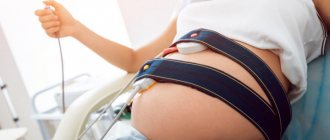
Detection of PPPL deviations occurs mainly during an obstetric examination of the patient.
Even visual inspection and palpation often allow an experienced specialist to suspect this disease. This is facilitated by the transverse oval stretched configuration of the abdomen, an increase in its circumference relative to gestation, and the low height of the uterine fundus. Palpation reveals the head and pelvic part of the fetus in the lateral uterine sections. In this position, during auscultation, it is possible to listen to the PL heartbeat in the umbilical region of the mother’s body.
The final diagnosis in most cases is made based on the results of ultrasound examination (ultrasound). It is informative enough to determine violations of the PL location, identify the threat of termination of pregnancy and conduct dynamic observations.
Why is the transverse position of the fetus dangerous for the mother and fetus?
Diagnosing an abnormal position of the fetus in the early stages of pregnancy should not be a cause for panic, since there is a high probability of its change to longitudinal. If this does not happen, the main danger is that when the baby is positioned transversely in the uterus, most pregnancies end in premature birth. Therefore, women with this diagnosis are carefully monitored and hospitalized in advance.
In the absence of timely medical care, the transverse position of the fetus can lead to the following complications:
- premature rupture of amniotic fluid;
- massive bleeding;
- neglected transverse position - loss of small parts, driving of the shoulder into the small pelvis, limited movements of the child;
- uterine rupture;
- infection with subsequent development of chorioamnionitis, sepsis, diffuse peritonitis;
- hypoxia or asphyxia of the fetus;
- birth of a twin fetus - occurs when the uterus is hypertonic and intense contractions, when the fetus is bent in the chest area, the birth of a live baby in this case is unlikely.
A set of exercises according to Grishchenko and Shuleshova
- Starting position lying on your side. It must be located on the side where the fetal legs are located. Pull your legs towards you and stay in this position for 5–10 minutes. Then turn over to the other side, tighten your legs in the same way and lie down for another 5-10 minutes.
- Lie on your right side, first bend and then straighten. Repeat the exercise 5-10 times. Then turn over to the other side and repeat the exercise 5-10 times.
- Starting position sitting on a hard surface. Bend your leg at the knee and pull it towards you. It is necessary to do the exercise on the side where the fetal legs are located. Bend your leg and make a semicircle with it, pulling it towards your stomach. Take a deep breath and inhale and slowly return your leg to the starting position.
Gymnastics with the transverse position of the fetus
Until 34 weeks of gestation, the position of the fetus is unstable, so there is a high probability that the baby will be placed longitudinally. For this purpose, expectant mothers are prescribed corrective gymnastics during the period 30-31 weeks. This is a group of exercises that help correct the incorrect position of the fetus.
Most often recommended:
- lie on the floor, bend your legs and rest your heels on the floor, raise your pelvis above head level;
- “cat” - get on all fours, while inhaling, raise your tailbone and head, and bend your lower back, while exhaling - the opposite movements;
- take a knee-elbow position, the pelvis is placed above the head - hold for up to 20 minutes.
These exercises are performed for 1-1.5 weeks three times a day. During this period, the fetus must acquire the correct position. If this happens, the stomach must be secured with a bandage.
Correct position while sleeping is also important. The baby is more accustomed to being head down, so the mother should sleep on the side on which the baby's head is located.
Corrective gymnastics is contraindicated in the following cases:
- abnormal placement of the placenta;
- multiple births;
- umbilical cord abnormalities;
- scar on the uterus;
- presence of bloody discharge from the vagina;
- abnormalities of amniotic fluid;
- severe chronic diseases of women;
- increased uterine tone;
- uterine fibroids.
"Coup" plan
If your due date is approaching and your baby is still in the wrong position, don't panic. You should never panic at all, especially if you are pregnant. There is an action plan!
Step 1. Corrective gymnastics...
... will help “persuade” the baby to take the correct position before childbirth. It is carried out after 24 weeks or at certain times in the third trimester. General contraindications to any set of exercises: threat of miscarriage, placenta previa. But there are other features of pregnancy in which doing gymnastics can be dangerous. Before performing any (!) exercises, be sure to consult your doctor!
With breech presentation
- Lie on your side, but not on a soft surface. Lie on one side for 10 minutes, turn to the other, lie down for another 10 minutes. Turn from side to side 3-4 times. Such simple exercises should be performed 2-3 times during the day.
- Lie on your back with your pelvis raised. To do this, place pillows under your legs and lower back. The legs should be 20–30 cm higher than the head. You can spend 10–15 minutes in this position 2–3 times a day.
- Take a knee-elbow position. Stay like this for 15–20 minutes. Repeat 2-3 times a day.
What happens: When performing such exercises, the motor activity of the fetus is stimulated, and it gets more opportunity to turn.
In transverse (oblique) position
- Lie on your side in accordance with the position of the fetus: the head on the left - on the right side, on the right - on the left. The legs are bent at the knee and hip joints. Lie down for 5 minutes.
- Take a deep breath, turn to the opposite side. Lie down for 5 minutes.
- Straighten the leg (in the 1st position - the right one, in the 2nd position - the left one), the other leg remains bent.
- Grab your knee with your hands and move it to the side opposite to the position of the fetus. Bend your torso forward. With your bent leg, describe a semicircle, touching the anterior abdominal wall, take a deep, extended exhalation and, relaxing, straighten and lower your leg.
What happens: A slight mechanical “pushing” of the baby by the muscles into the correct position.
Step 2: Additional steps
- In the transverse position, it is recommended to sleep on the side where the fetal head is located.
- With a breech presentation, turning the baby head down stimulates swimming (after consulting a doctor!).
Step 3. Visit to an osteopath
After the 35th week, a doctor in a hospital setting can rotate the fetus (in transverse and oblique cases, less often in breech presentation). During the entire “operation,” the condition of the mother and child is monitored. The procedure has contraindications and a high risk of complications and injuries, so it is performed in extreme cases.
Step 4. Consolidate the result
As soon as the efforts have been crowned with success and the little “striker” has decided to take the correct position, it is important to help him “get a foothold.” To do this, purchase a prenatal bandage, wear it throughout the day and do a special exercise (consult a doctor!).
Sit on the floor, spread your knees to the sides and press them as close to the floor as possible. Press your feet together. Stay in this position for 10–15 minutes. You can do this several times a day.
What happens: stretching of the ligaments and muscles of the pelvis, which promotes the insertion of the head into the pelvis.
Childbirth with a transverse fetal position
If the position of the fetus does not change, then natural childbirth is not recommended and most often a planned caesarean section is prescribed.
Natural childbirth is possible with a low baby weight, but even then it is necessary to monitor the dilation of the cervix. If the dilatation does not allow birth on its own, then an emergency caesarean section is prescribed.
In the case of premature birth with a transverse position of the fetus, a decision is usually made on an emergency caesarean section.
Surgical intervention in the case of a transverse position of the fetus is quite justified, because there is a significant risk of complications during natural childbirth.
The transverse position of the fetus entails many difficulties, but they can be easily avoided if you follow all the instructions of your obstetrician-gynecologist.
External rotation of the fetus
External fetal rotation is a dangerous method used in exceptional cases.
External fetal rotation is a very dangerous, traumatic procedure, during which the doctor presses on the abdomen with his hands in order to rotate the fetus. Because the doctor cannot see the exact location of the fetus and its limbs, this manipulation is not considered a safe or effective way to turn the baby around. This manipulation is carried out only in a hospital setting, since complications such as uterine rupture, bleeding or placental abruption very often occur. Today, this operation is prohibited in many European countries. In Russia it is not prohibited, but is carried out very rarely and in exceptional cases. A much safer method is a caesarean section.
Breech presentation of the fetus
Ultrasound machine HM70A
Expert class at an affordable price.
Monocrystal sensors, full-screen display mode, elastography, 3D/4D in a laptop case. Flexible transformation into a stationary scanner with a cart.
Currently, breech presentation of the fetus belongs to the section of pathological obstetrics. The frequency of breech presentation of the fetus has remained constant over the past few decades and averages 3% - 5%.
Reasons for the formation of breech presentations
The reasons for the formation of breech presentation of the fetus are varied, numerous and not yet fully understood. These include the following:
- Obstacles to the establishment of the fetal head at the entrance to the pelvis (uterine fibroids, anatomical narrowing of the pelvis or abnormal shapes of the pelvis, tumors of the pelvic organs, an increase in the size of the fetal head due to hydrocephalus or cephalocele, placenta previa or its low location.
- Pathological hypertonicity of the lower segment of the uterus and decreased tone of its upper sections. In this case, the fetal head is pushed away from the entrance to the pelvis and takes a position in the upper part of the uterus. Such disturbances in the contractile activity of the uterus can be caused by dystrophic changes in the myometrium due to inflammatory processes, repeated curettage, multiple pregnancies and complicated childbirth. The scar on the uterus, including after cesarean section, also has a negative effect on the contractile activity of the myometrium.
- Increased fetal mobility due to polyhydramnios, small size of the fetal head due to anomalies of its development (anencephaly, microcephaly), delayed fetal development, prematurity.
- Limitation of fetal mobility with a bicornuate and saddle-shaped uterus or with septa in the uterus, with oligohydramnios, and the umbilical cord entwined around various parts of the fetal body.
On the other hand, it is not at all necessary that if the presence of the listed factors is confirmed, a breech presentation of the fetus will necessarily form. In a number of cases, it can be quite difficult to establish the obvious cause of breech presentation of the fetus.
There are breech and leg presentations. Breech presentations, in turn, are divided into purely breech (incomplete) presentation, when the buttocks of the fetus are facing the entrance to the small pelvis, and its legs are extended along the body, and mixed breech presentation, in which the buttocks of the fetus along with the legs are facing the entrance to the small pelvis, bent at the hip and knee joints.
The types of leg position include:
full - both legs of the fetus are presented; incomplete - one leg of the fetus is presented; knee - the knees of the fetus are presented.
If, with a small size of the fetus in a purely breech presentation, the normal size of the mother's pelvis, childbirth through the birth canal is possible without complications, then with a mixed and leg presentation, the prognosis for the health and life of the newborn is significantly worse. Foot presentation of the fetus is the most unfavorable due to the frequent occurrence of complications during childbirth such as fetal asphyxia, prolapse of umbilical cord loops and severe injury to the fetus.
During the physiological course of pregnancy, the fetus is finally established with the presenting part above the entrance to the pelvis only by 34 - 35 weeks. Before this period, it can change its position many times, even during the day.
Therefore, the formulation of the diagnosis “breech presentation of the fetus” is advisable precisely at 34–35 weeks of pregnancy and not earlier. An earlier diagnosis may be erroneous and will mislead the pregnant woman, her relatives and other consultants in related specialties observing the patient, and create unnecessary emotional tension.
Diagnosis of breech presentations
Diagnosis of breech presentation is primarily based on data from external obstetric and vaginal examination. To clarify the diagnosis, ultrasound is used. With the help of echography, it is possible to determine not only the breech presentation itself, but also, in a number of cases, its type. Three-dimensional echography also provides invaluable assistance in diagnosing breech presentation of the fetus.
.
It is important to determine the position of the fetal head and the degree of its extension. Excessive extension of the fetal head, which is not detected during pregnancy, can lead to such serious complications during childbirth as injury to the cerebellum, cervical spinal cord and other injuries.
The condition of the fetus can also be determined by the results of functional assessment using Dopplerography and CTG.
The course of pregnancy with breech presentation is more often than with cephalic presentation, accompanied by various complications. The most typical among them are: threat and premature termination of pregnancy, gestosis and fetoplacental insufficiency. These complications are often accompanied by hypoxia and delayed fetal development, an abnormal amount of amniotic fluid, and entanglement of the umbilical cord.
The frequency of congenital malformations with breech presentation is almost 3 times higher than with cephalic presentation. Among them there are malformations of the central nervous system, cardiovascular system, gastrointestinal tract, and musculoskeletal system.
According to Doppler ultrasound, a more frequent and more pronounced disturbance of uteroplacental blood flow is noted. In more than half of the observations, signs of chronic fetoplacental insufficiency are revealed.
In patients classified as a high-risk group for the formation of breech presentation of the fetus, prevention of gestosis, premature birth, post-term pregnancy and fetoplacental insufficiency should be carried out.
A pregnant woman needs a gentle regime, a full night's sleep, and daytime rest. Particular attention is paid to a balanced, balanced diet to prevent the development of a large fetus.
If a breech presentation of the fetus is detected from 34 to 35 weeks, the use of corrective gymnastics is recommended
, based on changing the tone of the muscles of the anterior abdominal wall and uterus to transfer breech presentation to cephalic presentation. The essence of this gymnastics is as follows: a pregnant woman, lying on a hard hard surface, alternately turns on her right and left side 3-4 times every 10 minutes. Exercises are repeated 3 times a day before meals for 7-10 days. However, the use of such exercises cannot guarantee the transfer of breech presentation to cephalic presentation.
A pregnant woman with a breech presentation of the fetus must be hospitalized in an obstetric hospital no later than 38 weeks for a full examination, determination of the due date, selection of the optimal method of delivery and preparation for childbirth.
As part of the examination of pregnant women in the hospital, the following activities are carried out: the patient’s medical history and previous diseases are studied, the number and nature of the course of previous pregnancies and births are determined; assess the general condition of the pregnant woman, her psychosomatic status, obstetric complications; specify the duration of pregnancy; determine the type of breech presentation of the fetus, assess the degree of “maturity” of the cervix and the body’s readiness for childbirth; determine the size and shape of the pelvis.
In addition, with the help of ultrasound, the condition of the fetus is determined, the estimated weight of the fetus is calculated, taking into account that with a weight of more than 3500 g, the fetus is considered large in a breech presentation. Echography also makes it possible to identify abnormalities in fetal development, assess the amount of amniotic fluid, and identify tumor-like formations of the uterus and uterine appendages.
An important place in diagnosis is occupied by placentography (location of the placenta, structure of the placenta, correspondence of the degree of maturity of the placenta to the gestational age, thickness of the placenta). With the help of Doppler sonography, not only the nature of the uteroplacental, fetal-placental and fetal blood flow is clarified. This technique, combined with color Doppler mapping, makes it possible to identify umbilical cord pathology and suspect umbilical cord entanglement around various parts of the fetal body.
It is important to establish the type of breech presentation of the fetus, as well as the degree of extension of the fetal head. It is also advisable to determine the sex of the fetus, since male fetuses tolerate the stress of childbirth much worse. More accurate information can be obtained using three-dimensional echography.
Choosing a method of delivery
Choosing a method of delivery requires a very careful and individual approach. Thus, in particular, expanding the indications for caesarean section for breech birth does not yet guarantee a favorable outcome of childbirth. During the operation, the fetus may receive birth trauma. To a significant extent, the risk of fetal injury during cesarean section increases with a premature or large fetus, an extended position of its head, untimely release of amniotic fluid, and insufficient surgical access. The optimal caesarean section rate is 60%-70%.
It should be emphasized that in the vast majority of cases, breech presentation itself is not an indication for cesarean section. However, quite often there is a combination with various complicating factors. Taking into account that breech birth is classified as pathological, in these situations its course and outcome are significantly complicated, which forces the issue to be resolved in favor of a cesarean section.
A planned caesarean section for breech presentations, even without accompanying complications, is indicated for: foot presentation of the fetus; posterior view of breech presentation; extension position of the fetal head.
The dangers of leg presentation
lies in the fact that after the discharge of amniotic fluid, the legs, and then the buttocks and torso of the fetus begin to quickly move forward along the birth canal while the cervix is not yet sufficiently smoothed and dilated. In this case, the fetal head, as a denser and larger part, is not able to pass through an insufficiently opened or spasmodic cervical pharynx, which leads to asphyxia and injury to the fetus or its death.
During childbirth, the initial extension position of the head is further aggravated, the biomechanism of childbirth is disrupted, which leads to significant injury to the fetus.
With the posterior type of breech presentation, the biomechanism of labor is also disrupted, its progress is significantly slowed down, which leads to asphyxia and injury to the fetus.
It is necessary to identify in advance a group of pregnant women with a breech presentation of the fetus who have indications for performing a cesarean section on a planned basis. These indications include: anatomically narrow pelvis and abnormal pelvic shapes; extension position of the fetal head; foot presentation of the fetus; posterior view of breech presentation of the fetus; mixed breech presentation in first-time mothers; fetal weight more than 3500 or less than 2000g; placenta previa and its low location; umbilical cord presentation; scar on the uterus; cicatricial changes in the cervix, vagina and perineum; elimination of a history of genitourinary and enterogenital fistulas; pronounced varicose veins in the vagina and vulva; severe gestosis; hemolytic disease of the fetus; delayed fetal development; severe fetoplacental insufficiency; severe concomitant diseases; large uterine fibroids; abnormalities of the uterus; lack of biological readiness of the body for childbirth during full-term pregnancy; lack of effect from preparing the cervix for childbirth; post-term pregnancy in combination with an immature cervix; the age of the first-time mother is over 30 years; complicated obstetric history (infertility, recurrent miscarriage, birth of a sick, traumatized child, premature birth with death of newborns, stillbirth); the onset of this pregnancy after the use of assisted reproduction methods.
The presentation of the fetal scrotum deserves special attention. Touch during vaginal examination, mechanical irritation that occurs during the advancement of the fetus, the birth of the scrotum with high buttocks and legs, thermal and painful irritation causes premature breathing and aspiration of amniotic fluid, which often contains meconium. It has been noted that boys born in a breech presentation through the natural birth canal often subsequently experience infertility due to testicular trauma during childbirth. Unfortunately, with breech presentations, it is not always possible to reliably determine the sex of the fetus using echography before birth. However, if a male fetus is identified and there are other aggravating circumstances for breech presentation, then it is advisable to resolve the issue of delivery by cesarean section as planned. In the case of vaginal delivery, a protracted course of the second stage of labor should be avoided. It is necessary to remove the fetus as quickly and carefully as possible, followed by appropriate care for the newborn.
A favorable obstetric situation in which childbirth can be carried out through the natural birth canal includes: satisfactory condition of the pregnant woman and the fetus; full proportionality of the pelvis of the mother and fetus; sufficient biological readiness of the body for childbirth; the presence of a pure breech or mixed breech presentation; bent fetal head.
If the issue of conducting childbirth through the natural birth canal has been decided, then the pregnant woman should undergo a complex of prenatal preparation, including antispasmodics, sedatives and restorative drugs, and vitamins. The prescription of these drugs is necessary to improve the function of the fetoplacental complex, prevent labor anomalies and postpartum hemorrhage.
Childbirth with a breech presentation of the fetus differs in a certain way from those with a cephalic presentation and is classified as pathological. In this regard, such births should be treated as a high-risk category for the development of perinatal pathology, using preventive measures to prevent possible complications.
One of the important tasks in the first stage of labor with breech presentation is maintaining the integrity of the amniotic sac until the cervix is completely or almost completely open.
For this purpose, the woman in labor must observe bed rest, lying on the side corresponding to the position of the fetus (on the side of the back of the fetus).
Childbirth is carried out under constant monitoring of the condition of the fetus and contractile activity of the uterus using cardiotocography (CTG). When the cervix is dilated by 4 cm, to prevent labor anomalies, intravenous drip administration of antispasmodics (no-spa 4-6 ml in 400 ml of 5% glucose solution) is started. Every 2-3 hours, fetal hypoxia is prevented by intravenous administration of drugs that improve microcirculation and uteroplacental blood flow.
During childbirth during breech presentation, in order to prevent birth stress for the mother and fetus and to prevent abnormal contractile activity of the uterus, anesthesia is mandatory, which begins in the active phase of labor when the cervix is opened by 3-4 cm. For this purpose, it is recommended to use epidural anesthesia, which It not only has a pronounced analgesic effect, but also helps regulate labor, relax the pelvic floor muscles and protect the fetus from injury. Management of childbirth using epidural anesthesia requires careful monitoring of the contractile activity of the uterus.
In childbirth with a breech presentation of the fetus, the frequency of complications exceeds that with a cephalic presentation. Cervical dilatation occurs more slowly, even with a intact amniotic sac. The buttocks stand above the entrance to the pelvis for a long time. A belt of adhesion is not formed, separating the waters into front and rear. These circumstances can lead to the development of the most typical complications for the first stage of labor with a breech presentation of the fetus. These complications include: untimely rupture of amniotic fluid; prolapse of the umbilical cord loop and small parts of the fetus; anomalies of labor; protracted labor; acute fetal hypoxia; premature detachment of a normally located placenta; Chorioamnionitis.
In case of untimely rupture of amniotic fluid, which occurs in 40%-60% of cases, due to the lack of differentiation between the anterior and posterior waters, they are completely poured out, which in turn is a prerequisite for the prolapse of the umbilical cord loop or small parts of the fetus, creating conditions for infection of the fetus and the development of chorioamnionitis during childbirth.
After the amniotic fluid has leaked, it is necessary to clarify the obstetric situation by performing a vaginal examination and exclude or confirm the prolapse of umbilical cord loops and small parts of the fetus. In the latter case, the tactics of labor management should be reconsidered in favor of cesarean section.
If the cervix is completely ready for childbirth, prenatal rupture of amniotic fluid, and the condition of the fetus is quite satisfactory, you can wait 2-3 hours until labor develops independently. Otherwise, labor induction should begin. If the cervix after the rupture of amniotic fluid is immature or not mature enough, then labor induction cannot be started. In this case, the issue of caesarean section is decided.
If at the time of the rupture of amniotic fluid the cervix was mature, and labor did not begin on its own within 2 hours, then labor induction begins. If there is no effect from labor induction within 2-3 hours or if the condition of the fetus worsens, the issue of delivery by cesarean section should be resolved.
Anomalies of labor
, occurring in 25-30% of cases, may be caused by immaturity of the cervix, untimely rupture of amniotic fluid, malformations of the uterus, initial disturbance of uterine tone, uterine fibroids, irrational management of labor, and the formation of a clinically narrow pelvis. If labor is weak, labor stimulation is performed by intravenous drip administration of drugs that increase the contractile activity of the uterus. Labor stimulation is carried out when the cervix is opened by more than 5 cm. If the opening is less and the contractile activity of the uterus develops, the birth must be completed by caesarean section in the interests of the fetus. If there is no effect from labor stimulation within 2-3 hours or the condition of the fetus worsens, then further labor stimulation is not advisable, and it is also necessary to resolve the issue of delivery in favor of a cesarean section.
Discoordination of labor activity poses a particular danger to the fetus during breech presentation. Further conservative management of labor in this situation should be considered unacceptable due to the increasing severity of hypoxia, increasing duration of labor and anhydrous interval.
Thus, the indications for performing an emergency caesarean section during childbirth with a breech presentation of the fetus are:
presentation or prolapse of umbilical cord loops and small parts of the fetus; immature cervix with prenatal rupture of amniotic fluid; development of weakness of labor when the cervix is opened by less than 5 cm; lack of effect from labor induction or labor stimulation for 2-3 hours; discoordination of labor; acute fetal hypoxia; premature detachment of a normally located placenta.
In the second stage of labor, when the cervix is fully dilated, the pelvic end of the fetus should be on the pelvic floor. From this moment the attempts begin.
Until the fetus is born up to the navel, labor is expectant, since forcing labor and pulling on the pelvic end leads to disruption of the fetal position, throwing back of the arms and extension of the fetal head.
The woman in labor lies on her back with her legs bent at the hip and knee joints, which rest on the supports. This position allows you to maintain good pushing activity, which is an important condition for maintaining the expulsion period during breech presentation of the fetus. To intensify efforts and reduce the angle of the pelvis, it is recommended to press your thighs to your stomach with your hands. This is especially important at the end of the expulsion period, as the reduced pelvic angle allows for easier passage of the head.
In the second stage of labor, monitoring the condition of the fetus is important. Heartbeats are heard through each push. Physiological in breech presentation is the release of meconium, which is squeezed out of the intestines as the fetus moves through the birth canal.
When the pelvic end is cut through, the perineum is dissected, which reduces the obstruction from the vulvar ring for the developing fetus, reduces the risk of fetal injury and asphyxia, helps speed up the second stage of labor, prevents rupture of the perineum, and facilitates manual assistance.
From the moment of birth of the fetus to the navel, the most critical stage of the second stage of labor begins. After the birth of the pelvic end, the birth canal remains poorly stretched for the passage of the subsequent head. When the fetal head is inserted into the entrance to the small pelvis and begins to pass through the birth canal, the umbilical cord loops are pressed against the walls of the pelvis. The danger increases at the moment of birth of the subsequent head. The time for pressing the umbilical cord should not exceed 3-5 minutes. If the birth of the head is delayed, if this period lasts longer, fetal injury may occur and asphyxia may develop. Pressing the umbilical cord for more than 10 minutes threatens the death of the fetus. Another danger when the birth of the head is delayed is the possibility of placental abruption due to a decrease in the volume of the uterus after the birth of the fetal body. In this regard, appropriate techniques and aids are used to carefully remove the child and successfully complete the birth.
The course and management of the succession and postpartum periods are practically no different from that with cephalic presentations. It is important to prevent postpartum hemorrhage by intravenous administration of drugs that increase uterine tone. You should carefully examine the birth canal using mirrors to identify possible trauma and then restore its integrity.
Management of newborns
Children born in breech presentation require special attention from neonatologists, as they are at high risk for various complications, primarily neurological. After the birth of the child, during examination, signs of intracranial injury, cerebrovascular accident, spinal injury, and hip dysplasia should be excluded. If a child was born with asphyxia or with symptoms of aspiration of amniotic fluid, then appropriate resuscitation measures are required. In the early neonatal period, children born in a breech presentation should be carefully examined. An ultrasound examination of the brain should be performed, cerebral blood flow should be assessed using Doppler, an echographic examination of internal organs should be performed, and, if indicated, an X-ray examination of the hip joints. An examination by a neurologist is also advisable.
Injuries to the birth canal in women during breech birth occur much more often than with cephalic presentation of the fetus. The most common among them are ruptures of the cervix, vulva, vagina and perineum, as well as pelvic injuries.
A higher incidence of maternal morbidity after childbirth with breech presentation is due to more frequent untimely rupture of amniotic fluid, the development of labor anomalies, prolonged labor and anhydrous interval, infection during pregnancy and childbirth, more extensive trauma to the birth canal, and traumatic surgical interventions. with increased blood loss.
Prevention of adverse birth outcomes in breech fetuses
- Identification of risk groups for the formation of breech presentation of the fetus.
- Preservation of the physiological course of pregnancy.
- Drug prevention, timely detection and treatment of threatened miscarriage, gestosis, fetoplacental insufficiency.
- Prevention of post-term pregnancy and large fetus.
- Use of corrective gymnastics.
- Careful consideration of risk factors for possible complications when choosing a method of delivery.
- Appropriate early selection of pregnant women for planned caesarean sections.
- Effective preparation of the body for childbirth.
- Rational management of labor, prevention of untimely rupture of amniotic fluid, abnormal contractile activity of the uterus and bleeding.
- Timely diagnosis of complications during childbirth and revision of their management tactics.
- Gentle delivery using appropriate manual aids and procedures.
- Rational management of the postpartum period.
- Thorough examination of newborns using clinical, instrumental and laboratory diagnostic methods.
Ultrasound machine HM70A
Expert class at an affordable price.
Monocrystal sensors, full-screen display mode, elastography, 3D/4D in a laptop case. Flexible transformation into a stationary scanner with a cart.
Management of childbirth
Hospitalization is scheduled for the 36th week; in the maternity hospital, the pregnant woman is examined and prepared for a caesarean section . Independent childbirth with an external-internal rotation is possible only in cases of extreme prematurity or twin births when the second child is in transit.
Indications for abdominal delivery:
- scar on the uterus;
- chronic hypoxia, FPN;
- prenatal breaking of waters;
- pathological localization of the placenta;
- post-term pregnancy;
- neoplasms of the uterus.
In the diagonal position, the woman in labor is prescribed bed rest on her side, from below which the fetal head/pelvis is palpated. If a limb/umbilical cord falls out, its repositioning is prohibited; the woman is operated on as an emergency.
Important
The neglected transverse position requires immediate abdominal delivery in the interests of the woman, regardless of the condition of the child. If symptoms of infection of the uterus or membranes appear, a cesarean section ends with the removal of the uterus.
Conditions for producing external-internal rotation:
- indwelling urinary catheter;
- full dilatation of the cervix;
- written consent of the woman in labor;
- estimated fetal weight less than 3600 g;
- live fruit;
- availability of a fully operational operating room;
- correspondence of head size to pelvis indicators.
Sozinova Anna Vladimirovna, obstetrician-gynecologist
20,806 total views, 4 views today
( 48 votes, average: 4.40 out of 5)
Abdominal injuries during pregnancy: consequences for the fetus and mother
22 week of pregnancy: fetal development, mother’s well-being, ultrasound and tests