Published 06/21/2019
Cervical cancer is the leading cause of death in women aged 30 to 35 years. At the same time, a woman often cannot detect the pathology on her own, experiencing minor complaints.
The cervix has a special anatomical structure. It is designed in such a way that the gynecologist can only see gross changes in the cervix, which, as a rule, are an indicator of an advanced process. A more detailed examination of the cervix for pathology is possible only under high magnification, which is precisely provided by a device called a colposcope.
Colposcopy of the uterus
Colposcopy of the uterus is a diagnostic procedure that is performed using a colposcope (a special optical instrument with a lighting device) and is aimed at examining the walls of the vagina and the visible vaginal part of the cervix.
During the examination, the doctor may:
- perform a biopsy of certain areas of the cervix for subsequent histological analysis;
- collect the necessary material for subsequent laboratory diagnostics of bacteriological and cytological smears;
- enlarge the image 2, 5, 7, 10, 15 or more times for a more detailed study of the structure of tissues.
In our clinic, this diagnostic procedure is performed using an expert-class colposcope. Its results will allow the doctor to identify pathology at the initial stage of its development, draw up a detailed picture of the disease and prescribe an effective course of treatment.
Advantages of video colposcopy
Modern video colposcopic systems perform video recording in high resolution, with automatic color balancing, which allows you to obtain an image absolutely identical to that available during direct visual inspection. The presence of such video recording opens up certain opportunities for gynecologists that were previously unavailable:
- a more detailed examination of areas that raise doubts regarding initial pathological structural changes;
- recording the examination results for subsequent assessment of dynamics during treatment therapy;
- the ability to involve colleagues in the study, including remotely (necessary in the most diagnostically difficult cases).
Indications for colposcopy
Indications for colposcopy may include the following diseases:
- true erosion and pseudo-erosion of the cervix;
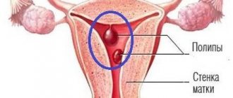
- cervical polyps;
- cervical endometriosis;
- inflammatory diseases of the cervical canal and vagina (vaginosis, colpitis, cervicitis, gonorrhea, trichomoniasis, etc.);
- suspected carriage of human papillomavirus;
- endometrial hyperplasia;
- uterine fibroids.
Indications for this study may include the following conditions:
- discharge and itching in the vagina;
- identifying signs of cell atypia in a cytological smear;
- intermenstrual bleeding;
- pain during or after sexual intercourse;
- discharge after sexual intercourse;
- frequent and prolonged pain in the lower abdomen;
- detection of genital warts on the external genitalia;
- detection of ruptures after childbirth;
- monitoring the condition of women with uterine erosion;
- monitoring the condition of women at risk for the development of cervical cancer;
- monitoring the condition of women after treatment of cervical pathologies.
When is it necessary to undergo examination by a gynecologist?
It is better to carry out female examination as planned, but in some cases it is necessary to contact a specialist as early as possible, for example:
- when planning pregnancy;
- for menstrual irregularities;
- in case of manifestations of sexually transmitted infections;
- if attempts to get pregnant within a year do not bring results;
- for pain in the mammary glands, discharge from the nipples;
- if consultation is necessary on the selection of a contraceptive method, etc.
It is difficult to overestimate the importance of a regular comprehensive gynecological examination - a woman’s general well-being, the quality of her sexual life, fertility and the health of her future children depend on reproductive health.
Don’t put off your health until tomorrow, call and make an appointment with a gynecologist right now! Our doctors will help you with maximum tact in resolving any issues related to women's health.
Why is cervical colposcopy performed?
The main purpose of colposcopy is aimed at determining the boundaries, malignancy or benignity of the pathological focus. During the research process the following can be determined:
- border of pathology;
- differentiation of the tumor process;
- identification of the inflammatory process;
- the need to perform a tissue biopsy from the pathological focus;
- exact area of tissue for biopsy.
After analyzing all the data obtained, the results of colposcopy allow the doctor to draw up a plan for further effective treatment.
Types of colposcopy of the uterus
Depending on the colposcope model used and the research methodology, the following types of colposcopy are distinguished:
- simple (overview);
- extended;
- chromocolposcopy;
- fluorescent;
- with color filters;
- colpomicroscopy.
Depending on the type of colposcope, the following can be performed:
- simple colposcopy;
- photocolposcopy (the results are reflected in photographs);
- video colposcopy (results can be recorded on various media and the patient herself can observe the examination process).
Composition of a colposcope
Colposcopes come in two types: those with a fixed focal length lens whose magnification can be changed by changing the power of the eyepieces, and those with a fixed focal length lens but with multiple magnifications where the dial setting can be changed or a button pressed. .
Increase
Multiple magnifications are preferred, starting at ×5 or ×7.5, ×20 or ×30. The lowest magnification gives a bird's eye view and is great for locating or zooming in on an area of interest. The magnification commonly used for detailed examination is ×15. The modern colposcope is based on the original prototype, but differs from the original in that the magnification varies from 6 to 40 times, as opposed to the original 7.5 times. 10x magnification is suitable for everyday use. Higher magnification allows you to recognize some minor points, but it is not necessary to use it for an accurate diagnosis.
The most important accessories for a colposcope are photographic equipment and special spare parts for teaching. Also, simple low-power devices without any accessories are always available on the market, as well as more complex ones with electrical control, a zoom lens with fine tuning and a camera. For routine use, a simple colposcope is fine, but for training, both the camera and viewing tube are essential. All colposcopes have lenses, eyepieces, filters, a light source and a stand.
Lens
This in turn affects the focal length and therefore the working distance. The best focal length is between 250mm and 300mm. This allows you to easily operate and manipulate tools without interfering with your vision.
Eyepieces
Eyepieces must have the following good characteristics: the axes of the eyepieces can be straight or inclined at an angle of 45° to the optical axis of the device, they must have rubber cups installed on them, they must have independent focusing elements that adapt to human vision. Also one very important feature is the convergent line of sight for fatigue-free working.
Filters
Most colposcopes are equipped with green filters. They absorb red light and enhance the image of blood vessels that appear black. The contrast between normal and abnormal epithelium is also enhanced.
Light source
The light source must be at least 30,000 lux, must be constantly centered, and must have a rheostat to vary the light intensity. Some equipment has automatic brightness adjustment depending on the magnification used. Most colposcopes are equipped with a halogen lamp with a power of 6 to 12 W and 20 to 75 W. A recent innovation is the light emitting diode (LED), which is a better light source. They generate light through semiconductor processes rather than heating a wire filament, as in a halogen lamp. The advantages of an LED light source are that its light output is five to seven times higher than that of a lamp; this light uses a different optical system, significantly improving contrast, and with it overall visibility improves. The service life of an LED is also many times higher (up to 10,000 hours) than a halogen lamp. It lasts for approximately 10 years with daily use of approximately 4 hours.
Simple colposcopy
During a simple colposcopy, the doctor examines the surfaces of the walls of the vagina and cervix under magnification. Then, using a tampon, the discharge is removed and the examination is repeated.
Using a simple colposcopy, the doctor can roughly assess:
- shape and size of the cervix and external os;
- relief and color of the mucous membrane;
- the severity of the vascular pattern;
- boundaries of flat and columnar epithelium.
This procedure is performed without prior preparation of the patient. It takes little time, is absolutely safe and painless.
To obtain a more detailed clinical picture, our clinic may prescribe other more informative types of colposcopies.
Extended colposcopy
Extended colposcopy provides more accurate and detailed results. It is carried out using various tests and dyes. For the standard procedure, 3% solutions of acetic acid and Lugol with glycerin are used as dyes. When conducting this type of examination, the woman undergoes special training, and before it begins, secretions are removed from the surface of the vagina and cervix using a sterile swab.
When performing an extended colposcopy, the doctor can evaluate:
- appearance and color of the cervix;
- the severity and condition of the vascular pattern;
- relief of the mucous layer of the cervix;
- the junction of the epithelium of the cervix and the cervical canal;
- condition and presence of glands;
- mucosal reaction to vinegar solution;
- reaction of the mucous membrane to Lugol's solution (Schiller test);
- type of epithelium;
- clarity or blurring of the boundaries of formations.
Also, for extended colposcopy, color filters, Chrobok's test (with a probe) and staining of the mucous membrane with methylene blue, eosin, hematoxylin (Derazhne test), fluorochrome or trichlortetrazole can be used. These types of this procedure make it possible to draw up a more detailed picture of the disease, determine the malignancy of the tumor and more clearly visualize the contours of blood vessels.
Historical facts
In 1924, Hinselmann was asked to write a chapter based on the etiology, symptoms and diagnosis of uterine cancer in the third edition of the Handbook of Gynecology. Hinselmann faced this challenge most successfully. He performed gynecological examinations and often encountered early detection of cervical cancer, which he believed could only be improved by optical means. He believed it was necessary to "provide an intense light source for the magnified image without sacrificing binocular vision."
Content:
- Historical facts
- Composition of a colposcope
- Indications for the use of colposcopy
- Differences between simple and extended colposcopy
- Preparing for the examination
- Methodology
- Complications after extended colposcopy
- Contraindications for the study
- Colposcopy and pregnancy
- Study protocol
By 1925, he reported the construction of the first colposcope. To this end, he connected a light source to Lake's binocular biological microscope. He connected the optical system to a stand that allowed movement in each direction and also supported a small screw for fine adjustment. After the invention of the colposcope, Hinselmann was able to state that “with regard to so-called early problems, colposcopy makes it possible to detect earlier cases.” Even a tiny tumor the size of a dot did not disappear and was visible even with an initial magnification of 7.5 times. Thus, Hinselmann discovered a completely new invention and proved its significance in the field of medicine.
Fluorescence colposcopy
Fluorescence colposcopy allows to detect the malignancy of a cervical tumor in 98% of cases. This type of colposcopy is performed using a special dye and UV rays.
After treating the cervix with fluorochrome, the cervix is examined under UV rays and the doctor evaluates the resulting glow:
- Normally, the unchanged mucous membrane has a violet or dark blue glow;
- in the early stages of a malignant tumor – the mucous membrane has a crimson, light green or bright yellow glow;
- in the later stages of a malignant tumor, the glow goes out.
The effectiveness of the technique
Today, such manipulation is rightfully considered accurate in terms of studying endometriosis, polyposis, oncological processes and precancerous conditions.
Normally, the epithelial area is light pink, shiny and smooth. Benign changes are characterized by true erosion, ectopia.
Best materials of the month
- Coronaviruses: SARS-CoV-2 (COVID-19)
- Antibiotics for the prevention and treatment of COVID-19: how effective are they?
- The most common "office" diseases
- Does vodka kill coronavirus?
- How to stay alive on our roads?
What time of day is best to perform a colposcopy?
Colposcopy can be performed at any time of the day. The most favorable moment for its implementation is the days of the first half of the menstrual cycle. It is best to carry out this procedure 2-3 days after the end of menstrual bleeding.
Colposcopy is not performed during menstruation, because bleeding and rejection of areas of the uterine cavity mucosa interfere with a detailed examination of the vaginal walls and the visible part of the cervix. Such a procedure will be uninformative and meaningless.
It is undesirable to perform colposcopy during the first 2-3 days after ovulation, because during this period of the cycle a large amount of mucus accumulates in the cervical canal, which distorts the results and interferes with the procedure. In the second half of the menstrual cycle, performing this diagnostic procedure is also undesirable, because after its implementation, the mucous membrane of the cervix will take a longer time to recover.
Preparing for colposcopy
After prescribing colposcopy, the doctors of our clinic will definitely familiarize the patient with the simple rules of preparation for this diagnostic procedure, since the accuracy of their implementation may depend on the reliability of the results obtained.
Preparation for colposcopy should begin 4-5 days before the procedure. The set of these measures includes the following recommendations:
- 4-5 days before the procedure, stop using any medications that are inserted into the vagina;
- 2-3 days before the procedure you should stop douching;
- 2 days before the procedure, you should use only warm water for washing and refrain from using any detergents;
- 1-2 days before the procedure, you must stop using lubricants;
- 1-2 days before the procedure, you should abstain from vaginal intercourse (if it is impossible to exclude sexual intercourse, you should use a condom).
No other specific preparation is required for cervical colposcopy.
Stages of cervical colposcopy
Colposcopy is performed on an outpatient basis or in a hospital after completing preparation for this procedure.
Colposcopy includes the following steps:
- the woman is examined on a gynecological chair using a regular gynecological speculum, which remains in the vagina throughout the entire subsequent procedure;
- the vaginal walls and cervix are dried with sterile cotton swabs to remove mucus;
- the colposcope is installed at the gynecological speculum a few centimeters from the entrance to the vagina;
- the doctor, using a colposcope, sequentially examines the walls of the vagina and the visible part of the cervix and selects areas for testing or biopsy;
- using a clamp with a tampon soaked in a 3% solution of acetic acid, the doctor treats the cervix;
- Normally, under the influence of acetic acid, the walls of blood vessels undergo spasm and completely disappear after 30-60 seconds;
- in the presence of cancerous degeneration of cells after treatment with acetic acid, the disappearance of the vascular pattern is not observed;
- after assessing the results of treating the epithelium with vinegar, the doctor performs a Schiller test, which is carried out by applying a 3% Lugol’s solution with glycerin to the cervix;
- Normally, under the influence of this test, the entire surface of the mucous membrane turns dark brown;
- in the presence of cancerous degeneration of cells, after testing, unpainted areas are revealed on the surface of the cervix;
- after performing the Schiller test, other additional research techniques can be performed (fluorescence colposcopy, chromocolposcopy and colpomicroscopy);
- if necessary, the doctor uses special forceps to perform a tissue biopsy from suspicious areas;
- a sample of tissue taken for histological examination is placed in a test tube and sent to the laboratory;
- After completion of the procedure, the colposcope is moved back and the gynecological speculum is removed from the vagina.
After colposcopy, the woman receives a doctor’s report, an invitation to the next appointment, and can go home. The results of the biopsy (if performed) will be ready in 1-2 weeks.
Abnormal colposcopic patterns
The presence of pathological changes may be indicated by detection during extended colposcopy:
- acetowhite epithelium;
- iodine-negative zones;
- signs of leukoplakia;
- punctuation;
- mosaics;
- atypical transformation zone;
- signs of invasive cancer.
All these cases require an individual approach and additional diagnostic procedures, including biopsy, PAP test, etc. Mild lesions are characterized by the detection of dense acetowhite epithelium with clear boundaries, delicate mosaics and punctures. With pronounced changes, rapid whitening of the treatment area is observed with the appearance of dense acetowhite epithelium and a rim around the open glands. Also in such situations, rough mosaic and punctation, signs of tuberosity, are observed.
Separately, mixed colposcopic patterns are distinguished, which are difficult to unambiguously classify. These include such “findings” as genital warts, inflammatory processes, atrophic changes in the mucous membrane, endometriosis, polyps, etc. In such cases, treatment is also prescribed, which can be either conservative or surgical, or additional examination.
Acetowhite epithelium
The appearance of areas of white or acetowhite epithelium when exposed to 3% acetic acid is one of the most important signs of pathology. Most researchers agree that the vast majority of areas in which the development of malignant processes is observed turn white with varying intensity under the influence of acetic acid.
Detection of acetowhite epithelium is typical for all degrees of CIN and makes it possible to diagnose the presence of changes at the earliest stages of development. CIN refers to cervical dysplasia, which is accompanied by the formation of cells with varying degrees of atypia in the covering epithelium. Untreated dysplasia can lead to cervical cancer, and each case of its development is the result of missed opportunities and ignoring the importance of regular colposcopy.
It is important to differentiate white epithelium from areas of dyskeratosis, i.e. leukoplakia. Some blanching of the mucous membrane can also be observed with:
- disturbances in the structure of epithelial cells;
- inflammatory process;
- papillomavirus infection;
- some other benign changes.
The more intense the whitening of the tissue and the longer it persists after exposure to acetic acid, the more serious and profound the changes.
Iodine-negative zones
A typical sign of pathology is the identification of so-called silent iodine-negative areas of the mucosa after applying Lugol's solution. In most cases, they are represented by keratinized epithelium and require a more detailed examination by performing a biopsy.
Leukoplakia
Leukoplakia is a pathology accompanied by keratinization of the epithelium of the vagina, vulva or cervix. Colposcopically, it looks like a white spot of arbitrary shape with clear boundaries, which can be noticeable even before treatment of the mucous membrane with solutions. Most often, leukoplakia is observed in the area of the transformation zone and can have different sizes. The true size of the modified area is determined after treatment with Lugol's solution, since it does not turn brown. The main danger of leukoplakia is the inability to determine, without the use of invasive methods, what changes are located under the layer of keratinized cells. Therefore, if it is detected, a targeted biopsy is indicated.
punctuation
Puncture refers to multiple red dots on the background of the epithelium, caused by its atypical vascularization, i.e., the formation of blood vessels. The degree of violation is determined based on the size and uniformity of the points. They are clearly visible after exposure to acetic acid, and sometimes have the appearance of papillae.
Mosaic
This term refers to the zone of the mucous membrane, which, after treatment with acetic acid, is delimited by red lines into yellowish-white segments. This area is devoid of open and closed glands. Based on the intensity of the mosaic, the degree of tissue damage is determined. Thus, with a rough mosaic, the mucous membrane takes on the appearance of a paved pavement, but more often a delicate mosaic is observed - the segments do not protrude above the level of the surrounding tissues and have a marble pattern. Most often, the mosaic is iodine-negative.
Atypical transformation zone
It is formed on the basis of normal ST, but differs in the presence of:
- atypical blood vessels;
- acetowhite epithelium;
- keratinized glands;
- leukoplakia;
- mosaics;
- punctuation;
- areas not stained with iodine.
This indicates the presence of significant changes in the structure of the epithelium and the likelihood of developing dysplasia.
Invasive cancer
The most distressing “finding” during colposcopy may be the discovery of signs of invasive carcinoma:
- atypical transformation zone with uneven tuberosity and the presence of a limited area rising above the level of surrounding tissues (plus tissue);
- ulcers;
- atypical vessels, characterized by fragility, which leads to bleeding.
As a rule, oncology develops against the background of benign changes in the cervix, which were not diagnosed and eliminated in time.
Thus, the possibilities of colposcopy are truly wide, and its accessibility, low cost and ease of implementation are its indisputable advantages. Therefore, it is important not to ignore preventive gynecological examinations and doctor’s recommendations to perform colposcopy. Often this is what makes it possible to diagnose severe pathologies in the early stages, carry out the necessary treatment and save the patient’s life.
0 0 votes
Article rating
Is it painful to have a cervical colposcopy?
Colposcopy is not accompanied by pain, since the colposcope is not inserted into the vagina. During the study, some women may feel discomfort from the cold gynecological speculum and slight tingling after treating the cervix with a solution of acetic acid.
The biopsy that may be performed during this test may cause short-term discomfort or mild pain. The painlessness of this manipulation largely depends on the skill of the doctor.
In our clinic, the colposcopy procedure is performed only by experienced gynecologists and patients do not experience pain.
Progress of gynecological examination
The process of vulvoscopy is in many ways similar to a regular examination by a gynecologist. A woman needs to undress to the waist and sit comfortably on a chair.
The doctor uses special instruments and a colposcope, which is located a short distance from the examination area.
- Colposcopy does not cause discomfort if the woman follows the recommendations of the gynecologist.
- After a visual examination and installation of a gynecological speculum, the doctor begins to examine the surface of the vulva, vaginal walls, and cervix. Video colposcopy allows you to display an image on a monitor screen, which gives the doctor a greater overview and recording of the results on a digital medium.
- During extended vulvoscopy, anesthesia can be used, due to the high sensitivity of the vulva during biopsy.
- If the results of a simple microscopic diagnosis do not provide a complete clinical picture, the doctor uses vinegar and Lugol to determine the contours of the pathological tissues for an accurate biopsy.
After completion of the diagnosis, the results of extended colposcopy are sent for detailed interpretation to a specialist or to a histological laboratory. The doctor gives recommendations on the woman’s behavior after colposcopy.
What happens after colposcopy of the uterus?
After colposcopy of the cervix for 3-5 days, a woman may experience slight dull and nagging pain in the lower abdomen and bloody, pink, dark brown or greenish discharge from the genital tract. The appearance of discharge is explained by the healing of the scar left on the cervix after cutting off sections of tissue for biopsy, or by the removal from the vagina of remnants of dyes used for colposcopy.
These symptoms are normal, go away on their own and do not require seeing a doctor. After completing the procedure, the doctor must warn the woman about the possibility of pain and discharge and recommend:
- use sanitary pads for 3-5 days;
- do not use vaginal tampons;
- After the biopsy, for 10 days do not take medications that thin the blood (Aspirin, Ibuprofen, etc.), do not visit the sauna or bathhouse, do not take a bath, do not have sex, and avoid heavy physical activity.
In some cases, colposcopy may be complicated by infection of the genital tract or bleeding after the biopsy. A woman should seek medical help if symptoms such as:
- profuse bleeding from the genital tract;
- temperature increase;
- chills;
- intense pain in the abdomen or lower back;
- discharge from the genital tract with an unpleasant odor.
FAQ
Despite the fact that before the procedure the doctor talks in detail about everything that will happen, questions always remain about the examination and diagnosis. Most often, patients are concerned about the following.
Is it painful to have a colposcopy or not?
In general, the procedure is painless, especially when performed by a highly qualified doctor. Mild discomfort or tingling may occur when using solutions, but this is uncommon. Therefore, to the question whether colposcopy is painful or not, the answer is negative. If during the examination you feel severe pain, you must immediately inform your doctor.
How long does the procedure take?
On average it does not take much time. Depending on the individual case, the procedure takes 20–30 minutes.
How to prepare for cervical colposcopy?
A diagnostic test such as colposcopy does not require special preparation. But patients are not recommended to have sexual intercourse several days before the procedure, and the use of vaginal creams and douching are also undesirable.
How often can and should you do a colposcopy of the cervix and which doctor should you contact?
A referral for a diagnostic examination is given by a gynecologist. He also recommends a preventative care plan, which may include a colposcopy. It all depends on your individual situation and medical history (if any).
If there are no serious problems, then the procedure, as a preventive measure, is recommended to be performed once a year.
Are there any consequences after the colposcopy procedure?
Over the next 3-5 days, you may experience a brownish discharge due to the use of iodine or another solution. Therefore, patients are advised to bring panty liners with them to use after the procedure.
In very rare cases, slight spotting and slight discomfort in the lower abdomen may occur. Sometimes the doctor may recommend taking painkillers.
What to do after the procedure?
If nothing particularly bothers you, then there is no need to take any special action. But it is highly desirable during the period while the discharge and/or slight malaise persists:
- abstain from sexual intercourse;
- do not use tampons;
- do not take a bath (shower only) or go to the pool;
- do not douche.
The doctor will tell you more about everything at your appointment.
Is it possible to have a colposcopy during menstruation?
Testing during menstruation is not recommended because the results may be inaccurate. In this regard, the doctor is often asked the question on what day (first, last) of the cycle during menstruation can a colposcopy be done. The best time for diagnosis is the first days after the end of menstruation. The doctor agrees on this with the patient in advance.
Is it possible to have a colposcopy during pregnancy?
In general, the procedure is safe for the fetus throughout the entire period of pregnancy. And the question of whether such a diagnosis is needed or not is decided by a gynecologist.
. If there are no prerequisites and compelling reasons, the examination is not prescribed. After childbirth, colposcopy can be performed after 1.5 months.
Why is colposcopy prescribed and performed for pregnant women and at what stages (early, late) during pregnancy is it required? Here everything depends on the patient’s medical history (whether she had diseases of the reproductive system), on how the pregnancy proceeds, on the doctor’s decision. If you are afraid, worried and worried, then do not hesitate to ask the doctors of our center in St. Petersburg questions about why colposcopy during pregnancy, whether it is safe for the child, etc.
Initial examination
At the initial consultation, the doctor conducts a simple examination of the patient and collects the necessary information, asking questions about the state of health, complaints, medical history (if any), symptoms, etc.
At the initial appointment, you can also ask any concerning questions: why a colposcopy is being done, which doctor will perform the procedure, what preparation should be done, are there any contraindications, what are the indications for the examination, etc.
Diagnostics
If examination and analysis of the information received does not make it possible to make an accurate diagnosis, then the doctor gives an appointment for examination. The doctor also gives a referral for colposcopy of the cervix if the previous transcript showed poor results. If you have previously undergone this examination, then you need to tell your doctor about it and it is advisable to bring its results.
In addition to colcoscopy, the following may be prescribed: smears, ultrasound and additional examinations.
Repeated appointment
At the second visit, a direct examination is carried out and, if necessary, the diagnostic and treatment plan is adjusted if the results of other tests are already known.
Control reception
Depending on each individual case, the doctor may schedule an additional visit after the end of the course of treatment to again perform a colposcopy procedure to check for relapses and to assess the patient’s health condition.
Where can I have a cervical colposcopy in St. Petersburg?
To undergo the procedure, we invite you to visit us at the medical office. We employ highly professional diagnosticians, have modern equipment and take an individual approach to the treatment and monitoring of each patient. In addition to this, we have some of the most affordable prices for cervical colposcopy and other types of diagnostics and treatment in St. Petersburg.
The interpretation of colposcopy of the cervix shows that everything is fine with you and there is nothing to worry about, or there are still problems in gynecology. The timing of the analysis results is individual, depending on the specific situation.
Colposcopy after childbirth
Colposcopy after childbirth is a relevant and necessary procedure, since the cervix after this important event in a woman’s life can undergo various scarring and other changes. Also, this diagnostic study can be indicated in the presence of various diseases of the cervix, erosion, ectopia, etc. in the prenatal period.
Colposcopy in the postpartum period is recommended by the doctors of our clinic 6 weeks after the date of birth. If necessary, a cervical biopsy may be performed during this procedure.
The results of colposcopy will convince you of the absence of pathologies or will allow you to promptly identify the disease and carry out its effective treatment.
Contraindications to manipulation
This kind of procedure is completely safe, because it does not harm the body in any way. I often prescribe it even during the period of pregnancy. There are no special contraindications to performing the research technique.
The only limitation is that the procedure is never performed in the early postpartum period (usually the first months after childbirth). The procedure is not recommended immediately after destructive or surgical therapy of the uterus. But these contraindications are only temporary.
There is only one absolute contraindication – intolerance to acetic acid or the iodine element.