Heart Attack Symptoms
Because cardiogenic shock typically occurs in people who have had a severe heart attack, it is important to know the signs and symptoms of a heart attack. These include:
- Pressing, bursting or squeezing pain in the chest lasting more than 15 minutes;
- Pain radiating to the shoulder, arm, back or teeth and lower jaw;
- Increased frequency of attacks of chest pain;
- Prolonged pain in the upper abdomen;
- Dyspnea;
- Sweating;
- A looming feeling of fear;
- Fainting;
- Nausea and vomiting.
If you see your doctor right away when these signs or symptoms appear, you can prevent the possibility of developing cardiogenic shock. Prompt treatment of a heart attack increases the chances of survival and reduces damage to the heart. Don't ignore these symptoms if they last more than five minutes. Call an ambulance immediately. If you cannot call an ambulance, ask someone to take you to the nearest hospital.
Classification
Classification of cardiogenic shock according to the severity of the patient’s condition involves three forms:
| Clinical manifestations | 1st degree (mild) | 2nd degree (moderate) | 3rd degree (severe) |
| Duration of shock | less than 5 hours | from 5 to 8 hours | more than 8 hours |
| Blood pressure in mm Hg. Art. | at the lower limit of normal 90/60 or up to 60/40 | upper at 80-40, lower at 50-20 | not defined |
| Tachycardia (beats per minute) | 100–110 | up to 120 | dull tones, threadlike pulse |
| Typical symptoms | weakly expressed | left ventricular failure predominates, pulmonary edema is possible | pulmonary edema |
| Response to treatment | good | slow and unstable | absent or short-term |
Causes
Cardiogenic shock occurs when the heart loses its ability to pump enough blood to the rest of your body. The most common cause of cardiogenic shock is damage to the left ventricle, the main pumping chamber of the heart, due to lack of oxygen due to a heart attack.
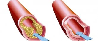
A heart attack occurs when one or more of the arteries that carry oxygenated blood to your heart (coronary arteries) becomes blocked. Sometimes, over time, a narrowing of the coronary arteries occurs due to the deposition of cholesterol on their walls. The formation of these deposits, called plaques, in the arteries throughout the body is called atherosclerosis.
During a heart attack, one of these plaques may rupture, forming a blood clot at the site of the rupture, blocking blood flow through the arteries. Without oxygenated blood entering the heart, the heart muscle weakens and cardiogenic shock develops.
In rare cases, cardiogenic shock develops when the right ventricle of the heart is damaged. From the right ventricle of the heart, blood travels to the lungs, where it is enriched with oxygen before being sent to the rest of your body. Damage to the right ventricle causes the heart to lose its ability to pump blood effectively to the lungs, causing the body to not receive enough oxygen.
Although a heart attack is the most common cause, other conditions that cause cardiogenic shock include inflammation of the heart muscle (myocarditis) or infection of the heart valves (endocarditis). Causes include drug overdose or poisoning from substances that affect the pumping function of your heart.
Cardiogenic shock.
Cardiogenic shock is an acute violation of the perfusion of body tissues caused by significant damage to the myocardium and disruption of its contractile function. The main causes of cardiogenic shock include myocardial infarction (MI), myocarditis, cardiomyopathies, toxic myocardial lesions, cardiac tumors, severe heart defects, trauma, pericardial tamponade, pulmonary embolism, severe cardiac arrhythmia.
Most often, a practicing physician has to deal with cardiogenic shock in patients with acute coronary syndrome (ACS), primarily with ST-segment elevation MI. Cardiogenic shock is the leading cause of death in patients with MI.
Typically, cardiogenic shock develops in the first hours after the onset of the first symptoms of MI and much less often in a later period. The risk of developing this formidable complication and its severity are largely determined by the extent of the infarction - the size of the myocardium affected by ischemia and necrosis. Therefore, most often, cardiogenic shock develops with MI of the anterior wall of the left ventricle, the apex of the heart and the anterior part of the interventricular septum, that is, with occlusion of the left coronary artery, which supplies most of the myocardial mass with blood, as well as with damage to all three main coronary arteries (which causes involvement more than 40% of the mass of the left ventricular myocardium into the infarction zone). Cardiogenic shock with right ventricular myocardial infarction is much less common.
The biggest problem with cardiogenic shock is the following vicious circle: severe depression of systolic function and a decrease in blood pressure (BP) cause ineffective coronary perfusion, as a result, coronary blood flow is further deteriorated, and myocardial ischemia and necrosis progressively worsen, which further impairs the pumping function of the left ventricle . If the mass of necrotic myocardium is 40-50% or more, then, as a rule, cardiogenic shock becomes areactive (torpid), that is, one in which the introduction of inotropes and vasopressors has no effect. Mortality in this group of patients approaches 100%.
Are there any positive trends in solving the problem of cardiogenic shock in recent years? What opportunities can modern medicine offer for the routine management of patients with this complication? We have selected the main evidence and practical recommendations on this pressing issue and present a summary of them in this article.
Cardiogenic shock yesterday and today
The incidence of cardiogenic shock in patients with ACS is difficult to accurately determine, since different authors use different definitions and criteria for diagnosing shock. According to rough estimates ten years ago (GUSTO-I, 1996; GUSTO-III, 1999; RJ Goldberg et al., 1999), cardiogenic shock developed in 7-10% of patients with ACS. Later, various population studies showed a slightly lower incidence of cardiogenic shock in ACS - from 3.2 to 8.6% (TRACE, 2003; NRMI, 2005; J. Fang et al., 2006; GRACE, 2007). In some of these studies, the positive dynamics in the risk of cardiogenic shock noted over the past few years was clearly associated with the implementation of modern evidence-based recommendations for the management of patients with ACS [2, 3], primarily with the wider use of coronary revascularization methods, especially surgical , as well as hemodynamic support using intra-aortic balloon counterpulsation.
The most valuable thing about the achievements of modern cardiology is that they are expressed not only in a lower risk of cardiogenic shock, but also in a decrease in mortality from this complication. More recently, cardiogenic shock was actually a death sentence. Before the introduction into practice of modern methods of treating MI (urgent revascularization, intra-aortic balloon counterpulsation), the development of cardiogenic shock doomed almost all patients to death - about 85-95% (E. Braunwald, 1988). As a number of studies in recent years have shown (NRMI, 2005; J. Fang et al., 2006; AMIS Plus, 2008), mortality in cardiogenic shock can be reduced to 30-40%, although in practice the real mortality figures even in developed countries of the world are still remain at the level of 50-60%. The latest European Society of Cardiology (ESC) guidelines for the management of patients with heart failure (HF) (2008) [4] indicate that the in-hospital mortality rate for people with cardiogenic shock is 40-60%.
Thus, the results of the American National Registry of Myocardial Infarction (NRMI) showed that in-hospital mortality among patients with cardiogenic shock that developed in connection with ST-elevation MI decreased from 60.3% in 1995 to 47. 9% in 2004 (p < 0.001). This increase in patient survival was associated with a doubling in the number of percutaneous coronary interventions (PCI) used for urgent coronary revascularization (27.4% in 1995 and 54.4% in 2004, p < 0.001). Similar data were obtained in the Swiss AMIS Plus registry - a decrease in mortality in cardiogenic shock over the past 10 years was noted from 62.8 to 47.7% (p = 0.01), which is associated with a significant increase in the number of PCI for ACS (from 7. 6 to 65.9%, p=0.01) [5].
In 1999, the SHOCK study [6] showed that urgent revascularization (angioplasty or coronary artery bypass grafting) for ACS complicated by cardiogenic shock, although it does not lead to a significant reduction in early (1-month) mortality compared with the drug treatment group, but statistically significantly improves the prognosis in the long term. The mortality rate of such patients during the first six months after MI decreased from 63.1 to 50.3% (p = 0.027), and within 1 year - from 66.4 to 53.3% (p < 0.03). The greatest benefits of revascularization were observed in the subgroup of patients younger than 75 years. Based on the results of this study, it was strongly recommended to increase the use of surgical methods of early coronary artery revascularization in patients with cardiogenic shock due to ACS [2].
Thrombolysis is less effective for revascularization in cases of cardiogenic shock because low perfusion pressure prevents adequate delivery of fibrinolytic to the coronary arteries. However, thrombolytic therapy, according to the GUSTO-I study, reduces the incidence of cardiogenic shock to 7.2%, and the mortality rate for this complication to 55%. Other studies have also confirmed a significant improvement in survival of patients with cardiogenic shock with the use of thrombolysis compared with conservative therapy (without revascularization). Therefore, thrombolysis in conditions of unavailability of PCI and urgent coronary artery bypass grafting is the optimal treatment method, especially considering that thrombolysis can be carried out in a much shorter time, including at the prehospital stage, which can be life-saving for patients with cardiogenic shock. For the realities of domestic medicine, this is currently a more acceptable treatment strategy, although not optimal, of course, compared to PCI and coronary artery bypass surgery.
Along with this, it was confirmed that in patients with MI who suffered cardiogenic shock and survived early, long-term clinical outcomes do not differ significantly from those in patients without a history of cardiogenic shock (SHOCK, 2006; M. Singh et al., 2007) . Therefore, measures that improve the early (in-hospital) survival of patients with cardiogenic shock actually equalize the further risks of patients, regardless of the presence of a history of shock.
AMIS Plus Registry: 10 Years of Progress in the Treatment and Prevention of Cardiogenic Shock
To illustrate the above data, I would like to talk in more detail about the results that were obtained within the Swiss registry for MI (Acute Myocardial Infarction in Switzerland, AMIS Plus) [5].
The register included more than 23.6 thousand patients with ACS (with and without ST segment elevation) from 70 clinics in Switzerland. Data were analyzed from January 1, 1997 to December 31, 2006. The authors assessed changes in the incidence of cardiogenic shock and mortality from this complication during the study period and determined the dependence of these changes on the nature of patient treatment.
It turned out that over the past 10 years, the incidence of cardiogenic shock in patients with ACS has decreased from 12.9 to 5.5% (p = 0.001). This happened due to a decrease in the risk of developing cardiogenic shock during hospital treatment (from 10.6 to 2.7%, p < 0.001), while the frequency of registration of already developed cardiogenic shock at the time of admission to the hospital did not change significantly - about 2-2.3%.
Although cardiogenic shock was 2 times more common in patients with ACS with ST elevation than without ST elevation, the reduction in the risk of its development over the past decade was approximately the same in both cohorts of patients.
In-hospital mortality in patients with cardiogenic shock also decreased significantly - from 62.8 to 47.7% (p = 0.01), and this also applied to patients admitted with cardiogenic shock (from 73.8 to 46.6%, p = 0.009), and those in whom cardiogenic shock developed after hospitalization (from 60.9 to 48.9%, p = 0.094), although, as can be seen, in the latter subgroup of patients the differences did not reach statistical significance.
Analysis of AMIS Plus data [5] indicates that positive trends in the dynamics of the risk of cardiogenic shock and associated mortality in patients with ACS correlate with the improvement of inpatient care for such patients, in particular with the widespread introduction of surgical revascularization methods. It should be noted that in the studied clinics during the study the number of PCIs performed significantly increased (from 7.6 to 65.9%, p = 0.01) and the use of intra-aortic balloon counterpulsation, while the number of thrombolysis decreased and the number of aortocoronary operations shunting has remained virtually unchanged.
The analysis showed that the reduction in both in-hospital mortality in patients with ACS in general and in-hospital mortality in the subgroup of patients with cardiogenic shock was significantly affected by the wider use of PCI.
In addition, the therapeutic treatment of patients with cardiogenic shock has significantly improved: during the studied period of time, the use of acetylsalicylic acid (from 80.4 to 89.2%), clopidogrel (from 11.7 to 65.5%), increased in this cohort of patients. inhibitors of IIb/IIIa glycoprotein platelet receptors (from 11.8% in 1999 to 35.6% in 2006), lipid-lowering drugs (from 14.3% in 1999 to 77.8% in 2006) , β-blockers (from 32.7 to 40%).
The AMIS Plus registry [5] also studied risk factors for the development of cardiogenic shock. It turned out that independent predictors of this complication were older age, ST segment elevation, tachycardia, and low systolic blood pressure. In contrast, the use of lipid-lowering drugs and PCI were significantly associated with a lower risk of cardiogenic shock.
Practical recommendations for the management of patients with cardiogenic shock
Diagnostics
Quick diagnosis of cardiogenic shock allows you to take the necessary measures in a timely manner and prevent the death of the patient. Therefore, it is very important to know the signs that most likely indicate the development of this complication.
To assess the severity of acute HF in patients with MI, the classifications of T. Killip (1967) and JS Forrester (1977) are used [4]. Both classifications imply the division of patients into 4 groups (stages, classes) depending on the severity of systemic hemodynamic disorders and pulmonary congestion. The differences between them are that the JS Forrester classification takes into account not only clinical signs, as in the T. Killip classification, but also some indicators of central hemodynamics (wedge pressure in the pulmonary artery, cardiac index). According to T. Killip's classification, the state of cardiogenic shock corresponds to a decrease in blood pressure < 90 mm Hg. Art. and the presence of signs of peripheral vasoconstriction (oliguria, cyanosis, sweating); according to the JS Forrester classification - signs of reduced perfusion of body tissues in combination with high “wedge” pressure in the pulmonary artery.
In the ESC guidelines for the management of ST-elevation MI, updated at the end of 2008 [1], cardiogenic shock is defined as a decrease in systolic blood pressure < 90 mm Hg. Art., an increase in the filling pressure of the ventricles of the heart (and, accordingly, the “jamming” pressure of the pulmonary artery) >20 mm Hg. Art., a decrease in cardiac index < 1.8 l/min/m2. The diagnosis of cardiogenic shock is established if other possible causes of hypotension (hypovolemia, vasovagal reflex, electrolyte imbalance, side effect of pharmaceuticals, myocardial tamponade) are excluded. But it should be remembered that cardiogenic shock accounts for about 80% of all shock conditions that complicate the course of myocardial infarction, that is, this pathology is the most likely when it comes to significant hypotension and hypoperfusion in heart attack patients.
According to the new ESC guidelines for the management of patients with heart failure (2008) [4], cardiogenic shock does not have clear diagnostic criteria, although in typical cases it can be considered when blood pressure decreases below 90 mmHg. Art. (or mean blood pressure drops by more than 30 mm Hg), and diuresis is absent or sharply reduced (<0.5 ml/kg/h). Within a short time, clinical signs of organ hypoperfusion and pulmonary congestion appear. Heart rhythm disturbances often develop.
Thus, for the initial diagnosis of cardiogenic shock (at the patient’s bedside), it is enough to detect threatening clinical signs, measure blood pressure and exclude other probable causes of hypotension. A decrease in the volume of urine output, which can be assessed after bladder catheterization, quickly confirms whether the diagnostic search is moving in the right direction. In addition, invasive testing of central hemodynamic parameters is extremely important both for diagnosing shock and for monitoring the effectiveness of treatment. For this purpose, catheterization of the right heart and pulmonary artery (balloon “floating” Swan-Ganz catheter) is recommended. In this case, the pressure in the right atrium and ventricle, pulmonary artery, pulmonary artery wedge pressure, and cardiac output are measured. These data allow you to most accurately assess the state of cardiac function and make it possible to notice its minimal deterioration or improvement, as well as exclude many other possible causes of hypotension.
It is important to recall that right ventricular myocardial infarction sometimes manifests as cardiogenic shock, but the approaches to treating both conditions are significantly different. Therefore, if cardiogenic shock is suspected, differential diagnosis with infarction of the right ventricular wall is also necessary. To this end, identification of distension of the jugular veins (especially during inspiration), confirmation of the absence of moist rales in the lungs, ECG signs of right ventricular myocardial infarction (for example, ST segment elevation in lead V4, characteristic changes in the recording of the ECG chest leads on the right), ultrasonographic evidence may help. right-sided infarction.
Prevention and treatment
To date, the only approach that has clearly confirmed the possibility of reducing the risk of cardiogenic shock in patients with ACS is the earliest possible revascularization of the coronary vessels. Clinical trials and large registries show that the currently recommended strategy of urgent reperfusion is the best way to reduce the number of patients with cardiogenic shock.
Treatment of cardiogenic shock is a difficult task, but not hopeless. As shown above, some therapeutic interventions for ACS have already proven their significant benefits not only in preventing cardiogenic shock, but also in improving survival if this complication develops. This applies primarily to the strategy of early revascularization, the use of which is associated with better outcomes for patients with manifest cardiogenic shock. A reduction in in-hospital mortality in patients with cardiogenic shock using PCI was convincingly shown in the AMIS Plus registry (2008) [5], and a positive effect on more long-term outcomes (6 months) - in the SHOCK study (1999) [6]. Based on the data obtained in the SHOCK study, the developers of American guidelines for the treatment of MI (American College of Cardiology, ACC; American Heart Association, AHA) classified emergency revascularization for cardiogenic shock as class I recommendations [2].
In the 2008 ESC guidelines for the management of patients with STEMI (1), early revascularization with PCI is the recommended strategy in the event of cardiogenic shock (grade I recommendation, level of evidence B). If PCI cannot be performed or is available only after some delay, immediate coronary artery bypass grafting may be indicated in patients with cardiogenic shock, especially if there are other indications for cardiac surgery (mitral regurgitation, left ventricular wall rupture, etc.). If both PCI and coronary artery bypass grafting are not possible in the near future, early revascularization with thrombolysis is necessary.
Early and effective elimination of myocardial ischemia and prevention of the formation of necrosis or significant limitation of its size ensures rapid restoration of systolic function of the heart and thereby breaks the vicious circle of “depression of cardiac output → decreased perfusion → additional deterioration of the myocardium.”
In addition, to interrupt this same vicious circle, hemodynamic support measures are very important to maintain blood pressure at a level that ensures adequate perfusion of vital organs, primarily the myocardium itself (90-100 mm Hg). For this purpose, inotropic drugs, vasopressors, and intra-aortic balloon counterpulsation are used.
The ESC guidelines for the management of patients with STEMI (2008) [1] and for the management of patients with HF (2008) [4] for the treatment of cardiogenic shock, in addition to early revascularization, recommend the use of oxygen therapy, mechanical ventilation depending on the level of blood gases, assessment hemodynamic conditions using cardiac catheterization, administration of inotropes (dopamine, dobutamine), the use of intra-aortic balloon counterpulsation and mechanical devices to ensure left ventricular function.
To date, there is no evidence that inotropes improve survival in patients with cardiogenic shock. But inotropes can either improve the patient's clinical condition and bring him out of shock, or at least stabilize his hemodynamics until more effective methods (intra-aortic balloon counterpulsation, surgery, special mechanical devices) can be used. The most popular inotrope is dopamine, especially since in low doses it effectively improves renal perfusion without significantly affecting systemic hemodynamics. Dobutamine has its benefits - although it is a slightly weaker inotrope than dopamine, it reduces pulmonary artery pressure. But it should be remembered that the administration of inotropes, especially dopamine in high doses, increases the risk of developing tachycardia and arrhythmia, so they should be used with caution in patients with an accelerated heart rate and require ECG monitoring. A combination of low doses of dopamine with higher doses of dobutamine is often used - this allows, with minimal risk of side effects, to improve systemic hemodynamics and especially renal perfusion.
The main criteria for the effectiveness of inotropic therapy are an increase in systolic blood pressure above 90 mmHg. Art., increase in cardiac index >2 l/min/m2, decrease in pulmonary artery “wedge” pressure to 20 mm Hg. Art., increased diuresis. It is important that the heart rate does not exceed 100 beats/min. If tachycardia or cardiac arrhythmias develop, the dose of inotropes should be reduced if possible.
If inotropes in standard doses are not effective enough, the following options for changing tactics are possible: increasing the dose of the inotrope used, a combination of two inotropes, adding vasopressors, using intra-aortic balloon counterpulsation, special mechanical devices to support left ventricular function.
Vasopressors (norepinephrine) are not recommended as first-line therapy for acute heart failure, but may be indicated in the case of cardiogenic shock if inotropes in combination with adequate fluid therapy are not sufficiently effective in stabilizing hemodynamics. In this case, the use of vasopressors must be done with great caution, since cardiogenic shock is usually accompanied by peripheral vasoconstriction and high peripheral vascular resistance. In addition, norepinephrine promotes lactic acidosis, increases pressure in the pulmonary artery (can provoke pulmonary edema) and is characterized by the fact that tolerance to it quickly develops. Therefore, even if you had to resort to norepinephrine, it is important to stop it as soon as possible. The use of epinephrine for the treatment of cardiogenic shock is not recommended; indications for its administration should be limited to cases of cardiac arrest only.
A method of temporary hemodynamic support that significantly improves the survival of patients with cardiogenic shock is intra-aortic balloon counterpulsation. However, it should be noted that the evidence demonstrating the clinical benefits of the method is somewhat contradictory. However, the SHOCK study showed a decrease in mortality in patients with cardiogenic shock when using counterpulsation, including in combination with thrombolytic therapy [6].
If the patient does not respond to standard therapy, the use of mechanical devices to maintain left ventricular function is indicated, although the evidence base for this strategy requires further research. Like intra-aortic balloon counterpulsation, the use of these devices, which partially or completely replace the pumping function of the left ventricle, allows you to gain time and maintain the patient’s hemodynamics until it stabilizes or until it becomes possible to solve the problem in a more radical way.
If cardiogenic shock is caused by a rupture of the myocardial wall, avulsion of the papillary muscle, mitral regurgitation or other serious changes in the structure of the heart, then the only chance to save the patient is immediate surgical intervention.
Among medications, the administration of inhibitors of platelet glycoprotein receptors IIb/IIIa may also be useful. These drugs have demonstrated certain advantages in the prevention of the “no-reflow” phenomenon after revascularization and are therefore most indicated for patients for whom adequate reperfusion is life-saving, that is, primarily for patients with cardiogenic shock. For example, in the CAPTURE study (1996), the use of absiximab in combination with urgent coronary angioplasty, the combined end point (death + recurrent MI + repeated urgent angioplasty) was 8.5% compared with 20.4% in the comparison group (p = 0.05 ). The EPIC, EPILOG, and EPISTENT studies confirmed similar benefits of combining abciximab with PCI.
Some innovative approaches are also being studied, for example, the use of nicorandil, NO synthase inhibitors, Na+/K+ pump inhibitors, glucose-insulin-potassium mixture, etc. But the feasibility of their use in patients with cardiogenic shock still needs to be confirmed in fairly large randomized studies.
Before removing the patient from a state of cardiogenic shock, beta-blockers, calcium antagonists, angiotensin-converting enzyme inhibitors, cardiac glycosides, and glucocorticosteroids should not be used without specific indications. Infusion therapy for cardiogenic shock should be very careful and carried out in small volumes (in contrast to the strategy for treating right ventricular myocardial infarction, when the patient is indicated for rapid volume-replenishment therapy).
Literature
1. Van de Werf F., Bax J., Betriu A. et al. Management of acute myocardial infarction in patients presenting with persistent ST-segment elevation: The Task Force on the management of ST-segment elevation acute myocardial infarction of the European Society of Cardiology. Eur Heart J 2008; 29: 2909-2945.
2. Antman EM, Anbe DT, Armstrong PW et al. American College of Cardiology/American Heart Association Task Force on Practice Guidelines (Writing Committee to Revise the 1999 Guidelines for the Management of Patients With Acute Myocardial Infarction). ACC/AHA guidelines for the management of patients with ST-elevation myocardial infarction – executive summary: a report of the American College of Cardiology/American Heart Association Task Force on Practice Guidelines (Writing Committee to Revise the 1999 Guidelines for the Management of Patients With Acute Myocardial Infarction). Circulation 2004; 110: 588-636.
3. Van de Werf F., Ardissino D., Betriu A. Et al. Task Force on the Management of Acute Myocardial Infarction of the European Society of Cardiology. Management of acute myocardial infarction in patients presenting with ST-segment elevation. The Task Force on the Management of Acute Myocardial Infarction of the European Society of Cardiology. Eur Heart J 2003; 24: 28-66.
4. Task Force for Diagnosis and Treatment of Acute and Chronic Heart Failure 2008 of the European Society of Cardiology, Dickstein K., Cohen-Solal A., Filippatos G. et al.; ESC Committee for Practice Guidelines, Vahanian A., Camm J., De Caterina R. et al. ESC Guidelines for the diagnosis and treatment of acute and chronic heart failure 2008: the Task Force for the Diagnosis and Treatment of Acute and Chronic Heart Failure 2008 of the European Society of Cardiology. Developed in collaboration with the Heart Failure Association of the ESC (HFA) and endorsed by the European Society of Intensive Care Medicine (ESICM). Eur Heart J 2008; 29 (19): 2388-442; Eur J Heart Fail 2008; 10 (10): 933-89.
5. Jeger RV, Radovanovic D, Hunziker PR et al.; for the AMIS Plus Registry Investigators. Ten-Year Trends in the Incidence and Treatment of Cardiogenic Shock. Ann Intern Med 2008; 149 (9): 618-26.
6. Hochman JS, Sleeper LA, Webb JG et al.; for The SHOCK Investigators. Early revascularization in acute myocardial infarction complicated by cardiogenic shock. SHOCK Investigators. Should We Emergently Revascularize Occluded Coronaries for Cardiogenic Shock. N Engl J Med 1999; 341 (9): 625-34.
7. Davies CH Revascularization for cardiogenic shock. QJ Med 2001; 94: 57-67.
8. Topalian S., Ginsberg F., Parrillo JE Cardiogenic shock. Crit Care Med 2008; 36 (1 Suppl): S66-74.
9. Sanborn TA, Feldman T. Management strategies for cardiogenic shock. Curr Opin Cardiol 2004; 19 (6): 608-12.
10. Lee KW, Norell MS Cardiogenic shock complicating myocardial infarction and outcome following percutaneous coronary intervention. Acute Card Care 2008; 10 (3): 131-43.
11. Mann HJ, Nolan PE Jr. Update on the management of cardiogenic shock. Curr Opin Crit Care 2006; 12 (5): 431-6.
12. Duvernoy CS, Bates ER Management of cardiogenic shock attributable to acute myocardial infarction in the reperfusion era. J Intensive Care Med 2005; 20 (4): 188-98.
Scientific and practical medical journal.
Complications
If not treated promptly, cardiogenic shock becomes a fatal condition. Another serious complication of cardiogenic shock is organ damage.
If the heart cannot pump enough oxygenated blood to the rest of your body, damage to the liver, kidneys and other organs develops. When the liver and kidneys are damaged, cardiogenic shock is worsened because the kidneys release chemicals into the blood that support muscle function and the liver produces proteins that help blood clot. With long duration of cardiogenic shock, permanent organ damage can develop.
Diagnostic methods
Diagnosis of cardiogenic shock requires immediate action. Doctors will check for signs and symptoms of shock and then order additional tests to determine the cause of your condition. Methods for diagnosing cardiogenic shock include:
- Blood pressure measurement. People with shock often have low blood pressure. If a person in shock is taken to hospital by ambulance, blood pressure is measured before admission to hospital.
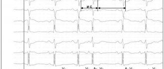
- Electrocardiogram (ECG). This is the first test to diagnose a heart attack. It is often done at the same time as a survey about symptoms. The test involves recording the electrical activity of the heart using electrodes attached to the skin. The pulses appear as "waves" displayed on a monitor or printed on paper. Because the heart muscle cannot conduct electrical impulses normally when damaged, an ECG can help determine whether you are having a heart attack or are in the process of having one.
- X-ray of the chest organs. A chest x-ray will help your doctor evaluate the size and shape of your heart and its blood vessels.
- Blood tests. Blood tests can determine if you have kidney or liver damage, detect signs of heart infection, and also determine if you are having a heart attack. Another type of blood test (arterial blood gas test) is also ordered to determine the amount of oxygen in the blood.
- Echocardiogram. This test uses sound waves to produce images of the heart. During echocardiography, sound waves are sent to the heart from a wand-shaped device (transducer) placed on the chest. Sound waves bounce off the heart and back through the chest and are processed electronically to produce a video image of the heart. An echocardiogram can help identify areas of damage to your heart and problems with the heart's pumping function.
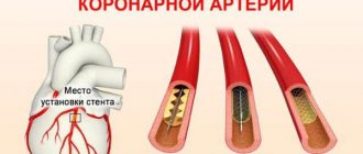
- Catheterization of coronary arteries (angiography, coronary angiography). This test will look for narrowing and blockage of the coronary arteries. Liquid contrast dye is injected into the arteries of the heart through a long, thin tube (catheter) that is passed through an artery in the leg or arm to the arteries of the heart. As the contrast agent fills the arteries, they become visible on X-rays, revealing areas of blockage.
Additionally, during catheterization, your doctor may remove the blockage in the artery through coronary angioplasty and stenting. Angioplasty uses tiny balloons that are inserted through a blood vessel into the coronary artery to widen the blocked area. After angioplasty, a mesh tube (stent) is placed inside the artery to maintain sufficient lumen and prevent re-narrowing in the future.
Clinical manifestations
Symptoms of cardiogenic shock indicate the manifestation of impaired blood circulation:
- the skin is pale, the face and lips have a grayish or bluish tint;
- cold, sticky sweat is released;
- hands and feet are cold to the touch;
- varying degrees of impairment of consciousness (from lethargy to coma).
When measuring blood pressure, low numbers are revealed (the upper one is below 90 mm Hg), the typical difference with the lower pressure is less than 20 mm Hg. Art. The pulse on the radial artery cannot be determined, on the carotid artery it is difficult.
When pressure drops and vasospasm occurs, oliguria (low urine output) occurs, leading to complete anuria.
The ambulance is required to deliver the patient to the hospital as quickly as possible after providing assistance.
Medications
Medicines to treat cardiogenic shock are prescribed to improve blood flow through the heart and improve the pumping function of your heart.
- Aspirin. Aspirin reduces blood clotting and maintains blood flow through the narrowed artery. Take aspirin on your own while waiting for emergency services to arrive, only if your doctor tells you to do so if you are experiencing symptoms of a heart attack.
- Thrombolytics. These drugs help dissolve a blood clot that is blocking blood flow to the heart. The sooner you are given a thrombolytic drug during a heart attack, the better your chance of survival and less damage to your heart. Thrombolytics are prescribed if emergency catheterization of the heart arteries and stenting cannot be performed.
- Antiplatelet agents. Doctors in the emergency room may give you other drugs that are similar to aspirin to prevent new blood clots from forming. These include drugs such as clopidogrel (Plavix) and other platelet glycoprotein IIb/IIIa receptor blockers.
- Other blood thinning drugs. You will likely be prescribed other medications. These include heparin, to reduce the likelihood of dangerous blood clots. Heparin is given intravenously or subcutaneously for the first few days after a heart attack.
- Inotropic drugs. Dopamine or epinephrine is prescribed to improve and maintain heart function.
Surgical interventions
Surgery to treat cardiogenic shock aims to restore blood flow through the heart. They are performed in specialized cardiac centers. Such interventions include:
- Angioplasty and stenting. Typically, when blood flow through the blocked artery is restored, the signs and symptoms of cardiogenic shock improve. Emergency angioplasty works to clear blocked arteries, allowing blood to flow freely to your heart. Doctors insert a long, thin tube (catheter) that is threaded through an artery in your leg or arm to the blocked artery in your heart. This catheter is equipped with a special balloon. When installing a stent in the area of narrowing, the balloon is inflated to restore the lumen of the artery. In addition, a metal mesh stent may be inserted into the artery to keep it open for a long time and restore blood flow to the heart. Doctors install stents that slowly release drugs into the blood to keep the artery wide enough.
- Balloon pump. Depending on your condition, doctors may place a balloon pump in your heart's main artery (aorta). The balloon pump inflates and deflates to mimic your heart and keep your blood flowing.
Mechanisms of pathology development
The pathogenesis of the appearance of hemodynamic disturbances differs depending on the form of shock. There are 4 varieties.
- Reflex shock is caused by the body's reaction to severe pain. In this case, there is a sharp increase in the synthesis of catecholamines (substances similar to adrenaline). They cause spasm of peripheral blood vessels and significantly increase resistance to heart function. Blood accumulates at the periphery, but does not nourish the heart itself. The energy reserves of the myocardium are quickly depleted, and acute weakness develops. This variant of the pathology can occur with a small area of infarction. It has good treatment results if pain is quickly relieved.
- Cardiogenic shock (true) - associated with damage to half or more of the muscle mass of the heart. If even part of a muscle is excluded from work, this reduces the strength and volume of blood ejection. With significant damage, the blood coming from the left ventricle is not enough to nourish the brain. It does not enter the coronary arteries, the supply of oxygen to the heart is disrupted, which further impairs the ability of myocardial contraction. The most severe variant of the pathology. Reacts poorly to therapy.
- Arrhythmic form - impaired hemodynamics is caused by fibrillation or rare contractions of the heart. Timely use of antiarrhythmic drugs, the use of defibrillation and electrical stimulation allows one to cope with such pathology.
- Areactive shock - most often occurs with repeated heart attacks. The name refers to the body's lack of response to therapy. In this form, hemodynamic disturbances are accompanied by irreversible tissue changes, accumulation of acid residues, and slagging of the body with waste substances. With this form, death occurs in 100% of cases.
Depending on the severity of shock, all described mechanisms take part in the pathogenesis. The result of the pathology is a sharp decrease in the contractility of the heart and severe oxygen deficiency of the internal organs and brain.
Operations
If drug treatment and the listed surgical interventions are ineffective, the doctor will recommend surgery:
- Coronary artery bypass grafting. Bypass surgery involves sewing veins or arteries to bypass the area of blockage or narrowing of the coronary artery. This restores blood flow to the heart. The doctor will suggest this surgery after the heart has recovered from a heart attack.
- Surgeries to repair heart damage. Sometimes the cause of cardiogenic shock is a rupture of one of the chambers of the heart or damage to the heart valve. To correct these problems, the doctor will suggest surgical treatment.
Prevention
The best way to prevent cardiogenic shock is to prevent heart attacks. Lifestyle changes used to treat heart disease can help prevent a heart attack. These lifestyle changes include:
- Control of high blood pressure (treatment of hypertension). One of the most important steps to reduce your risk of heart attack and cardiogenic shock is to control your blood pressure. Regular exercise, managing stress, maintaining a healthy body weight, and limiting salt and alcohol intake will help control high blood pressure. In addition to recommendations for lifestyle changes, your doctor may prescribe medications to treat hypertension, such as diuretics, angiotensin-converting enzyme (ACE) inhibitors, or angiotensin receptor blockers.
- To give up smoking. Quitting smoking reduces the risk of having a heart attack. Several years after quitting smoking, a smoker's risk of developing a stroke is the same as a non-smoker's.
- Maintaining normal body weight. Excess weight contributes to the risk factors for heart attack and cardiogenic shock: high blood pressure, cardiovascular disease and diabetes. Losing just 10 pounds of weight will lower your blood pressure and cholesterol levels.
- Reduce cholesterol and saturated fat in your diet. Eating less cholesterol and fat, especially saturated fat, will reduce your risk of heart disease.
- Regular exercise. Exercise reduces your risk of having a heart attack. Exercise lowers blood pressure, increases high-density lipoprotein (HDL) levels, and improves the overall health of your blood vessels and heart. They will help you lose weight, control diabetes and reduce stress. Get regular exercise such as walking, jogging, swimming or cycling for 30 minutes every day.
How to provide first aid to a patient
Help for cardiogenic shock from loved ones or passers-by may consist of calling an ambulance as soon as possible and providing a full description of the symptoms (pain, shortness of breath, state of consciousness). The dispatcher can send a specialized cardiology team.
Laying the patient down with his legs elevated is necessary to improve blood supply to the brain
As first aid, you should remove or untie your tie, unfasten your tight collar, belt, and give Nitroglycerin for heart pain.
Goals of first aid:
- elimination of pain syndrome;
- maintaining blood pressure with medications at least at the lower limit of normal.
To do this, the ambulance administers intravenously:
- painkillers from the group of nitrates or narcotic analgesics;
- drugs from the group of adrenergic agonists are used carefully to increase blood pressure;
- with sufficient pressure and swelling of the lung, fast-acting diuretics are necessary;
- oxygen is given from a cylinder or pillow.
The patient is urgently taken to the hospital.