- home
- Oncology
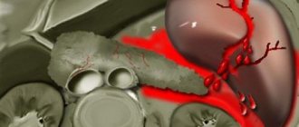
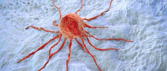
- Spleen tumor
Quite a lot of attention is paid to the morphology of focal lesions of the spleen. There are malignant and benign tumors of the spleen
.
The first include plasmacytoma and sarcomas, which, depending on the underlying tissue, can be of four forms: fibrosarcoma, lymphosarcoma, reticulosarcoma and angiosarcoma. Such lesions of the spleen are extremely rare. Among benign tumors,
lymphangiomas and hemangiomas are more common.
To determine the type of spleen tumor, its localization to the main structures of the organ and indications for surgery, as well as choosing the correct surgical treatment tactics, you must send me a complete description of the abdominal ultrasound, MSCT data of the spleen with contrast, and indicate your age and main complaints to your personal email address. Then I will be able to give a more accurate answer to your situation.
Symptoms, signs and clinical manifestations of a spleen tumor.
In patients with focal formations of the spleen, the most common complaints are the presence of a tumor-like formation in the upper floor of the abdominal cavity, a feeling of heaviness or fullness in the left hypochondrium and epigastrium, an enlarged abdomen and its asymmetry, decreased appetite, and weight loss.
Due to compression of neighboring organs, pain often appears, body weight decreases significantly, and weakness increases. Compression of the left renal artery sometimes leads to hypertension, dysuric disorders, edema of the lower extremities, symptoms of intoxication are often associated, and dyspeptic disorders may develop.
It is believed that for benign tumors and true cysts
clinical manifestations develop gradually, gradually. Patients cannot indicate the exact timing of the onset of signs and symptoms of the disease, which is due to the slow growth of benign formations.
Thus, the clinical picture of tumors and cysts of the spleen is extremely poor and nonspecific. Benign tumors and true cysts are particularly rare in their manifestations. More often, symptoms appear in formations with a complicated course. It becomes obvious that the leading place in the diagnosis of spleen tumors belongs to instrumental non-invasive research methods.
Complications
In the absence of medical care, splenic infarction, which usually ends with volvulus, leads to the development of a severe state of shock with a sharp drop in blood pressure, an increase in heart failure, depression of consciousness, and coagulopathic conditions. The most dangerous complication of splenic volvulus is organ rupture, accompanied by massive bleeding into the abdominal cavity with a high risk of death. Thrombotic occlusion of the venous vessels of the splenic pedicle can be complicated by thrombosis of the portal vein and its branches. Contamination of the linear parenchyma with bacterial flora results in the formation of an abscess or generalization of infection with the involvement of the peritoneum in the pathological process and the development of peritonitis. In the chronic version of the pathology, gradual obliteration of the splenic artery occurs, followed by tissue necrosis. Against the background of congestive venous splenomegaly, segmental portal hypertension can form.
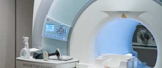
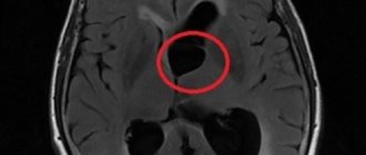
Diagnosis of spleen tumors
Currently, with the widespread introduction of non-invasive diagnostic methods, focal formations of the spleen are often detected accidentally during ultrasound or CT (NMRI) studies during a routine examination.
When a tumor-like formation is detected in the spleen, the specialists of instrumental research methods must set the following tasks for the surgeon:
- Clarification of the localization and size of the pathological focus in the spleen;
- Presumptive determination of the morphological nature of the formation (primarily, the likelihood of a malignant and parasitic nature);
- Determining the condition of adjacent organs (signs of compression, germination, primary lesion);
- Determination of the characteristics of the blood supply to the spleen (the level of division of the splenic artery into lobar branches, their number).
In recent years, spiral computed tomography (SCT) with intravenous bolus contrast enhancement using non-ionic contrast agents (Ultravist, Visipaque, Omnipaque) has become widespread. First, a non-contrast scan of the abdominal cavity is performed, and then a SCT study with BCU (intravenous administration of 100 ml of contrast agent at a rate of 3 ml/sec) with various scanning time delays (from 17 to 40-80 sec.).
The use of this technique makes it possible to clearly distinguish unchanged tissue zones that accumulate contrast agents well from areas of tissue decay and fluid accumulations. In addition, it is possible to obtain a more complete understanding of the angioarchitecture of the spleen itself and the adjacent great vessels, which largely contributes to a high differential diagnosis between cysts and tumor lesions. The diagnosis of SCT is usually confirmed morphologically in 95% of cases.
Thus, it is desirable to include ultrasound, duplex scanning, CT and MRI in a modern preoperative diagnostic program, and in this order, since each method, complementing the previous one, solves narrower, specific problems. Of course, none of them is absolute in identifying focal lesions of the spleen.
Noteworthy is the frequency of diagnostic errors when identifying spleen formations, which reaches 75-80% even when using modern research methods. Therefore, an integrated approach is needed here using all modern diagnostic methods.
Only a comprehensive examination in a specialized hospital makes it possible to determine the pathological focus, its topographic and anatomical characteristics and, ultimately, the optimal treatment tactics. Using this diagnostic scheme, in recent years we have been able not only to identify a lesion in the spleen, but also to accurately localize it in almost 94% of cases.
The final diagnosis is established only during surgery using histological examination.
Indications for surgical treatment of splenic tumor
Based on the data obtained during the examination, the surgeon needs to decide on the nature and scope of the upcoming intervention.
The choice of treatment method, especially determining the indications for surgery for tumors and cysts of the spleen, is the most difficult issue. As already noted, a significant part of splenic formations with an uncomplicated course do not manifest symptoms and are often discovered by chance. Very often a dilemma arises here: to observe or operate. What should play a decisive role in solving this difficult issue?
In our opinion, when solving this problem, the presumptive morphological nature of the lesion and signs of a complicated course come first. Next, the answers to the challenges facing diagnostic methods fall into the balance in favor of the operation. These include the size and localization of the pathological focus, assessment of the blood supply to the spleen, and anatomical relationships with adjacent organs. But these data are more conducive to the choice of the upcoming volume and nature of the operation.
The size of the formation cannot serve as a guide for the choice of treatment tactics, but only determines the indications for a specific type of operation, especially if one of the methods of preoperative examination suspected the presence of an abscess or parasitic cyst. In practice, there are often cases when, after identifying a mass in the spleen, long-term follow-up is carried out, while in almost a third of cases, malignant tumors at the preoperative stage were interpreted as benign, and only a morphological study of the removed specimen made it possible to finally establish the diagnosis. Therefore, focal formations in the spleen, regardless of their size, serve as an indication for surgical treatment
.
Methods of surgical treatment of tumors and cysts of the spleen
Watch a video of operations for a spleen tumor performed by Professor K.V. Puchkov. You can visit the website “Video of operations of the best surgeons in the world.”
All methods of surgical treatment can be reduced to the following:
- Organ removal – splenectomy (open or laparoscopic approach)
- Organ-preserving – resection of the spleen or splenectomy with autotransplantation of splenic tissue into the greater omentum (open or laparoscopic approach)
- Percutaneous punctures for cystic formations of the spleen.
Splenectomy
Indications for splenectomy (removal of the spleen) can be divided into two groups: surgical and hematological.
Surgical indications:
- Injuries to the spleen, open, closed (one- and two-stage ruptures)
- Spleen abscess
- Splenic cysts (non-parasitic, parasitic)
- Tumors of the spleen (benign - hemangiomas, lymphangiomas, endotheliomas, malignant - fibrosarcomas, lymphosarcomas, etc.)
- Portal hypertension with splenomegaly and hypersplenism
Hematological indications:
- ITP (Werlhof's disease)
- Aplastic anemia
- Microspherocytic anemia (Minkowski-Choffard disease)
- Acquired autoimmune hemolytic anemias
- Polycythemia (erythremia)
- Chronic leukemia
- Non-Hodgkin's lymphoma
- For the purpose of diagnosis and treatment of lymphogranulomatosis.
The essence of splenectomy is the ligation and intersection of the vessels leading to the spleen, and the removal of the organ itself. The key to the success of the operation is sufficient access to the vascular pedicle and control over it throughout the operation. These parameters are most adequately met by the laparoscopic approach.
Laparoscopic splenectomy is an alternative to open surgery and, with appropriate manual skills of the surgeon and sufficient material and technical support of the clinic, can significantly reduce the frequency of intra- and postoperative complications, reduce postoperative hospital stay and improve the quality of life of patients.
Spleen resection
In clinical practice, as a rule, atypical resection of the spleen is performed, in which the pathological formation is removed without taking into account the segmental structure of the organ. This operation should be performed only for benign diseases of the spleen. When performing atypical resections, it is possible to preserve a much larger volume of parenchyma than with anatomical resections. Organ-saving operations on the spleen should be carried out using modern devices for separating parenchyma (ultrasonic scissors, argon-enhanced plasma, dosed bipolar ligation, etc.), modern local hemostatic agents - PerClot (Italy) and highly qualified surgeons performing surgery.
Heterotopic autotransplantation of splenic tissue into the greater omentum during splenectomy.
Indications for surgical autotransplantation of splenic tissue:
- Impossibility of performing organ-preserving surgery for spleen injury.
- Forced splenectomy during operations on adjacent organs.
- Removal of the spleen due to benign formations.
Contraindications to autotransplantation of splenic tissue:
- A malignant process in the spleen or organ during which it was removed.
- Malignant blood disease.
- The presence of residual tissue foci (splenosis, accessory spleen) during splenectomy.
- Total damage to an organ by a pathological process.
- Age over 50 years.
- The critical condition of the patient and other reasons requiring a reduction in the scope of the operation.
Autotransplantation into the greater omentum is considered preferable due to the peculiarities of its blood supply and ease of manipulation; however, it is also possible to use the mesentery of the small intestine for this purpose. An experimental study demonstrated a similar course of engraftment of spleen fragments both in the greater omentum and in the mesentery of the small intestine. Scientific studies prove a significant degree of regeneration of transplanted spleen fragments, which is confirmed by histological methods.
It should be emphasized that autotransplantation of splenic tissue, which is often considered as an organ-preserving procedure, is a method of prosthetics of some organ functions, since it is necessarily preceded by splenectomy, and the structure of the graft differs in many respects from normal splenic tissue. As a rule, this tissue takes on up to 70% of the functions of the organ. In 20%, there is a lack of engraftment of the transplanted splenic tissue and its gradual resorption.
Percutaneous punctures for cystic formations of the spleen
Percutaneous therapeutic punctures and catheter drainage for fluid formations of the spleen, performed under local anesthesia, are especially justified in patients with severe concomitant diseases. In cases of suspected benign genesis of focal lesions, small sizes and “convenient” localization for puncture (subcapsular location), such manipulations are effective, low-traumatic organ-saving operations. As a rule, these interventions are carried out for small cysts up to 4-5 cm in diameter.
Causes
Twisting of the splenic pedicle usually occurs with dystopia of the organ in the small pelvis, iliac region, chest cavity (with diaphragmatic hernias), hernial sac of abdominal hernias. In extreme cases, the wandering spleen with an elongated neurovascular bundle moves freely throughout the abdominal cavity. According to experts in the field of gastroenterology, abdominal surgery and hematology, the following factors contribute to the occurrence of bloat:
- Congenital defect of the ligamentous apparatus . The risk of torsion is considered greatest when one or more ligaments holding the organ in the left upper abdominal cavity are missing. Congenital wandering spleen is a sporadic dysontogenetic developmental anomaly and can be combined with other defects.
- Weakening of the splenic ligaments . Frequently giving birth, women who generally have weakened muscle-ligamentous structures of the abdominal and pelvic cavities are most susceptible to volvulus. The pathological condition is often aggravated by a decrease in the muscle tone of the anterior abdominal wall, against the background of which splanchnoptosis is formed.
- Enlarged spleen. With splenomegaly, especially enlargement of the upper pole, an imbalance in the mass of the lienal parenchyma occurs, causing torsion of the organ. The likelihood of splenic volvulus increases with leukemia, lymphogranulomatosis, splenic tuberculosis, and other infectious, oncohematological, and autoimmune diseases.
Displacement of the spleen can also be detected when ligaments are torn during abdominal injuries or accidentally dissected during abdominal operations. In some patients, the additional lobe of the spleen, which has its own neurovascular pedicle, is subject to volvulus. In this case, the risk of torsion increases in proportion to the distance to which the additional lobule is displaced from its normal anatomical location.