Consultation with a dermatologist-venereologist – 1,750 rubles.
- Types of phlebitis
- Symptoms
- Diagnostics
- Treatment
Phlebitis is an inflammation of the vascular wall, namely the venous wall, with its gradual destruction. It occurs in both acute and chronic forms. The main danger of this disease is the development of a high risk of blood clots with subsequent blockage of blood vessels.
Provoking factors for the development of phlebitis most often are infections suffered by a person, acute inflammatory diseases, or the entry into the bloodstream of substances that cause irritation of the vessel wall.
What types of phlebitis are there?
There are several categories in the classification of phlebitis:
Depending on the location of the vein lesion, phlebitis is distinguished between superficial and deep . These two types are radically different in their symptoms and are easily differentiated.
In addition, phlebitis is divided depending on which vein wall is affected:
Endophlebitis is characterized by inflammatory changes in the inner wall of the vessel. The provoking factor is internal damage to the vein, such as during prolonged catheterization;
Periphlebitis - the lesion is predominantly localized on the outer wall of the vein. Occurs during the transition of the inflammatory process with a number of located organ tissues;
Panphlebitis is a common inflammation of all membranes.
How and why leg veins hurt
The causes of pain in the veins in the legs can be different:
- Varicose veins of the lower extremities;
- Thrombophlebitis;
- Venous thrombosis.
The veins on the top of the feet hurt in people who wear uncomfortable shoes. A vein in the leg hurts like a bruise if the integrity of its wall is damaged. In patients whose lower limbs are exposed to inadequate physical activity, the veins in the legs under the knee hurt.
How do veins hurt in your legs? Symptoms of chronic venous disease depend on the type of disease and the stage of the pathological process. In the initial stage of varicose veins, patients complain of a dull pain in the legs that appears by the end of the day. In the presence of acute deep vein thrombosis, acute pain appears in the lower leg, which intensifies with dorsiflexion of the foot or compression of the lower leg in the anteroposterior direction. In patients suffering from thrombophlebitis of the superficial veins of the lower extremities, pain appears along the inflamed, tense, dense vein. In the presence of a trophic ulcer, pain in the area of the skin defect is a constant concern.
Symptoms of phlebitis
As mentioned earlier, superficial and deep phlebitis have significant differences. Thus, with superficial phlebitis, local symptoms of inflammation come to the fore. At the site of the lesion, you can feel an area of tension, pain and compaction. The skin in this area becomes hyperemic, with a local increase in temperature. Sometimes this process is accompanied by a general deterioration in well-being, the development of weakness, and malaise.
In the case of deep phlebitis, the primary symptom is a significant increase in body temperature. Against this background, pain occurs in the affected area and pronounced swelling. The skin over this area does not turn red, but rather acquires a marble-white hue.
In severe inflammation, cerebral symptoms may occur in the form of headache, dizziness, and vomiting. An increase in blood pressure occurs. Without timely treatment, this disease can lead to liver or kidney failure.
Compression jersey medi
Thanks to the breathable and elastic material, compression jersey provides high wearing comfort. Modern medical compression hosiery is visually indistinguishable from model hosiery, but provides high medical effectiveness when used.
Here you can find more information about medi compression stockings.
The human body
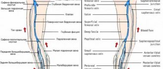
How do veins work?
Vienna
Product Tips
Ideal compression product
Compression hosiery
How to treat phlebitis?
First of all, the patient is provided with bed rest. The affected limb is fixed in an elevated position. Anticoagulants and anti-inflammatory drugs are prescribed. After relief of inflammation - physiotherapy.
Surgical treatment is carried out in case of complications, for example. thrombosis
To prevent the development of phlebitis, it is important to promptly treat infectious and inflammatory diseases, as well as follow the rules for performing intravenous injections.
Methods for the prevention and treatment of varicose veins
Many people underestimate the seriousness of the problem of varicose veins, so they consult a doctor already in the last stages of the development of the disease. If treatment is not provided on time, it can cause the development of complications, which are much more difficult to deal with.
First, you need to see a doctor to make an accurate diagnosis. Next, the specialist prescribes specific treatment based on the individual characteristics of the body and the symptoms of the disease. The most common treatment methods are:
- Using compression garments – tights, stockings or socks;
- Drug treatment with special drugs that have a vasoconstrictor, venotonic and angioprotective effect on venous vessels;
- Surgical intervention. Most often these are minimally invasive treatment methods: phlebectomy, sclerotherapy, EVLT, RFO.
Prevention of varicose veins includes regular physical activity (aerobics, walking, swimming, yoga), nutrition control, giving up bad habits and a comfortable, comfortable wardrobe.
Causes and risk factors
Subclavian vein thrombosis is associated with compression of the thoracic outlet. The subclavian vein originates from the first rib from the axillary vein, and at the level of the sternoclavicular junction with the jugular vein it forms the brachiocephalic vein. Compression of the vein walls in the area between the collarbone and the first rib leads to slower blood flow and thrombosis. It can be considered the venous equivalent of thoracic outlet syndrome.
One of the causes of thrombosis is muscle hypertrophy (increase in the volume or mass of skeletal muscles). As a result, the subclavian vein can become compressed between the ribs (in front of it), the muscle (behind it), and the collarbone (above it).
Another reason is that a person has a congenital small anatomical space between the collarbone and the first rib. In this case, compression of the subclavian vein is possible even without major muscle hypertrophy.
Other causes of thrombosis include:
- incorrect posture,
- bone pathology (in the subclavian region),
- collarbone fractures
- use of subclavian catheters
- incorrect sleeping position
- thoracic outlet syndrome
- Risk factors include excessive physical activity. People who undergo intense physical activity and athletes (wrestlers, weightlifters or bodybuilders) have a higher risk of developing thrombosis due to repeated damage to the subclavian vein from frequent mechanical compression of the vessels between the collarbone, first rib and joint.
The average age of patients with Paget-Schroetter syndrome is 30-40 years, and the male to female ratio is approximately 2:1. It is more common on the right side, probably due to the frequency of right hand dominance, and 60% to 80% of patients have this condition. who performed vigorous exercise involving the upper limbs.
Course of the disease
The course of the disease is divided into two stages: acute (lasts about three weeks; at the initial stage, symptoms appear during physical activity) and chronic (symptoms last for more than two months).
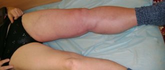
The acute phase is characterized by increasing swelling of the arm, pain, and a feeling of fullness. The patient's ability to work decreases. Gradually, tense saphenous veins develop, which take on the function of outflow of venous blood and contribute to the subsidence of the process.
The chronic phase is the consequences of thrombosis. With inadequate treatment, occlusion of the subclavian vein persists and chronic Paget-Schroetter syndrome develops. It is characterized by the development of a powerful collateral network of saphenous veins around the shoulder joint. Patients are concerned about the increase in volume of the sore arm, sometimes there is pain and increased fatigue.
Superficial periphlebitis of the lower extremities: diagnosis
The first step on the path to healthy legs and improvement of general condition will be a consultation with specialists such as a therapist, vascular surgeon, angiosurgeon or phlebologist. The specialist will collect an anamnesis of the disease, clarify the patient’s complaints and the time of onset of the first symptoms, examine the limb, palpate it and inquire about the presence of concomitant diseases. This will give the doctor the opportunity to make a preliminary diagnosis and prescribe a series of diagnostic tests. What might they include?
First of all, these are general clinical tests. A general blood and urine test is a mandatory diagnostic procedure that allows you to assess the condition of the patient’s body as a whole. This will be followed by instrumental studies. This may be Doppler ultrasound or duplex angioscanning. As a rule, the latest diagnostic method has been used recently. It is the most informative, since the scan determines the speed of blood flow, the presence and size of blood clots, as well as their location. The procedure is painless and safe, however, it has contraindications. In particular, ultrasound cannot be performed in case of violations of the integrity of the skin.
Expert opinion
If a blood test reveals a pathologically low content of total protein, an increase in ESR, a decreased level of hemoglobin and a decrease in the number of red blood cells, then this is an indirect sign of anemia, intoxication of the body, the course of an infectious-inflammatory process of an acute or chronic nature.
Vascular surgeon, phlebologist
Osipova Ekaterina Yakovlevna
The result of a urine test will also give the specialist an understanding of the state of the body. If in a patient it is characterized by protein loss, an increased number of leukocytes and erythrocytes, then with a fairly high degree of probability we can talk about periphlebitis. After reviewing the conclusion of instrumental vein diagnostics, the doctor will make a final diagnosis and select an individual treatment regimen that takes into account all the patient’s characteristics.
Treatment of periphlebitis: basic methods
Periphlebitis is an inflammation of the periphlebitis surrounding the vein.
Etiology and pathogenesis
Periphlebitis occurs as a result of the transition of the inflammatory process to a vein from the surrounding tissues with an abscess, burn wound, panaritium, phlegmon, tuberculosis, etc. With non-specific In the bedroom, the outer wall of the vein is swollen and infiltrated. Then swelling and infiltration spread to the middle part of the vein wall, destroying the muscle layer, and to the endothelium. A wall thrombus forms and thrombophlebitis develops. If the process ends successfully, then phlebosclerosis is formed.
Causes of periphlebitis
The most common reason why various types of phlebitis occur, including periphlebitis - varicose veins. It is also possible to develop periphlebitis as a result of an allergic reaction (allergic phlebitis) or as a complication after childbirth (painful phlebitis). In some cases, the disease may occur against the background of an infectious lesion or injury to the vein (violation of the integrity of the vascular wall).
Symptoms of periphlebitis
The disease is characterized by pain in the lower extremities; upon examination, redness is detected along the affected vein; Some compaction is detected by touch.
The main clinical symptoms - pain and hyperemia of the skin along the vein - are observed only in the initial stage of the disease and when the saphenous veins are affected. Upon palpation along the course of the affected vein, a dense, painful cord or infiltrate is detected, indicating the involvement of significant areas of subcutaneous tissue in the inflammatory process.
Treatment of periphlebitis
Treatment of periphlebitis is mainly conservative. Ointments and gels with heparin, troxevasin, as well as thermal physiotherapeutic procedures are prescribed. Anti-inflammatory therapy with NSAIDs is carried out, drugs are used that improve the rheological properties of blood and the trophism of the venous wall.
In physiotherapy, magnetotherapy is indicated first. The action of the magnetic field accelerates tissue regeneration, has anti-inflammatory, decongestant and immunomodulatory effects, reduces blood clotting, reducing the risk of parietal prevents thrombus formation, improves blood and lymph circulation, and causes an antispastic effect.
A new drug, the medicinal patch VAZOPLAST, has shown good effectiveness in the treatment of periphlebitis.
Treatment of periphlebitis with medicinal plaster VAZOPLAST (vasoplast)
The therapeutic plaster VAZOPLAST is intended for the treatment of diseases of the venous system.
The patch has analgesic, anti-inflammatory and muscle-relaxing effects, reduces swelling in the affected area, relieves fatigue and a feeling of heaviness in the legs, improves blood and lymph circulation This, especially venous and lymphatic outflow, improves the rheological properties of blood in the inflammation zone.
The VAZOPLAST patch helps improve vascular permeability, reduce thrombus formation and venous stagnation in the affected area, improves the condition of capillaries, increases elasticity veins and tone, has an angioprotective and venotonic effect.
More details about VAZOPLAST