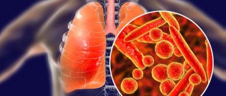
Exogenous allergic alveolitis is a diffuse inflammation of the lung tissue, leading to the development of pneumosclerosis, respiratory failure and cor pulmonale.
- Types of allergic alveolitis
- Pathogenesis
- Symptoms
- Diagnostics Differential diagnostics
- Laboratory diagnostics
Exogenous allergic alveolitis, or hypersensitive interstitial pneumonitis, develops with repeated aerogenic entry into the sensitized body of antigens (allergens) of microbial, animal, plant origin or various low-molecular chemical compounds.
First, sensitization of the body occurs in the form of Tn2 activation and the appearance of IgG antibodies to the causally significant antigen. The reaction can be caused by substances that can enter the alveoli. When a causally significant antigen enters a sensitized organism, the reaction occurs quickly (within 4-8 hours) similar to the Arthus phenomenon with the formation of immune complexes. The processes of deposition of immune complexes and the formation of chemoattractants, anaphylatoxins (C5a, C3) as a result of complement activation lead to acute damage to blood vessels and alveoli with the involvement of neutrophils and macrophages.
On the 2nd day after contact with the antigen, DTH develops with the involvement of CD4+ (Tn1), activated macros and CD8+ cytotoxic T lymphocytes. Subsequently, epithelioid-macrophage granulomas are formed, fibroblasts are activated, and interstitial fibrosis develops.
Some types of exogenous allergic alveolitis and sources of causally significant allergens
| Type of exogenous allergic alveolitis | Source of allergens and allergens |
| Alveolitis caused by medications | Medicines: penicillins and other antibiotics, sulfonamides, nitrofurans, gold salts |
| Alveolitis caused by low molecular weight compounds | Dithioisocyanates, heavy metal salts |
| Barn disease | Wheat, flour (allergens of grain weevil, fungi) |
| Coffee miller's disease | Coffee beans |
| Disease of those washing in the sauna | Damp wood (fungal allergens) |
| Lung disease associated with the use of humidifiers and air conditioners | Water and air containing allergens of fungi and bacteria, including allergens of actinomycetes |
| Cheesemakers' disease | Moldy cheese (penicillium fungi allergens) |
| Weavers cough | Cotton (mold allergens) |
| Tanner's lung | Maple bark (mold allergens) |
| Bird lover's lung | Feathers and droppings of pigeons, budgies (bird and fungal allergens) |
| Lung of a worker processing malt | Rotted barley, malt dust (allergens of Aspergillus fungi - A. fumigatusl A. clavatus) |
| Farmer's lung | Rotted hay (allergens of thermophilic actinomycetes and fungi) |
Pathogenesis
In the pathogenesis of the diseases under consideration, the immune complex mechanism of tissue damage is of primary importance. An allergen that enters the body leads to sensitization (addiction). In this process, antibodies to it are formed. Most often precipitating, which mainly relate to immunoglobulin G.
Antibodies form immune complexes with the allergen. Deposits of the latter can be observed using special methods on the walls of the alveoli and the smallest bronchioles, which is why an inflammatory process cannot but occur. In the pathological process described, increased permeability of the vascular wall is important, the cause of which is the inclusion of IgE-mediated allergic reactions. Vasoactive amines are released from basophilic granulocytes or mast cells in the body. Platelet-activating factor is also important, which is involved in the release of serotonin and histamine from platelets, which are vasoactive amines.
The vessels become more permeable (particles can pass through more easily) due to vasoactive amines and platelet-activating factor. The chemotaxis of neutrophilic and eosinophilic granulocytes increases, which causes and intensifies inflammatory processes.
Also in the pathogenesis there may be a delayed allergic reaction, in which so-called granulomas form in the lungs. Whether it appears or not depends on the allergen that triggered the pathogenesis. Often the cause is fungal spores or particles. The presence of immune insoluble complexes also leads to the formation of granulomas.
The development of granuloma is conventionally divided into stages that end with the formation of a scar, which indicates a diagnosis of pulmonary fibrosis. In some cases of the disease, allergens are fixed on lung tissue cells, which changes their antigenic properties. For this reason, a cytotoxic mechanism of tissue damage is triggered.
In the pathogenesis of exogenous allergic alveolitis, pseudoallergic mechanisms may be important. The classical or alternative pathway characterizes the activation of complement by allergens that originate from certain fungi. Also, activation may be due to the ingestion of extracts into the body, including from dust, the source of which is moldy hay.
4.Treatment
The first step should always be to completely avoid contact with the allergen. In many cases, this is enough (without any treatment) to normalize the condition and functioning of the respiratory system. In more complex cases, when stopping contact with the allergen is impossible or problematic, for example, because it requires a certain amount of time, a change of profession and/or place of residence, or when the symptoms become severe or threatening, drug therapy is prescribed.
Standard antihistamines and desensitizing agents are ineffective in this case; the method of choice is long-term, for a month or more, administration of glucocorticosteroid hormones in gradually decreasing (until complete withdrawal) dosages.
Sign up for a consultation
Symptoms
Phases of the disease:
- acute
- subacute
- chronic
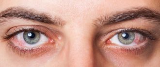
The acute course of the disease develops 4-12 hours after inhalation of the allergen. The patient develops a cough, clear sputum, wheezing, shortness of breath, low-grade fever, pain in muscles and joints.
The chronic phase of the disease is accompanied by progressive shortness of breath during exercise, pneumosclerosis, and increased respiratory and heart failure.
Prevention
Prevention of EAA is based on improving technological processes (sealing, mechanization of technologies, moving control panels outside the work premises, air humidification, etc.); upon entry to work - high-quality preliminary preventive medical examinations, in accordance with the order of the Ministry of Health and the MP of the Russian Federation “On approval of lists of harmful and (or) hazardous production factors and work, during the performance of which preliminary and periodic medical examinations (examinations) are carried out, and the Procedure for conducting preliminary and periodic medical examinations (examinations) of workers engaged in heavy work and work with harmful and (or) dangerous working conditions” No. 302 n dated 04/12/2011, periodic allergological examination of workers.
Additional medical contraindications to employment in contact with industrial substances of toxic-allergic action are total degenerative and allergic diseases of the upper respiratory tract; chronic diseases of the bronchopulmonary system; allergic diseases when working with allergenic aerosols; congenital anomalies (malformations) of the respiratory and heart organs.
The rule when determining the ability to work of patients with EAA is the following: the presence of the disease is an absolute contraindication to continuing to work in contact with the causative substance. The patient is recognized as permanently partially incapacitated, permanently disabled in his profession, in need of constant rational employment, in need of medical and social rehabilitation.
Diagnostics
To diagnose this disease (more precisely, a number of diseases of exogenous origin), a detailed history taking is important. It is necessary to find out in what conditions the patient lives and works, in what environment he spent a long time, etc. The doctor also collects the patient’s complaints and detects characteristic symptoms. All patients are sent for radiography.
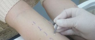
Confirmation of the diagnosis of exogenous allergic alveolitis is carried out using serological methods, as well as during allergic diagnostic tests. Using these methods, chronic or subacute forms of the disease are detected (especially important in cases where the doctor did not detect a possible allergen when collecting anamnesis).
Differential diagnosis
When making a diagnosis, it is necessary to take into account that a number of other pathologies may have similar symptoms and development:
- sarcoidosis
- infectious pneumonia
- idiopathic fibrosing alveolitis
- disseminated pulmonary tuberculosis
Radiography allows one to differentiate EAA from infectious pneumonia. In the latter cases, lobar, segmental or lobular darkening is detected. In sarcoidosis, the pathological process affects other organs in addition to the lungs. There is an increased amount of calcium in the urine, and the bronchopulmonary lymph nodes are larger than normal in size.
If a person has disseminated pulmonary tuberculosis, most likely he has previously had tuberculosis, or was for some time near a person with this disease. Tuberculin tests are carried out for differential diagnosis, they show a positive result. You can also take sputum for analysis to detect the pathogen in it.
Idiopathic fibrosing alveolitis differs from the disease in question by pneumofibrosis, which develops rapidly, and respiratory failure, which gradually worsens.
Idiopathic fibrosing alveolitis is characterized by rapid progression of pneumofibrosis with the development of respiratory failure.
Laboratory diagnostics
Bronchoalveolar lavage is obtained from patients using bronchoalveolar lavage during fiberoptic bronchoscopy. Immune complexes and circulating antibodies to the causally significant antigen are detected in the lavage fluid. The titer of IgO-, IgA-, IgE-antibodies is increased during the disease. A cytogram of lavage fluid is studied with a quantitative account of alveolar macrophages, neutrophils, eosinophils and basophils. Polycytosis in the acute phase exceeds the norm by 5-7 times.
Histological examination of lung biopsies reveals the exudative phase of inflammation in the form of abundant macrophage-lymphocytic infiltration, vasculitis and fibrin loss into the alveoli, in which lymphocytes accumulate. The formation of mixed granulomas is possible.
Etiology
Etiological factors causing the development of professional EAA can be divided into several groups [4–8]:
• microorganisms (bacteria, thermophilic actinomycetes, fungi, protozoa) and their metabolic products (endotoxins, proteins, glyco- and lipoproteins, polysaccharides, enzymes);
• biologically active substances of animal (whey proteins, animal hair, etc.) and plant origin (tree sawdust, moldy straw, coffee bean extracts);
• low molecular weight compounds (heavy metals and their salts, toluene diisocyanate, trimelytic anhydride, etc.), as well as many drugs (antibiotics, intal, nitrofurans, antimetabolites, antimitotic drugs, enzymes, hormones, etc.).
The disease called “farmer's lung” was first described by J. Campbell in 1932, who studied five farmers who developed acute respiratory symptoms after working with damp, moldy hay [13]. The leading causative agents leading to the development of “farmer's lung” are thermophilic actinomycetes - bacteria less than 1 micron in size, which have the morphological properties of fungi; they are widely found in soil, compost, and water (Table 1).
The most common species of thermophilic actinomycetes associated with EAA are Micropolysporafaeni, Thermoactinomyces vulgaris, Thermoactinomyces viridis, Thermoactinomycessaccharis, Thermoactinomycescandidum. These microorganisms reproduce at a temperature of 50–60 °C, i.e., in the conditions that are achieved during debate and decay of organic material [2, 8, 10, 11].
Cases of EAA have been described in grain growers, cane growers, cotton growers and cotton processors, tobacco growers, flour millers, cheese makers, furriers, shoemakers, rice grinders, hemp producers, workers in vegetable stores, granaries, coffee plantations, persons making malt, drugs, in the etiology of which an important role is played true fungi, thermophilic and other bacteria, bacterial products (endotoxins, glycoproteins), animal proteins, fish proteins, algae, plant dust.
It is possible to develop EAA in workers who come into contact with plastics, polyurethane, resins, and dyes. The most important are diisocyanates and phthalic anhydrite. There are known cases of EAA when exposed to salts of heavy metals (chromium, cobalt, gold, arsenic, copper, zinc), insecticides, especially when using copper sulfate for spraying fruit trees, vineyards, tomatoes, diisocyanate compounds (toluene diisocyanate, hexamethylene diisocyanate, diphenylmethane diisocyanate) , widely used in the automotive, rubber, paint and varnish industries, and in the production of polyurethanes (Table 2).
EAA has been described in workers producing drugs, as well as in patients when they use, for example, drugs from the pituitary gland of cattle (pituitrin, adiurecrin), used in the form of inhalation in the treatment of diabetes insipidus. Severe cases of EAA have been described in workers producing pepsin, trypsin, penicillin, streptomycin, amiodarone, and antimitotic drugs: methotrexate, azathioprine, 6-mercaptopurine [3, 4, 8].