Pneumonia is an infectious disease characterized by inflammation of the lungs. In severe cases of the disease, damage to other organs and systems occurs. Streptococcal pneumonia is diagnosed in 30% of patients. For the treatment of patients suffering from pneumonia, the Yusupov Hospital has created all the necessary conditions.
The therapy clinic rooms are equipped with ventilation and air conditioning, which allows you to create a comfortable temperature regime. To diagnose lung diseases, doctors use the latest, constantly updated equipment from leading companies in the world. Pulmonologists, adhering to European recommendations for the treatment of streptococcal infections, take an individual approach to the treatment of pneumonia in each patient. Doctors prescribe the most effective antibacterial drugs that act on streptococcus pneumoniae.
Patients with severe streptococcal pneumonia are hospitalized in the intensive care unit. Doctors monitor their condition around the clock using modern cardiac monitors and determine blood oxygen saturation. All patients receive oxygen therapy. If there are indications, artificial ventilation of the lungs is performed using stationary and portable expert-class ventilators.
Features of streptococcal pneumonia
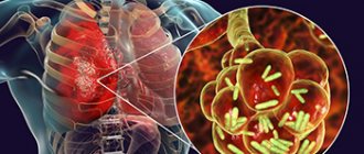
The most common causative agents of pneumonia are pneumococci (Streptococcus pneumoniae). Pneumonia can be caused by beta-hemolytic streptococci and peptostreptococci. The pathogen is spread by airborne droplets from patients and carriers. The disease often occurs during influenza epidemics in patients with chronic lung diseases. Streptococcus pneumoniae causes pneumonia in young children and people with weakened immune systems. Streptococci contribute to the development of purulent complications.
Streptococcal pneumonia is widespread due to the fact that pathogens are present in the normal human microflora. They are activated and become capable of causing an inflammatory process when the general and local resistance of the body decreases. This happens under the influence of the following factors:
- inadequate use of antibiotics;
- congenital and acquired immunodeficiency conditions;
- chronic respiratory diseases;
- in hazardous production;
- unfavorable living conditions.
Streptococcus pneumoniae enters the lungs in the following ways:
- bronchopulmonary;
- hematogenous;
- lymphogenous.
When the causative agent of streptococcal pneumonia enters the lung tissue, it causes inflammation, accompanied by the effusion of fluid into the alveoli.
Purpose of the study. Molecular genetic characteristics based on serogenotyping of pneumococcal isolates circulating among children with acute otitis media and sinusitis in Minsk.
Material and methods. The study included 66 clinical strains of pneumococcus isolated from children with acute otitis media, and 14 strains from children with sinusitis. The study used classical methods of bacteriology, as well as molecular genetic methods (real-time PCR, multiplex PCR) to confirm the species identity of the strains and carry out genotyping to determine the serotype of the strains.
Results. The serotype composition of pneumococcal strains causing otitis and sinusitis in children in Minsk was determined, and age-related characteristics were characterized. A comparative analysis of the serotype landscape of circulating strains with neighboring regions and globally distributed serotypes was carried out. Indicators of coincidence between circulating serotypes and those included in modern pneumococcal conjugate vaccines have been established.
Conclusion. The serotype landscape of pneumococcal strains isolated from children with otitis is characterized by high homogeneity and predominant distribution of vaccine serotypes (14th, 19F, 19A, 9V/9A, 6A/6B, 3rd, 23F, 18C/18F/18B/18A, 7F/7A), which provides high levels of compliance (in 76-97% of cases) with the spectrum of serotypes of conjugate vaccines. Regional geographic features include a wider distribution of serotypes 14 and 19A and a more limited distribution of serotype 3 than in neighboring regions and most countries globally. The serotype composition of pneumococcal strains isolated from children with sinusitis is characterized by high diversity with the circulation of both vaccine (6A/6B, 7F/7A, 14th, 19A, 19F and 23F) and non-vaccine (9N/9L, 1SA/1SF, 22F/22A, 31 and 33F/33A/37) serotypes, which determines significantly lower levels of compliance (in 36-50%) cases with the spectrum of serotypes of conjugate vaccines.
Streptococcus pneumoniae (pneumococcus) is a gram-positive bacterium that was discovered independently in 1881 by G. Sternberg in the USA and L. Pasteur in France when rabbits were infected with human saliva containing paired cocci lanceolate-shaped [1]. The main factor in the pathogenicity of pneumococcus is the polysaccharide capsule, which determines resistance to phagocytosis and host-specific immunity factors, provides the ability for long-term colonization in the nasopharynx and determines the invasiveness of pneumococcus. The antigenic diversity and unique chemical structure of the capsule determine the presence of at least 93 pneumococcal serotypes belonging to 48 serogroups [2]. Different serotypes of pneumococcus determine different specific clinical manifestations, levels of virulence, lethality, and the ability to induce an inflammatory response. The prevalence of individual serotypes varies widely depending on the form of pneumococcal disease (PI), age group and geographic region, although 20 major variants are most common [3, 4].
Pneumococcus is currently one of the main causative agents of bacterial infections (meningitis, community-acquired pneumonia, bacteremia, acute otitis media (AOM), sinusitis), especially in children. According to WHO, remaining one of the main causes of child mortality, pneumococcus, due to the high level of mortality of the infections it causes, annually claims up to 1 million children’s lives worldwide [5]. High incidence rates of various forms of PI, combined with a progressive increase in pneumococcal resistance to antibacterial drugs used for therapy, as well as the emergence and spread of multi-resistant strains [6, 7], make it urgent to study the molecular genetic structure of pneumococcal populations.
AOM is one of the most common infectious diseases in children worldwide [8], a leading reason for seeking medical help and prescribing antibiotics [9, 10]. On average, by the age of 3 years, 80% of children have already experienced at least one episode of AOM [11]. The disease often leads to purulent complications (mastoiditis, meningitis), and sometimes to hearing loss [9, 12]. S. pneumoniae is the main causative agent of AOM, found in 30-60% of cases [13]. The high social significance of PI in the past predetermined the need to develop preventive measures. In 2000, the first modern pneumococcal conjugate vaccine (PCV) became available in the United States, in which capsular polysaccharides from seven serotypes (4, 6B, 9V, 14, 18C, 19F, and 23F) were conjugated to a carrier protein that allowed the vaccine to create protective immunity from 2 months of age (unlike existing polysaccharide vaccines) [3]. WHO, due to the availability of reliable data on the high effectiveness of vaccination, recommended PCV7 for inclusion in national immunization calendars [5], and by 2009, the vaccine was included in the national immunization schemes of 35 countries around the world. However, the experience of introducing mass immunization with PCV demonstrates the substitution effect, which consists in changing the serotype composition of pneumococci: against the background of a general decrease in the incidence rate, there is an increasing role of non-vaccine serotypes, which is most clearly noted for 19A and 3rd serotypes [14, 15]. Later, new conjugate vaccines containing additional antigens (PCV10 and PCV13) appeared. Since PCV is included in the national calendars of preventive vaccinations of most neighboring countries (including Russia since 2014) and there are prerequisites for the start of mass immunization in Belarus, studying the prevalence of serotypes, antibiotic resistance of pneumococcus and changes in the serotype composition occurring under the pressure of vaccination is currently an important task.
The purpose of this study is molecular genetic characterization based on serogenotyping of pneumococcal isolates circulating among children of different ages with otitis and sinusitis.
Key words:
Streptococcus pneumoniae, acute otitis media, pneumococcal infection, pneumococcal conjugate vaccines, pneumococcal serotypes, sinusitis
Author(s):
Davydov A. V., Titov L. P., Klyuiko N. L., Gurinovich V. V.
Medical institution : Belarusian State Medical University, Republican Scientific and Practical Center for Epidemiology and Microbiology, Minsk City Children's Infectious Diseases Clinical Hospital
Symptoms of streptococcal pneumonia
Streptococcal pneumonia begins suddenly, with an increase in body temperature to 39°C. Signs of intoxication are rapidly increasing. Against the background of fever, cough, shortness of breath, and chest pain appear. Chills rarely occur. The cough is initially unproductive, but soon becomes wet, with the release of mucopurulent sputum. Intoxication and respiratory symptoms may be accompanied by the appearance of a scarlet-like rash.
During a physical examination, therapists determine the lag of one half of the chest in the act of breathing, shortening of the percussion sound over the focus of infiltration, wheezing and crepitus. In severe cases of streptococcal pneumonia, patients experience respiratory or cardiovascular failure, rapid heartbeat, cardiac arrhythmia, and asthma attacks. If such symptoms appear, the patient is urgently hospitalized in the intensive care unit.
On days 2-3 of the disease, pleurisy develops in 50% of adults and 60% of children and pleural empyema develops. Less common is purulent pericarditis and the formation of pulmonary abscesses in the area of the pneumonic focus. Sometimes the disease is complicated by purulent arthritis, osteomyelitis and glomerulonephritis.
Streptococcus pneumonia of newborns develops during the first 5-7 days of a child’s life. It is most often a manifestation of intrauterine sepsis caused by streptococcal infection. Streptococcal pneumonia in newborns occurs with severe respiratory disorders.
Pneumonia
- Content:
- Symptoms of the disease
- Causes of the disease
- Diagnostics
- Complications
- Treatment of the disease
- Risk group
- Prevention
- Diet and lifestyle
Pneumonia caused by Streptococcus pneumoniae
Find doctors
Pneumonia caused by Streptococcus pneumoniae is an acute inflammation of lung tissue, which is caused by an infectious agent - Streptococcus pneumoniae or pneumococcus. It can occur as a separate disease or as a complication of another pathology.
Symptoms of the disease
Symptoms of pneumonia caused by Streptococcus pneumoniae are practically no different from pneumonia of other etiologies. A special feature is the scale of pathological changes and the severity of the course. The pathogen often causes lobar pneumonia, rather than focal pneumonia (the entire lobe of the lung is affected, and not just part of it). The following symptoms are most often observed:
- Acute onset of illness
- Increase in body temperature to 40 or more
- Shortness of breath that tends to progress
- Cough with purulent sputum
- Chest pain
- Pale or bluish skin
- Heartbeat
- Feeling of wheezing in the chest
- Participation of additional respiratory muscles in breathing
- General weakness, lack of appetite, muscle pain.
Causes of the disease
This pneumonia is caused by the bacterium Streptococcus pneumoniae (pneumococcus). Belongs to the group of alpha-hemolytic streptococci, which are part of the normal microflora of the nasopharynx. A person who is a carrier of pneumococcus does not feel any pathological symptoms. The infection is transmitted by airborne droplets. Streptococcus pneumoniae is the leading causative agent of respiratory infections - tracheitis, bronchitis and bacterial pneumonia. Normally, protective mechanisms prevent pneumococcus from entering the respiratory tract (the work of the tonsils, the filtration of air flow in the nasal cavity, local immunity, the activity of the ciliated epithelium of the respiratory tract, sputum separation). If any protective factor is violated, pneumococcus can enter the alveoli of the lungs and cause inflammation. Predisposing factors:
- Tobacco smoking
- Chronical bronchitis
- Congestive heart failure
- Chronic lung pathology
- Endocrine system diseases
- Primary and secondary immunodeficiencies
- Alcoholism
- Prolonged immobilization
- Cystic fibrosis.
Diagnostics
Diagnosing pneumonia is not difficult. The disease can be suspected based on the patient’s complaints and the data of an objective examination of the patient (dull percussion sound over the lungs, moist rales when listening). Mandatory diagnostic methods are:
- An X-ray examination of the chest organs is mandatory if pneumonia is suspected; without X-ray confirmation, the diagnosis of pneumonia cannot be made
- Sputum microscopy (examination of sputum smears under a microscope to identify the causative agent of the disease)
- Sowing biomaterial on nutrient media and determining the sensitivity of the isolated microorganism to antibiotics is a very important analysis, as it makes it possible to identify the causative agent of pneumonia and prescribe adequate treatment
- General and biochemical analysis of blood and urine
- Blood oxygenation test (determining the level of oxygen saturation in the blood).
Additional diagnostic methods:
- CT scan
- Pleural puncture
- Bronchoscopy (examination of the inner surface of the bronchi using a bronchoscope; if necessary, a biopsy can be performed).
Complications
Most often, pneumococcal pneumonia occurs without complications, but with untimely and inadequate antibiotic therapy, severe complications are possible. The most common of them are:
- Abscess and infarction of the lung
- Purulent pleurisy
- Obstructive syndrome
- Acute respiratory failure
- Sepsis
- Endocarditis and pericarditis;
- Pulmonary edema.
Treatment of the disease
In the treatment of pneumonia they use:
- Antibiotics
- Drugs that thin mucus and activate coughing
- Corticosteroids
- Detoxification measures (intravenous administration of saline solutions)
- Oxygen therapy
- Physiotherapeutic procedures.
Diagnosis of streptococcal pneumonia
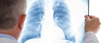
Pulmonologists diagnose streptococcal pneumonia after examining the patient, taking a medical history, analyzing the results of tests and examinations. The most informative examination method, which allows the doctor to determine the presence of pneumonia based on individual signs and judge the nature of the pathogen, is radiography in two projections or large-frame fluorography.
On an x-ray of the lungs, foci of inflammation appear as a local decrease in the airiness of the lung tissue of varying degrees of density and prevalence. In patients with streptococcal pneumonia, signs of focal, limited (polysegmental), subtotal and total darkening can be detected.
Foci of decreased airiness are characterized by infiltration of lung tissue. In severe cases of streptococcal pneumonia, necrosis of the lung tissue occurs, its disintegration, which on an x-ray image looks like heterogeneous infiltration with the formation of thick-walled cavities.
Pneumonia caused by pneumococcus (Streptococcus pneumoniae) is manifested by the following radiological signs:
- focal darkening (presence of small-sized infiltration of lung tissue);
- infiltration of an entire lobe, often involving the pleura in the process of inflammation (thickening, the presence of exudate in the pleural cavity);
- limited opacification syndrome, damage to several segments of the lobe.
In the case of pneumonia caused by pyogenic streptococcus, radiologists at the Yusupov Hospital determine on radiographs subtotal and total darkening of the lungs, cavities with or without a fluid level, thickening of the pleura and effusion into the pleural cavity.
If X-ray examination methods do not allow an accurate diagnosis to be made, doctors at the Yusupov Hospital perform computed tomography, which can reveal infiltration of the lung tissue and enlargement of the lymph nodes of the lung root. The procedure is used for the differential diagnosis of pneumonia and lung cancer or tuberculosis.
To determine the severity of the inflammatory process, doctors at the Yusupov Hospital prescribe a general and biochemical blood test. Streptococcal pneumonia is characterized by leukocytosis (a large number of leukocytes in the peripheral blood) with a shift in the leukocyte formula, an increase in the erythrocyte sedimentation rate. The amount of fibrinogen and the level of sialic acids increase, and C-reactive protein appears.
Bacteriological examination of sputum before prescribing antibiotics helps to detect pneumococcus pneumoniae and determine its sensitivity to antibiotics. The severity of the inflammatory process in the bronchi is determined using diagnostic bronchoscopy. If differential diagnosis is necessary, a lung biopsy is performed during the procedure to collect material for microscopic examination.
To determine the degree of involvement of other organs and systems in the pathological process, the following studies are carried out:
- electrocardiography – allows you to assess the condition of the myocardium;
- echocardiography - helps to detect pericardial effusion or bacterial colonies on the heart valves;
- study of external respiratory function indicators - allows you to assess the state of bronchial patency.
Streptococcus pneumoniae, DNA [real-time PCR]
Detection of the causative agent of streptococcal infection (Streptococcus pneumoniae), during which the genetic material (DNA) of pneumococcus is determined in a sputum sample using the polymerase chain reaction (PCR) method.
What is this analysis used for?
- For the diagnosis of infectious and infectious-allergic diseases of the respiratory system.
When is the test scheduled?
- For symptoms of typical pneumonia or exacerbation of chronic bronchitis.
Synonyms Russian
Pneumococcus, Weixelbaum diplococcus, Frenkel diplococcus.
English synonyms
Diplococcus pneumoniae.
Research method
Polymerase chain reaction (PCR).
What biomaterial can be used for research?
Sputum.
How to properly prepare for research?
It is recommended to drink a large amount of liquid (water) 8-12 hours before collecting sputum.
General information about the study
Streptococcus pneumoniae (pneumococcus) is a gram-positive diplococcus that is the main causative agent of community-acquired pneumonia. This microorganism can also cause meningitis and sepsis. There is evidence of the role of pneumococcus in the development and progression of bronchial asthma and chronic bronchitis.
People with a reduced immune status are most susceptible to streptococcal pneumonia and meningitis: children, the elderly, HIV-infected people, those suffering from alcoholism and drug addiction.
There are several laboratory methods for isolating pneumococcus: bacteriological (culture of sputum on nutrient media), sputum microscopy and detection of genetic material (DNA) of S. pneumoniae using the polymerase chain reaction method.
Quite often, a sputum sample turns out to be uninformative, and the result of its Gram stain is false negative. The result of the bacteriological method can be obtained only after a few days, which complicates diagnosis and delays the start of specific therapy. Unlike these methods, PCR has high sensitivity and specificity and allows you to obtain an accurate result as soon as possible after delivery of the sample to the laboratory. PCR does not require the presence of a large number of viable microorganisms in a biomaterial sample, since it is not the microorganism itself that is determined, but its genetic material. This advantage makes it possible to establish the etiology of the disease against the background of initiated antibacterial therapy. False positive results are also completely excluded.
Since S. pneumoniae is a representative of the normal microflora of the upper and lower respiratory tract of most people, the diagnosis of pneumococcal pneumonia requires not only the detection of this microorganism in sputum, but also the presence of additional clinical and laboratory signs of an infectious-inflammatory process in the respiratory organs. For example, S. pneumoniae, detected in the sputum of a patient without clinical signs of an infectious lung infection, without abnormalities in a blood test and without changes on an x-ray, should be considered as a normal representative of the microflora of the respiratory tract.
What is the research used for?
- For the diagnosis of infectious (pneumonia, chronic bronchitis) and infectious-allergic lung diseases.
When is the study scheduled?
In the presence of:
- symptoms of typical community-acquired pneumonia (fever, shortness of breath and chest pain, cough with orange-colored sputum (“rusty” sputum);
- symptoms of exacerbation of chronic bronchitis (cough with sputum against the background of increased body temperature, increased sweating, malaise).
What do the results mean?
Reference values: negative.
Positive result:
- pneumococcal pneumonia;
- exacerbation of chronic bronchitis.
Negative result:
- absence of pneumococcal infection;
- incorrect collection of biomaterial for research.
What can influence the result?
- The use of antibacterial drugs (cephalosporins (ceftriaxone), meropenem, macrolides (azithromycin), clindamycin, linezolid and streptogramins) before submitting the biomaterial for analysis can lead to a negative result.
- Most people are asymptomatic carriers of pneumococcus in the upper and lower respiratory tract, so the test result should be interpreted taking into account other clinical and laboratory signs of the disease.
Also recommended
- Complete blood count (without leukocyte formula and ESR)
- Leukocyte formula
- Erythrocyte sedimentation rate (ESR)
- Streptococcus pneumoniae, DNA [PCR]
- Total immunoglobulin E (IgE) in serum
Who orders the study?
General practitioner, pulmonologist, infectious disease specialist, pediatrician, allergist.
Literature
- Blaschke AJ. Interpreting assays for the detection of Streptococcus pneumoniae. Clin Infect Dis. 2011 May;52 Suppl 4:S331-7.
- Waterer G, Rello J. Why should we measure bacterial load when treating community-acquired pneumonia? Curr Opin Infect Dis. 2011 Apr;24(2):137-41.
- Korppi M. Bacterial infections and pediatric asthma. Immunol Allergy Clin North Am. 2010 Nov;30(4):565-74.
- Schwerk N, Brinkmann F, Soudah B, Kabesch M, Hansen G. Wheeze in preschool age is associated with pulmonary bacterial infection and resolves afterantibiotic therapy. PLoS One. 2011;6(11):e27913. Epub 2011 Nov 29.