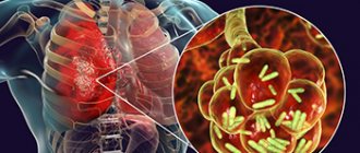
Pneumonia is an inflammatory disease of the lungs that occurs under the influence of various pathogens. Severe pneumonia develops when pneumonia is caused by bacterial-bacterial, bacterial-viral and bacterial-mycotic associations of microorganisms. Treatment of severe pneumonia in adults requires special approaches. Patients with severe pneumonia are hospitalized in the intensive care unit of the Yusupov Hospital.
Oxygen is supplied centrally to the wards. Resuscitation doctors constantly monitor the functioning of the respiratory and cardiovascular systems using cardiac monitors and determine the level of oxygen in the blood. All patients receive oxygen therapy. Patients with severe respiratory failure undergo artificial ventilation using stationary and portable devices.
The Yusupov Hospital employs candidates and doctors of medical sciences, doctors of the highest category.
Modern classification of community-acquired pneumonia:
- By pathogenesis:
a. Primary - pneumonia that developed as an independent disease
b. Secondary - pneumonia that develops against the background of another disease, such as influenza
- By localization:
a. Unilateral (left-sided and right-sided pneumonia)
b. Double-sided
- Based on the volume of tissues involved in inflammation, the following types of pulmonary pneumonia in adults and children are distinguished:
a. Focal - a small focus of inflammation (bronchopneumonia - with damage to the bronchi and respiratory sections)
b. Segmental - involving one or more segments of the lung
c. Lobar - with damage to the lobe of the lung (lobar pneumonia - with damage to the alveoli and part of the pleura)
d. Confluent - when lesions merge into larger ones
e. Total - damage to the entire lung
- Depending on the etiology and characteristics of the patient:
a.
Typical (in patients without severe immune disorders):- Bacterial
Viral pneumonia
- Fungal
- Mycobacterial
- Parasitic
b. In patients with severe immunocompromise :
- In patients with acquired immunodeficiency syndrome
- In patients with other diseases and pathological conditions
c. Aspiration pneumonia/lung abscess/pneumonia (one lung)
Obstructive pulmonary inflammation: causes of disease development
Obstructive pneumonia develops slowly over a long period of time. The disease is often preceded by an inflammatory process in the bronchi. Obstructive pulmonary inflammation can be caused by various factors:
- the entry of pathogenic microorganisms into the body is the most common cause;
- viral attack of the body;
- fungal infection;
- weakening of the immune system caused by stress, surgical interventions;
- various parasites;
- inhalation of toxic substances and large amounts of dust;
- active and passive smoking;
- hereditary predisposition;
- for bronchitis and bronchial asthma.
The list of factors causing the development of this disease is quite extensive, therefore, when diagnosing a patient with obstructive pneumonia, a pulmonologist seeks to establish its causes.
The Yusupov Hospital includes a therapy clinic, staffed by an experienced staff of therapists, pulmonologists and other specialists. When treating patients, they use modern and effective techniques that allow them to achieve significant results in treatment. Obstructive pneumonia occurs without complications if it is detected and treated in a timely manner, therefore, when the first signs appear, you should seek medical help.
Features of community-acquired pneumonia
Determining the pathogen is often a difficult task due to the difficulties in obtaining a substrate for diagnostics and often the lack of sufficient time for additional diagnostics. In addition, patients often develop a mixed infection.
The most common causative agent of bacterial infection (30–50% of cases) is the pneumococcus Streptococcus pneumoniae. The so-called atypical strains, undetectable by bacterioscopy and culture on conventional nutrient media, account for up to 30% of cases of the disease (Chlamydophila pneumoniae, Mycoplasma pneumoniae, Legionella pneumophila). 3–5% of cases of bacterial pneumonia are associated with rare pathogens (Haemophilus influenzae, Staphylococcus aureus, Klebsiella pneumoniae, other enterobacteria). Extremely rarely, community-acquired pneumonia can be caused by Pseudomonas aeruginosa (in the presence of bronchiectasis or in patients with cystic fibrosis).
Viral forms of CAP are caused by respiratory viruses: influenza virus, coronaviruses, rhinosyncytial virus, human metapneumovirus, human bocavirus. In most cases, respiratory viruses cause mild pneumonia with a self-limiting nature, however, in elderly and senile people, in the presence of concomitant bronchopulmonary, cardiovascular diseases or secondary immunodeficiency, the development of severe, life-threatening complications is possible.
Secondary bacterial pneumonia can be a complication of a primary viral infection of the lungs or an independent late complication of influenza.
How?
Irritation appears in places where toxic substances are deposited. Large grains (from 10 to 20 microns, or one to two hundredths of a millimeter) settle in the upper respiratory tract, smaller particles (from 5 to 10 microns) - in the trachea and bronchi, and “guests” less than 5 microns can reach the alveoli. After a foreign “guest” enters, damage (alteration) of epithelial cells occurs, which “helps” the development of bronchospasm. Then swelling of the bronchial mucosa with accumulation of exudate (purulent fluid) in the alveoli and so on until the epithelium is destroyed!
The amount of damage caused by damaging substances depends on the physical properties, concentration, exposure time, temperature and humidity, and the presence of pathogenic microbes.
Clinic
The main symptoms of pneumonia, which appear in both adults and children.
- Fever: occurs acutely, quickly reaches febrile levels
- Cough with copious discharge of purulent sputum; there may also be blood in the sputum (especially in the case of lobar pneumonia)
- Chest pain associated with breathing
- Shortness of breath, feeling of lack of air
- Manifestations of intoxication: severe weakness, fatigue, sweating, nausea, vomiting
- In the case of “atypical” pneumonia (Mycoplasma pneumoniae, Legionella pneumophila, Chlamydia pneumoniae, Pneumocystis jirovecii), there may be a gradual onset with a dry, unproductive cough, not without common symptoms - pain and sore throat, weakness, malaise, myalgia, headaches, pain in the abdomen - with minimal changes on the x-ray
- Elderly patients often have more severe general symptoms: drowsiness, confusion, anxiety, sleep disturbances, loss of appetite, nausea, vomiting, signs of exacerbation/decompensation of chronic diseases
Laboratory and instrumental diagnostics
Leukopenia below 3x10⁹/l or leukocytosis above 25x109/l is considered an unfavorable prognostic sign of septic complications.
The following changes are possible in the patient's blood test:
- Leukocytosis or leukopenia
- Neutrophil shift in leukocyte formula
- Increasing ESR
- Increased acute phase reactants (CRP, fibrinogen, procalcitonin test). Other changes in the biochemical blood test may indicate decompensation in other organs and systems
Determination of arterial blood gases/pulse oximetry is performed in patients with signs of respiratory failure, massive effusion, and the development of CAP due to chronic obstructive pulmonary disease (COPD). Decrease in PaO2 below 60 mm Hg. Art. is an unfavorable prognostic sign and indicates the need to admit the patient to the intensive care unit (ICU).
A bacterioscopy of the patient's sputum is carried out to identify the pathogen, and the sputum is cultured on nutrient media to determine sensitivity to antibiotics. For severe patients, it is recommended to collect venous blood for blood cultures (2 blood samples from 2 different veins, sample volume - at least 10 ml), PCR study, serological diagnostics to identify respiratory viruses, atypical pathogens of bacterial pneumonia.
X-ray examination of the lungs usually reveals focal infiltrative changes and pleural effusion. Chest CT may be a useful alternative to radiography in a number of situations:
- In the presence of obvious clinical symptoms of pneumonia and no changes on the radiograph
- In cases where, during examination of a patient with suspected pneumonia, atypical changes are revealed (obstructive atelectasis, pulmonary infarction due to pulmonary embolism, lung abscess, etc.)
- Recurrent pneumonia, in which infiltrative changes occur in the same lobe (segment), or prolonged pneumonia, in which the duration of existence of infiltrative changes in the lung tissue exceeds 4 weeks
Fiberoptic bronchoscopy with a quantitative assessment of microbial contamination of the obtained material or other invasive diagnostics (transtracheal aspiration, transthoracic biopsy) is performed if pulmonary tuberculosis is suspected in the absence of a productive cough, as well as with bronchial obstruction by bronchogenic carcinoma or a foreign body
If the patient has pleural effusion and there are conditions for safe pleural puncture, it is necessary to conduct a study of the pleural fluid: counting leukocytes with a leukocyte formula; determination of pH, lactate dehydrogenase activity, protein content; Gram staining of smears; culture to identify aerobes, anaerobes and mycobacteria.
Pathogenesis
The infectious agent that causes TP enters the body through the bronchogenic route. But in some cases, drift into the lungs is observed from other organs, then pneumonia is formed against the background of infectious-toxic shock.
The introduction of microorganisms into the lower respiratory tract from the bronchi occurs when local protective immune factors (immunoglobulin A) are imperfect and the mucociliary apparatus is insufficient.
Bronchopulmonary dysplasia, obstructive disease, and chronic bronchopulmonary diseases contribute to the severe course of pneumonia and the development of a toxic form. The neonatal period and immunodeficiency also increase the risk of developing TP.
The pathomorphological picture can be different and is often determined by the type of pathogen.
There are three stages in the development of toxic pneumonia:
- Bacterial shock. Against the background of a massive release of toxins and a sharp production of biologically active substances, a narrowing and then a sharp expansion of the capillary network occurs. Against this background, there arises:
- redistribution of the liquid part of the blood;
- tissue ischemia;
- acidosis;
- DIC syndrome is formed;
- microthrombosis;
- decreased blood clotting.
- Exit from toxicosis. On the background:
- inhibition of the proliferation of microorganisms;
- reducing the production of toxins;
- the functioning of the blood coagulation system is normalized;
- vessels;
- hearts;
- kidney
- Repair. Happens:
- healing of damaged tissues;
- restoration of lost functions.
The first stage of toxic pneumonia is often accompanied by the development of complications: “shock lung”, renal and cardiovascular failure.
The basis of the pathogenesis of TP is respiratory failure, damage to blood vessels, heart, kidneys, and nervous system. Respiratory failure develops by three mechanisms (mixed):
- parenchymal (due to damage to the lung parenchyma by toxins and waste products of microbes);
- mechanical (clogging of the respiratory tract with mucus, narrowing of the bronchial lumen, insufficiency of mucociliary clearance);
- ventilation (inhibitory effect of toxins on the respiratory center).
Respiratory failure leads to hypoxia of tissues and organs and disruption of their functions. The length of time organs remain in a state of hypoperfusion (especially the brain and heart) determines the further prognosis. During hypoxia, the body tries to compensate for the lack of oxygen in various ways.
Treatment
It is fundamentally important to determine the patient’s management tactics - whether he should be hospitalized or whether treatment can be carried out on an outpatient basis. For these purposes, you can use the CURB-65/CRB-65 scale, which involves assessing a number of indicators (presence - 1 point, absence - 0):
C —impaired consciousness
U - blood urea nitrogen more than 7 mmol/l (not taken into account in CRB-65)
R — respiratory rate ≥ 30 per minute.
B - systolic blood pressure < 90 mm Hg or diastolic blood pressure < 60 mm Hg. Art.
65 - age 65 years and older
Explanation:
0 points - the patient belongs to group I (mortality 1.2%) and is treated on an outpatient basis.
1–2 points correspond to group II (mortality 8.15%), patients in this group are subject to hospitalization with observation and dynamic assessment of the condition.
3 points and above - patients are subject to emergency hospitalization; mortality in this group can reach 31%.
Management of patients with CAP in outpatient settings
For mild pneumonia, antibacterial treatment of adults and children can be completed with stable normalization of body temperature for 3–4 days (total course duration 7–10 days). If there is clinical/epidemiological evidence of mycoplasma or chlamydial infection, the duration of therapy should be 14 days. An initial assessment of the effectiveness of therapy should be carried out 48–72 hours after the end of the course of treatment (re-examination). If symptoms persist or progress, it is necessary to reconsider the tactics of antibacterial therapy and re-evaluate the advisability of hospitalization.
According to the recommendations of the Russian Respiratory Society, outpatient patients are divided into 2 groups, differing in etiological structure and tactics of antibacterial therapy (see Table 1. Antibacterial therapy of CAP in outpatients):
Table 1.
Antibacterial therapy of CAP in outpatients
| Group | Most common pathogens | Drugs of choice | Alternative drugs | Patient characteristics |
| Mild CAP in patients under 60 years of age without concomitant pathology | S. pneumoniae M. pneumoniae C. pneumoniae H. influenzae | Amoxicillin orally or macrolides (azithromycin, clarithromycin) orally | Respiratory fluoroquinolones (levofloxacin, moxifloxacin) orally | |
| Mild CAP in patients over 60 years of age and/or with signs of concomitant pathology | S. pneumoniae C. pneumoniae H. influenzae S. aureus Enterobacteriaceae | Amoxicillin + clavulanic acid or Amoxicillin + sulbactam orally | Respiratory fluoroquinolones (levofloxacin, moxifloxacin) orally | Concomitant diseases affecting etiology and prognosis: COPD, diabetes, congestive heart failure, liver cirrhosis, alcohol abuse, drug addiction, exhaustion |
Sources and reference materials
Click on the selected document to download:
| # | File | file size |
| 1 | Clinical protocol. Pneumonia in adults | 458 KB |
| 2 | Federal clinical recommendations for vaccine prevention of pneumococcal infection in adults | 199 KB |
| 3 | Community-acquired pneumonia in adults - practical recommendations for diagnosis, treatment and prevention (a manual for doctors). RRO, MACMAH | 715 KB |
| 4 | Clinical pharmacology of drugs for the treatment of respiratory diseases | 4 MB |
| 5 | Rational antimicrobial pharmacotherapy | 4 MB |
Management of patients with CAP in a hospital setting
It is advisable to begin therapy for pneumonia in hospitalized patients with parenteral antibiotics. After 3–4 days, provided the temperature normalizes, intoxication and other symptoms decrease, it is possible to switch to oral forms of antibiotics until the course of treatment is completed (step-down therapy). Antibacterial drugs can be used both as part of combination therapy and as monotherapy. More detailed treatment regimens for CAP are presented in Table No. 2.
Table 2.
Treatment regimens for CAP depending on the course
| Group | Most common pathogens | Recommended treatment regimens | |
| Drugs of choice | Alternative drugs | ||
| EP of non-severe course | S. pneumoniae H. influenzae C. pneumoniae S. aureus Enterobacteriaceae | Benzylpenicillin IV, IM ± macrolide (clarithromycin, azithromycin) orally, or Ampicillin IV, IM ± macrolide orally, or Amoxicillin + clavulanic acid IV ± macrolide orally, or Cefuroxime IV, IV m ± macrolide orally, or Cefotaxime IV, IM ± macrolide orally, or Ceftriaxone IV, IM ± macrolide orally | Respiratory fluoroquinolones (levofloxacin, moxifloxacin) IV Azithromycin IV |
| Severe EP | S. pneumoniae Legionella spp. S. aureus Enterobacteriaceae | Amoxicillin + clavulanic acid IV + macrolide IV, or Cefotaxime IV + macrolide IV, or Ceftriaxone IV + macrolide IV | Respiratory fluoroquinolones (levofloxacin, moxifloxacin) IV + III generation cephalosporins IV |
Note:
- If the presence of “atypical” pneumonia is suspected, the use of macrolides is indicated.
- If there is a risk of infection with P. aeruginosa (bronchiectasis, use of corticosteroids, therapy with broad-spectrum antibiotics for more than 7 days during the last month, exhaustion), the drugs of choice are ceftazidime, cefepime, cefoperazone + sulbactam, meropenem, imipenem, piperacillin/tazobactam, ciprofloxacin, in monotherapy or in combination with aminoglycosides of the II–III generation.
- If aspiration is suspected, the following treatment options are indicated: cefoperazone + sulbactam, ticarcillin + clavulanic acid, carbapenems, piperacillin/tazobactam.
An initial assessment of the effectiveness of antibacterial therapy should be carried out 24–48 hours after the start of treatment. If signs of pneumonia persist in adults or children, treatment tactics should be reconsidered.
For mild CAP, the course of antibiotic therapy is about 7–10 days. For severe CAP of unspecified etiology, a 10-day course of antibiotic therapy is recommended. If there is evidence of mycoplasma or chlamydial etiology, therapy should last 14 days; in the case of CAP caused by staphylococci, gram-negative enterobacteria, legionella, the duration of therapy is 14–21 days.
In addition to antibiotics, the following are used in the treatment of CAP:
- Antiviral therapy: neuraminidase inhibitors (oseltamivir, zanamivir), active against influenza A and B viruses. Duration of use is 5–10 days.
- Mucolytics and expectorants - tablet or inhalation forms.
- Glucocorticosteroids: used for developed septic shock (hydrocortisone 200–300 mg/day for no more than 7 days).
- Oxygen therapy for PaO2 < 55 mm Hg. Art. or SpO2 < 88% (breathing air). It is optimal to maintain SpO2 within 88–95% or PaO2 within 55–80 mm Hg. Art.
- Infusion therapy - to correct hypovolemia.
- Low molecular weight heparins for the prevention of thromboembolism in severe patients.
- Physiotherapy and exercise therapy to reduce inflammation and improve sputum discharge.


