UDC: 616.281-007:616.283.1-089.843 V.E. Kuzovkov, Yu.K. Yanov, S.V. Levin St. Petersburg Research Institute of Ear, Throat, Nose and Speech (Director – Honored Doctor of the Russian Federation, Prof. Yu.K. Yanov)
Cochlear implantation (CI) is currently generally recognized in world practice and the most promising direction for the rehabilitation of persons suffering from high-grade sensorineural hearing loss and deafness, with their subsequent integration into the hearing environment.
In modern literature, the issues of classification of anomalies of the development of the inner ear are widely covered, including in relation to CI, and surgical techniques for performing CI for this pathology are described. The world experience of CI in persons with developmental anomalies of the inner ear spans more than 10 years. At the same time, there are no works on this topic in the domestic literature. At the St. Petersburg Research Institute of Ear, Throat, Nose and Speech, for the first time in Russia, CI began to be performed on people with developmental anomalies of the inner ear. Three years of experience in such operations, the presence of successful results of such interventions, as well as an insufficient amount of literature on this issue, served as the reason for carrying out this work.
Classification of developmental anomalies of the inner ear. Current state of the issue.
With the advent of the late 80's - early 90's. high-resolution computed tomography (CT) and magnetic resonance imaging (MRI), these techniques have become widely used for diagnosing hereditary hearing loss and deafness, especially when determining indications for CI. Using these progressive and high-precision techniques, new anomalies were identified that did not fit into the existing classifications of F. Siebenmann [5] and K. Terrahe [7]. As a result, RK Jackler [1, 2] proposed a new classification, expanded and modified by N. Marangos [4] and L. Sennaroglu [5]. However, it should be noted that MRI in particular currently reveals such fine details that the detected malformations can be difficult to classify [3, 4].
In his classification of developmental anomalies of the inner ear, based on conventional radiography and the first CT data, RK Jackler [1] took into account the separate development of the vestibular semicircular and vestibular cochlear parts of a single system. The author suggested that various types of anomalies appear as a result of a delay or disruption of development at a certain stage of the latter. Thus, the types of malformations detected are correlated with the time of disruption. Later, the author recommended classifying combined anomalies as category A, and suggested a connection between such anomalies and the presence of an expanded aqueduct in the vestibule (Table 1).
Table 1 - Classification of developmental anomalies of the inner ear according to RKJackler
| Category A | Cochlear aplasia or malformation |
POSSIBLE presence of an expanded aqueduct of the vestibule | |
| Category B | Normal snail |
|
Thus, items 1–5 of categories A and B represent isolated developmental anomalies. Combined anomalies falling into both categories should be classified as category A in the presence of an expanded vestibular aqueduct. In accordance with RK Jackler, S. Kösling [6] made the statement that isolated anomalies represent not only a deformation of one structural unit of the inner ear, but can be combined with anomalies of the vestibule and semicircular canals, as well as with vestibular dysplasia and dilated aqueduct vestibule.
The N. Marangos classification [3] includes incomplete or aberrant development of the labyrinth (Table 2, point 5).
Table 2 - Classification of developmental anomalies of the inner ear according to N. Marangos
| Category | Subgroup |
| A = incomplete embryonic development |
|
| B = aberrant embryonic development |
|
| C = isolated hereditary anomalies | X-linked hearing loss |
| D | Anomalies in hereditary syndromes |
Thus, four categories (AD) of inner ear abnormalities are described. The author considers the aqueduct of the vestibule to be dilated if the interosseous distance in the middle part exceeds 2 mm, while other authors give a figure of 1.5 mm.
L. Sennaroglu [4] differentiates 5 main groups (Table 3): anomalies of the development of the cochlea, vestibule, semicircular canals, internal auditory canal and aqueduct of the vestibule or cochlea.
Table 3 - Main groups and configurations of cochleovestibular anomalies according to L. Sennaroglu
| Main groups | Configuration |
| Cochlear abnormalities | Michel anomaly / cochlear aplasia / common cavity / incomplete separation type I / cochlear hypoplasia / incomplete separation type II / normal cochlea |
| Vestibular abnormalities | Vestibule: absent/hypoplasic/widening (including Michel anomaly and common cavity) |
| Anomalies of the semicircular canals | Absence/hypoplasia/increased size |
| Anomalies of the internal auditory canal | Absent/narrow/extended |
| Anomalies of the aqueducts of the vestibule and cochlea | Advanced/Normal |
Cochlear malformations (Table 4) were divided by the author into six categories based on the degree of severity, depending on the time of disruption of the normal course of embryonic development. This classification of cochlear malformations includes an incomplete separation of types I and II.
Table 4 - Classification of cochlear anomalies according to the time of disruption of intrauterine development according to L. Sennaroglu
| Cochlear malformations | Description |
| Michel anomaly (3rd week) | Complete absence of cochleovestibular structures, often - aplastic internal auditory canal, most often - normal aqueduct of the vestibule |
| Cochlear aplasia (end of 3rd week) | The cochlea is absent, normal, dilated or hypoplastic vestibule, and the system of semicircular canals, often - dilated internal auditory canal, most often - normal aqueduct of the vestibule |
| General cavity (4th week) | The cochlea and vestibule are a single space without internal architecture, a normal or deformed system of semicircular canals, or its absence; the internal auditory canal is more often widened than narrowed; most often – normal aqueduct of the vestibule |
| Incomplete separation type II (5th week) | The cochlea is represented by a single cavity without internal architecture; expanded vestibule; most often – an enlarged internal auditory canal; absent, enlarged or normal system of semicircular canals; normal aqueduct of the vestibule |
| Cochlear hypoplasia (6th week) | Clear separation of cochlear and vestibular structures, cochlea in the form of a small bubble; absence or hypoplasia of the vestibule and semicircular canal system; narrowed or normal internal auditory canal; normal aqueduct of the vestibule |
| Incomplete separation, type II (Mondini anomaly) (7th week) | Cochlea with 1.5 whorls, cystically dilated middle and apical whorls; the size of the cochlea is close to normal; slightly expanded vestibule; normal system of semicircular canals, expanded aqueduct of the vestibule |
Taking into account the above modern ideas about the types of cochleovestibular disorders, we use the classifications of RK Jackler and L. Sennaroglu, as the most consistent with the findings encountered in our own practice.
Taking into account the small number of patients operated on, one case of successful CI for an inner ear anomaly is presented below.
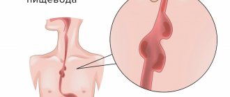
What is an ear?
The human ear consists of three main parts
: the outer ear, the middle ear and the inner ear.
ENT office
Diseases of the upper respiratory system and hearing organs are dealt with by an otorhinolaryngologist, otherwise an otolaryngologist, or an ENT doctor.
Find out when it's time to see a doctor in that unpronounceable specialty. The outer ear
can be seen in a mirror - it includes the pinna and the external auditory canal (1).
Its walls contain cells that produce earwax, which is designed to protect against dust and bacteria. The external auditory canal ends with the tympanic membrane
, located at an angle to it (2).
It, like a microphone membrane, transmits sound to the middle ear, which is located directly behind it - in the cranial cavity. Sound vibrations are amplified by the smallest bones of the human body - the malleus, incus and stirrup (4). The middle ear also contains the Eustachian tube
(3), which connects to the nasopharynx.
With its help, the pressure in the middle ear is equalized. Above the base of the eustachian tube is the inner ear
(5). Due to its shape resembling a snail shell, it is called a labyrinth. This fluid-filled formation provides the perception of sounds. Inside there is a canal, the walls of which are covered with receptors that capture vibrations of sound waves and transmit them to the auditory nerves.
Case from practice
In March 2007, the parents of patient K., born in 2005, came to the St. Petersburg Research Institute of ENT with complaints about the child’s lack of reaction to sounds and lack of speech. During the examination, a diagnosis was made: Chronic bilateral sensorineural hearing loss of IV degree, congenital etiology. Secondary receptive and expressive language disorder. Consequences of intrauterine cytomegalovirus infection, intrauterine damage to the central nervous system. Residual organic damage to the central nervous system. Left-sided spastic upper monoparesis. Aplasia of the first finger of the left hand. Hip dysplasia. Spasmodic torticollis. Pelvic dystopia of the hypoplastic right kidney. Delayed psychomotor development.
According to the conclusion of a child psychologist, the child’s cognitive abilities are within the age norm, intelligence is preserved.
The child received binaural hearing aids with heavy-duty hearing aids, without effect. According to the audiological examination, short-latency auditory evoked potentials were not recorded at a maximum signal level of 103 dB, and otoacoustic emissions were not recorded on both sides.
When conducting game audiometry in hearing aids, reactions to sounds with an intensity of 80-95 dB in the frequency range from 250 to 1000 Hz were revealed.
CT scan of the temporal bones revealed the presence of a bilateral anomaly of the cochlea in the form of incomplete division of type I (Table 4). Moreover, this statement is true for both the left and right ears, despite the seemingly different picture (Fig. 1).
After the examination, the patient underwent CI on the left ear using the classical approach through anthromastoidotomy and posterior tympanotomy, with the introduction of an electrode through a cochleostomy. For the operation, a special shortened electrode (Med-El, Austria) was used, having a working length of the active electrode of about 12 mm, specially designed for use in cases of anomaly or ossification of the cochlea.
Despite the intact auditory ossicles and stapedius muscle tendon, acoustic reflexes from the stapedius muscle were not recorded during the operation. However, when performing neural response telemetry, clear responses were obtained when 7 out of 12 electrodes were stimulated.
Postoperative transorbital radiography of the cochlea revealed that the active electrode of the implant is located in the common cavity (Fig. 4, arrow), taking the shape of an ideal circle.
During a control audiological examination one year after surgery, the patient was found to have reactions in the free sound field to sounds with an intensity of 15-20 dB in the frequency range from 250 to 4000 Hz. The patient’s speech is represented by one- and two-syllable words (“mom”, “give”, “drink”, “kitty”, etc.), a simple phrase of no more than two one- or two-syllable words. Considering that the patient’s age at the time of the re-examination was less than 3 years, the results of auditory-speech rehabilitation in this case should be considered excellent.
How does hearing work?
Sound is a wave that propagates in any elastic medium: water, air and various materials. The strength of sound vibrations is measured in decibels, and the frequency that a person perceives as the pitch of sound is measured in hertz. The human ear can perceive a limited range of the sound spectrum - from 20 Hz (very low bass) to 20 kHz. However, most adults are able to detect very high-pitched sounds around 16 kHz. When sound waves enter the ear canal, they hit the eardrum. It begins to vibrate, including the auditory ossicles in the process, which, in turn, transmit vibrations to the fluid of the inner ear. There they are perceived by hair cells, which translate the vibrations into electrical impulses transmitted by the auditory nerve to the brain.
Literature
- Jackler RK Congenital malformations of the inner ear: a classification based on embryogenesis//RK Jackler, WM Luxford, WF House/ Laryngoscope. – 1987. – Vol. 97, no. 1. – P. 1 – 14.
- Jackler RK The large vestibular aqueduct syndrome//RK Jackler, A. De La Cruz/ Laryngoscope. – 1989. – Vol. 99, No. 10. – P. 1238 – 1243.
- Marangos N. Dysplasien des Innenohres und inneren Gehörganges//N. Marangos/HNO. – 2002. – Vol. 50, no. 9. — P. 866 – 881.
- Sennaroglu L. A new classification for cochleovestibular malformations//L. Sennaroglu, I. Saatci/Laryngoscope. – 2002. – Vol. 112, No. 12. – P. 2230 – 2241.
- Siebenmann F. Grundzüge der Anatomie und Pathogenese der Taubstummheit// F. Siebenmann/Wiesbaden: JF Bergmann; 1904. – 76s.
- Stellenwert der MRT bei Verdacht auf Innenohrmissbildung//S. Kösling, S. Jüttemann, B. Amaya et al. / Fortschr Röntgenstr. – 2003. – Vol. 175, No. 11. – S. 1639 – 1646.
- Terrahe K. Missbildungen des Innen- und Mittelohres als Folge der halidomidembryopathie: Ergebnisse von Röntgenschichtuntersuchungen//K. Terrahe/Fortschr Röntgenstr. – 1965. – Vol. 102, No. 1. – P. 14.
The head of the department for the development and implementation of high-tech treatment methods is Doctor of Medical Sciences, Professor Anikin Igor Anatolyevich. Clinic phone number
Commission for the selection of patients for the provision of high-tech medical care Secretary of the commission - candidate of medical sciences Vladislav Evgenievich Kuzovkov, phone
Schedule of consultation appointments with clinic staff
What Causes Hearing Loss?
Partial or complete hearing loss can be caused by a variety of reasons. Congenital hearing loss
is one of the most common birth defects in humans.
It affects approximately one in 1,000 newborns. Hearing loss
also occurs as a result of ear injury, previous infections, or the natural aging process.
In addition, hearing loss
can occur as a result of exposure to sounds that are too loud and damage the hair cells in the inner ear. The longer the auditory analyzer is overloaded, the more pronounced the subsequent disturbances in its operation are. For example, ringing in the ears after an hour-long rock concert will go away by morning. However, prolonged exposure to loud sounds leads to permanent hearing damage.
In the maternity hospital after childbirth
During your stay in the maternity hospital, doctors examine the child, take blood for screening, check your hearing and give the first vaccinations. Find out more about how days go in the maternity hospital.
How to protect your hearing?
1. Limit your exposure to loud noises. Experts do not recommend exposing the hearing organs to sound loads above 80 dB
for more than two hours a day.
exposure to sound as high as 110 dB
to be dangerous to hearing. 2. Listen to “live” sounds. Try to be in nature more often, listen to soft music through speakers, and give up headphones for a while. This will allow the sensitive villi to recover from the loud sounds of the metropolis and constant wearing of headphones. 3. Contact an otolaryngologist for regular ear and hearing checks at least once a year. See your doctor immediately if you notice unexpected hearing loss, especially after a cold or injury to the ear and surrounding tissues.